

The neural correlates of texture perception: A systematic review and activation likelihood estimation meta-analysis of functional magnetic resonance imaging studies
- PMID: 37749852
- PMCID: PMC10636420
- DOI: 10.1002/brb3.3264
The neural correlates of texture perception: A systematic review and activation likelihood estimation meta-analysis of functional magnetic resonance imaging studies
Abstract
Introduction: Humans use discriminative touch to perceive texture through dynamic interactions with surfaces, activating low-threshold mechanoreceptors in the skin. It was largely assumed that texture was processed in primary somatosensory regions in the brain; however, imaging studies indicate heterogeneous patterns of brain activity associated with texture processing.
Methods: To address this, we conducted a coordinate-based activation likelihood estimation meta-analysis of 13 functional magnetic resonance imaging studies (comprising 15 experiments contributing 228 participants and 275 foci) selected by a systematic review.
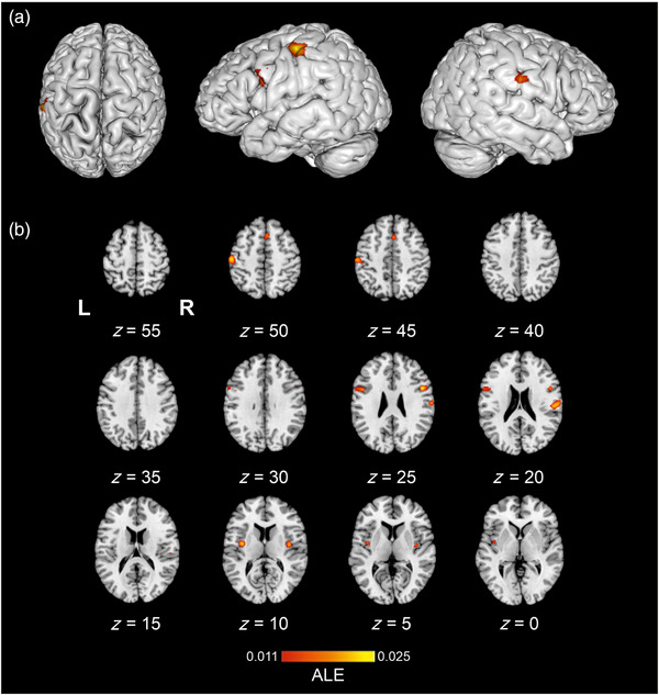
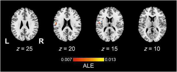
Results: Concordant activations for texture perception occurred in the left primary somatosensory and motor regions, with bilateral activations in the secondary somatosensory, posterior insula, and premotor and supplementary motor cortices. We also evaluated differences between studies that compared touch processing to non-haptic control (e.g., rest or visual control) or those that used haptic control (e.g., shape or orientation perception) to specifically investigate texture encoding. Studies employing a haptic control revealed concordance for texture processing only in the left secondary somatosensory cortex. Contrast analyses demonstrated greater concordance of activations in the left primary somatosensory regions and inferior parietal cortex for studies with a non-haptic control, compared to experiments accounting for other haptic aspects.
Conclusion: These findings suggest that texture processing may recruit higher order integrative structures, and the secondary somatosensory cortex may play a key role in encoding textural properties. The present study provides unique insight into the neural correlates of texture-related processing by assessing the influence of non-textural haptic elements and identifies opportunities for a future research design to understand the neural processing of texture.
Keywords: activation likelihood estimation meta-analysis; discriminative touch; functional magnetic resonance imaging; systematic review.
© 2023 The Authors. Brain and Behavior published by Wiley Periodicals LLC.
Conflict of interest statement
The authors declare no conflicts of interest.
Figures


Similar articles
-
Dual pathways for haptic and visual perception of spatial and texture information.Neuroimage. 2011 Jul 15;57(2):462-75. doi: 10.1016/j.neuroimage.2011.05.001. Epub 2011 May 7. Neuroimage. 2011. PMID: 21575727 Free PMC article.
-
Crossmodal interactions of haptic and visual texture information in early sensory cortex.Neuroimage. 2013 Jul 15;75:123-135. doi: 10.1016/j.neuroimage.2013.02.075. Epub 2013 Mar 16. Neuroimage. 2013. PMID: 23507388
-
ALE meta-analysis reveals dissociable networks for affective and discriminative aspects of touch.Hum Brain Mapp. 2016 Apr;37(4):1308-20. doi: 10.1002/hbm.23103. Epub 2016 Feb 1. Hum Brain Mapp. 2016. PMID: 26873519 Free PMC article.
-
[Human Brain Representations of Haptic and Visual Textures].Brain Nerve. 2015 Jun;67(6):691-700. doi: 10.11477/mf.1416200203. Brain Nerve. 2015. PMID: 26062584 Review. Japanese.
-
Shared and distinct functional networks for empathy and pain processing: a systematic review and meta-analysis of fMRI studies.Soc Cogn Affect Neurosci. 2020 Sep 24;15(7):709-723. doi: 10.1093/scan/nsaa090. Soc Cogn Affect Neurosci. 2020. PMID: 32608498 Free PMC article.
Cited by
-
Active touch in tactile perceptual discrimination: brain activity and behavioral responses to surface differences.Exp Brain Res. 2025 Mar 6;243(4):84. doi: 10.1007/s00221-025-07034-7. Exp Brain Res. 2025. PMID: 40047968 Free PMC article.
References
-
- Avanzini, P. , Abdollahi, R. O. , Sartori, I. , Caruana, F. , Pelliccia, V. , Casaceli, G. , Mai, R. , Lo Russo, G. , Rizzolatti, G. , & Orban, G. A. (2016). Four‐dimensional maps of the human somatosensory system. Proceedings of the National Academy of Sciences of the United States of America, 113(13), E1936–E1943. 10.1073/PNAS.1601889113/SUPPL_FILE/PNAS.1601889113.SM04.GIF - DOI - PMC - PubMed
Publication types
MeSH terms
LinkOut - more resources
Full Text Sources
