. 2023 Oct 18;67(10):e0035023.
doi: 10.1128/aac.00350-23. Epub 2023 Sep 26.

Exploring avibactam and relebactam inhibition of Klebsiella pneumoniae carbapenemase D179N variant: role of the Ω loop-held deacylation water

Affiliations

Exploring avibactam and relebactam inhibition of Klebsiella pneumoniae carbapenemase D179N variant: role of the Ω loop-held deacylation water

T A Alsenani et al. Antimicrob Agents Chemother. .

Abstract

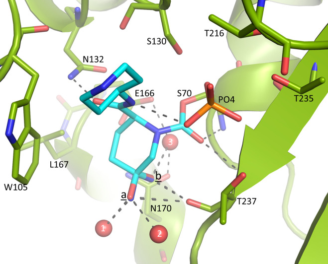
Klebsiella pneumoniae carbapenemase-2 (KPC-2) presents a clinical threat as this β-lactamase confers resistance to carbapenems. Recent variants of KPC-2 in clinical isolates contribute to concerning resistance phenotypes. Klebsiella pneumoniae expressing KPC-2 D179Y acquired resistance to the ceftazidime/avibactam combination affecting both the β-lactam and the β-lactamase inhibitor yet has lowered minimum inhibitory concentrations for all other β-lactams tested. Furthermore, Klebsiella pneumoniae expressing the KPC-2 D179N variant also manifested resistance to ceftazidime/avibactam yet retained its ability to confer resistance to carbapenems although significantly reduced. This structural study focuses on the inhibition of KPC-2 D179N by avibactam and relebactam and expands our previous analysis that examined ceftazidime resistance conferred by D179N and D179Y variants. Crystal structures of KPC-2 D179N soaked with avibactam and co-crystallized with relebactam were determined. The complex with avibactam reveals avibactam making several hydrogen bonds, including with the deacylation water held in place by Ω loop. These results could explain why the KPC-2 D179Y variant, which has a disordered Ω loop, has a decreased affinity for avibactam. The relebactam KPC-2 D179N complex revealed a new orientation of the diazabicyclooctane (DBO) intermediate with the scaffold piperidine ring rotated ~150° from the standard DBO orientation. The density shows relebactam to be desulfated and present as an imine-hydrolysis intermediate not previously observed. The tetrahedral imine moiety of relebactam interacts with the deacylation water. The rotated relebactam orientation and deacylation water interaction could potentially contribute to KPC-mediated DBO fragmentation. These results elucidate important differences that could aid in the design of novel β-lactamase inhibitors.

Keywords: antibiotic resistance; protein crystallography; β-lactamase; β-lactamase inhibitor.


Conflict of interest statement

The authors declare no conflict of interest.

Figures

Fig 1
Fig 1
The chemical structures of avibactam and relebactam. The unique R1 groups and atom numbering of the scaffold atoms of each inhibitor are indicated.
Fig 2
Fig 2
Differential scanning fluorimetry/thermal shift assay of WT KPC-2 and KPC-2 D179N in the presence or absence of 0.5 mM avibactam or 0.5 mM relebactam.
Fig 3
Fig 3
Avibactam complex crystal structure with KPC-2 D179N. (A) Avibactam (cyan-colored carbons) is covalently bound to S70 in the active site of KPC-2 D179N. The protein is colored green, and the Ω loop is colored yellow. Relevant active site residues are labeled with N179 in bold. (B) Unbiased |Fo|-|Fc| omit map contoured at 3.00 σ showing avibactam covalently bound (zoomed-in view rotated 90° from view in A). The map was obtained after removing avibactam from the model and subsequent 10 cycles of crystallographic refinement prior to the map calculation. (C) Interactions of avibactam in the active site of KPC-2 β-lactamase. Hydrogen bonds are represented by black dashed lines, and water molecules are shown in red spheres. Relevant active site residues are represented in the stick model and are labeled.
Fig 4
Fig 4
Superimposition of the avibactam-bound structures of KPC-2 D179N and WT KPC-2 β-lactamases. (A) Superimposition showing avibactam-bound active sites of WT KPC-2 (gray color; PDB accession number 4ZBE) (41) and KPC-2 D179N (green color) with avibactam with cyan-colored carbon atoms (avibactam in both structures shown in ball-and-stick). (B) The view is zoomed-out and rotated compared to panel (A) to include the Ω loop (shown in the stick model). The rotations of R164 away and residue D163 toward the site of the D179N mutation are indicated by the red and purple arrows, respectively.
Fig 5
Fig 5
Relebactam bound in the active site of KPC-2 D179N. (A) Unbiased omit |Fo|-|Fc| map is obtained after removing relebactam from the model; refinement prior to map calculation to remove phase bias and contour levels is as in Fig. 3. Relebactam is shown in a stick model with cyan carbon atoms; a nearby phosphate ion is labeled “PO4.” The alternate conformations for the oxygen and nitrogen atom substituents attached to the tetrahedral carbon 5 atom (labeled “5”) of the main piperidine ring are labeled “a” and “b.” (B) Same as panel (A) but the view is rotated ~90°.
Fig 6
Fig 6
Close-up view of relebactam interactions in the active site of KPC-2 D179N. Hydrogen bonds are represented by black dashed lines. Crystallographically observed water molecules interacting with relebactam are shown as red spheres (labeled “1,” “2,” and “3”); the interacting phosphate is labeled “PO4.”
Fig 7
Fig 7
Comparison of relebactam-bound WT KPC-2 (PDB ID: 6QW9) and KPC-2 D179N. (A) Superimposition of relebactam in the active site of WT KPC-2 [PDB ID: 6QW9 (46); in gray] and relebactam-bound KPC-2 D179N (green with relebactam shown with cyan carbon atoms). Relebactam is represented in ball-and-stick in both structures. Relebactam in the WT KPC-2 complex is observed as two intermediates, a sulfated acyl-intermediate (light gray carbon atoms) and a desulfated acyl-intermediate (dark gray carbon atoms) with occupancies of 0.65 and 0.35, respectively. The atom numbers for the main scaffold atoms of relebactam are shown in black and gray numbers for the KPC-2 D179N and WT KPC-2 complexes, respectively (atom numbering for the desulfated intermediate in WT KPC-2 is followed by an apostrophe). The two conformations of W105 in the WT KPC-2 complex and the oxygen and nitrogen atoms on the ring of relebactam in the KPC-2 D179N complex are indicated by labels “a” and “b.” The large reorientation of the piperidine ring of relebactam in the WT KPC-2 structure compared to the KPC-2 D179N complex is indicated by the red arrow showing the movement of the piperidine ring carbon “5” atom. The hydrogen bond made by the amide oxygen of relebactam with N132 in both complexes is indicated by a dashed line. The deacylation water held in place by E166 and N170 in the WT KPC-2 and KPC-2 D179N structures is labeled “wtW” and “W 3,” respectively. (B) Zoomed-out view to include the Ω-loop residues. The rotation of R164 away and residue D163 toward the site of the D179N mutation are indicated by the red and purple arrows, respectively.
Fig 8
Fig 8
Schematic of the relebactam inhibition pathway of KPC-2. The relebactam intermediate structure captured by protein crystallography is the imine hydrolysis intermediate. Note that the mass spectrometry accuracy is mentioned to be about 1 Da (43). The different possible states in the relebactam inhibition pathway are labeled A through G.

References

    1. Bush K, Bradford PA. 2019. Interplay between β-lactamases and new β-lactamase inhibitors. Nat Rev Microbiol 17:295–306. doi: 10.1038/s41579-019-0159-8 - DOI - PubMed
    1. van Heijenoort J. 2001. Formation of the glycan chains in the synthesis of bacterial peptidoglycan. Glycobiol 11:25R–36R. doi: 10.1093/glycob/11.3.25r - DOI - PubMed
    1. Bonomo RA, Burd EM, Conly J, Limbago BM, Poirel L, Segre JA, Westblade LF. 2018. Carbapenemase-producing organisms: a global scourge. Clin Infect Dis 66:1290–1297. doi: 10.1093/cid/cix893 - DOI - PMC - PubMed
    1. Doi Y, Iovleva A, Bonomo RA. 2017. The ecology of extended-spectrum beta-lactamases (ESBLs) in the developed world. J Travel Med 24:S44–S51. doi: 10.1093/jtm/taw102 - DOI - PMC - PubMed
    1. Shapiro AB. 2017. Kinetics of sulbactam hydrolysis by beta-lactamases, and kinetics of beta-lactamase inhibition by sulbactam. Antimicrob Agents Chemother 61:e01612-17. doi: 10.1128/AAC.01612-17 - DOI - PMC - PubMed