Review
. 2023 Dec;96(1152):20230407.
doi: 10.1259/bjr.20230407. Epub 2023 Oct 24.

Cardiac imaging with photon counting CT

Affiliations
Review

Cardiac imaging with photon counting CT

Thomas Flohr et al. Br J Radiol. 2023 Dec.

Abstract

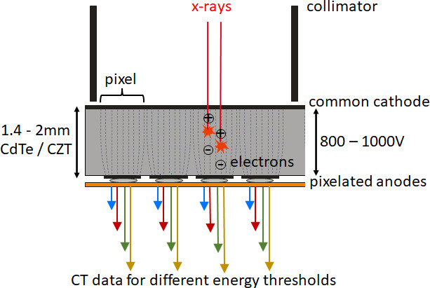
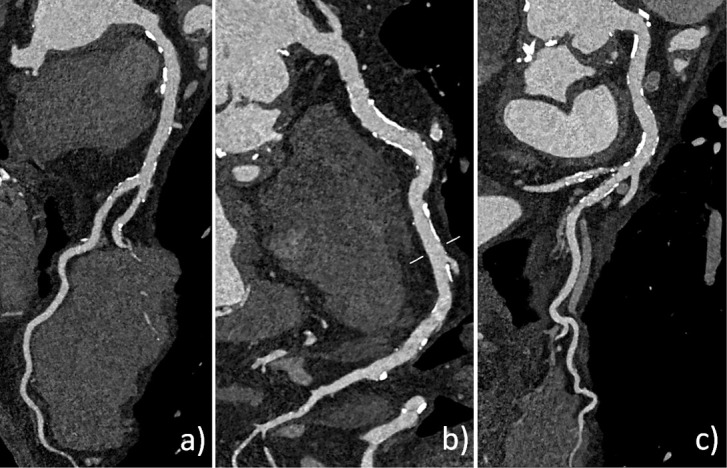
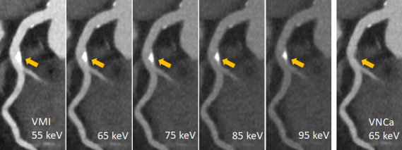
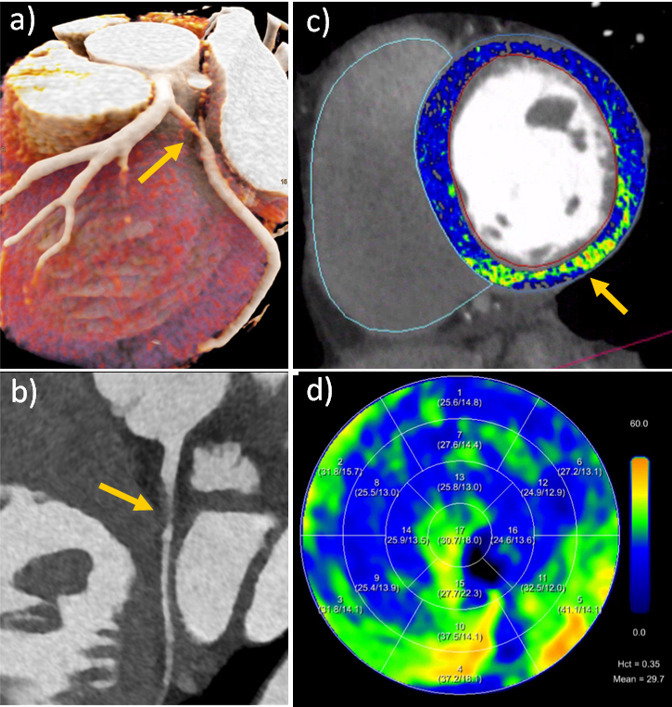
CT of the heart, in particular ECG-controlled coronary CT angiography (cCTA), has become clinical routine due to rapid technical progress with ever new generations of CT equipment. Recently, CT scanners with photon-counting detectors (PCD) have been introduced which have the potential to address some of the remaining challenges for cardiac CT, such as limited spatial resolution and lack of high-quality spectral data. In this review article, we briefly discuss the technical principles of photon-counting detector CT, and we give an overview on how the improved spatial resolution of photon-counting detector CT and the routine availability of spectral data can benefit cardiac applications. We focus on coronary artery calcium scoring, cCTA, and on the evaluation of the myocardium.


Figures

Figure 1.
Figure 1.
Schematic representation of a CdTe/CZT photon-counting detector. The detector pixels are formed by a strong electric field between the common cathode and the pixelated anodes (indicated by dashed lines) without demarcation by separating layers. In the detector structure shown here, there are four pixels between two collimator blades which serve to suppress scattered radiation. Four data streams per pixel (indicated by colored arrows) correspond to four different threshold energies for counting (and thus four different energy ranges of the registered X-rays). CdTe, cadmium telluride; CZT, cadmium zinc telluride.
Figure 2.
Figure 2.
Clinical example of CAC scoring with PCD-CT. (a) Non-contrast CAC scoring scan of a 73-year-old male patient (120 kVp, 70 keV, 3 mm slice, Qr36, 61 bpm). Agatston score 322. (b) VNI image from a contrast-enhanced scan (120 kVp, 70 keV, 3 mm slice, Qr36, 57 bpm). Agatston score 330. Note the reduced image noise in the VNI image because of the higher radiation dose of the contrast-enhanced scan. CAC, coronary artery calcium; PCD-CT, photon-counting detector CT; VNI, virtual non-iodine.
Figure 3.
Figure 3.
Ultra-high-resolution cCTA of an 82-year-old male patient on a clinical PCD-CT. Heart rate during data acquisition: 66 bpm. (a) Standard reference reconstruction (0.6 mm slice, Bv40, QIR4). (b) UHR reconstruction (0.2 mm slice, Bv56, QIR4). Note the improved visualization and larger perfused diameter (arrow) of the proximal LAD in the UHR image, which appears almost occluded in the standard reconstruction. cCTA, coronary CT angiography; PCD-CT, photon-counting detector CT.
Figure 4.
Figure 4.
Ultra-high-resolution cCTA of a 62-year-old male patient on a clinical PCD-CT, prior to transcatheter aortic valve replacement. Heart rate during data acquisition: 74 bpm. (a) Standard reference reconstruction (0.6 mm slice, Bv40, QIR4). A moderate stenosis (60% in diameter) is seen in the proximal RCA (arrow). (b) UHR reconstruction (0.2 mm slice, Bv60, QIR4). Note the reduced calcium blooming, leading to a re-classification to a mild stenosis (38% in diameter). (c) Invasive catheter coronary angiography confirmed a mild stenosis in the proximal RCA (arrow). cCTA, coronary CT angiography; PCD-CT, photon-counting detector CT.
Figure 5.
Figure 5.
Ultra-high-resolution cCTA of a 68-year-old male patient on a clinical PCD-CT (retrospective ECG-gating). Heart rate during data acquisition: 72 bpm. UHR reconstruction (0.2 mm slice, Bv60, QIR4). (a, b and c) curved multiplanar reformations of the LAD, RCA, and CX allow reliable exclusion of significant coronary stenosis despite a very high CAC score of 5220. CAC, coronary artery calcium; cCTA, coronary CT angiography; PCD-CT, photon-counting detector CT.
Figure 6.
Figure 6.
Spectral cCTA of a 73-year-old patient on a clinical PCD-CT (0.6 mm slice, Qr40, QIR3, 55bpm). Calcified plaque in the LAD (arrow). VMIs at 55, 65, 75, 85 and 95 keV: note the visually perceived reduction of Ca-blooming with higher keV at the expense of reduced iodine contrast. VNCa at 65 keV: the calcified plaque is removed from the contrast filled vessel without reducing the iodine contrast. cCTA, coronary CT angiography; PCD-CT, photon-counting detector CT.
Figure 7.
Figure 7.
Spectral cCTA and ECV-evaluation of a 46-year-old female patient on a clinical PCD-CT (144 × 0.4 mm, 0.6 mm slice thickness, Bv40, QIR 3, 57 bpm). (a, and b) Cinematic rendering and curved planar reformation from cCTA show a spontaneous dissection of the proximal CX artery (arrow). (c) Overlayed color map from late enhancement shows a focal area of increased ECV-fraction (arrow) in the inferior wall, corresponding to the CX dissection in this left dominant coronary system. (d) Polarmap of the ECV-distribution. cCTA, coronary CT angiography; ECV, extracellular volume; PCD-CT, photon-counting detector CT.

References

    1. von Ballmoos MW, Haring B, Juillerat P, Alkadhi H. Meta-analysis: diagnostic performance of low-radiation-dose coronary computed tomography angiography. Ann Intern Med 2011; 154: 413–20. doi: 10.7326/0003-4819-154-6-201103150-00007 - DOI - PubMed
    1. Seitun S, Clemente A, Maffei E, Toia P, La Grutta L, Cademartiri F. Prognostic value of cardiac CT. Radiol Med 2020; 125: 1135–47. doi: 10.1007/s11547-020-01285-w - DOI - PubMed
    1. Kwan AC, Pourmorteza A, Stutman D, Bluemke DA, Lima JAC. Next-generation hardware advances in CT: cardiac applications. Radiology 2021; 298: 3–17. doi: 10.1148/radiol.2020192791 - DOI - PMC - PubMed
    1. Dodd JD, Leipsic JA. Evolving developments in cardiac CT. Radiology 2023; 307(): e222827. doi: 10.1148/radiol.222827 - DOI - PubMed
    1. Serruys PW, Hara H, Garg S, Kawashima H, Nørgaard BL, Dweck MR, et al. . Coronary computed Tomographic angiography for complete assessment of coronary artery disease: JACC state-of-the-art review. J Am Coll Cardiol 2021; 78: 713–36. doi: 10.1016/j.jacc.2021.06.019 - DOI - PubMed