. 2023 Nov 8;8(21):e165974.
doi: 10.1172/jci.insight.165974.

Dimethyl fumarate modulates the dystrophic disease program following short-term treatment

Affiliations

Dimethyl fumarate modulates the dystrophic disease program following short-term treatment

Cara A Timpani et al. JCI Insight. .

Abstract

New medicines are urgently required to treat the fatal neuromuscular disease Duchenne muscular dystrophy (DMD). Dimethyl fumarate (DMF) is a potent immunomodulatory small molecule nuclear erythroid 2-related factor 2 activator with current clinical utility in the treatment of multiple sclerosis and psoriasis that could be effective for DMD and rapidly translatable. Here, we tested 2 weeks of daily 100 mg/kg DMF versus 5 mg/kg standard-care prednisone (PRED) treatment in juvenile mdx mice with early symptomatic DMD. Both drugs modulated seed genes driving the DMD disease program and improved force production in fast-twitch muscle. However, only DMF showed pro-mitochondrial effects, protected contracting muscles from fatigue, improved histopathology, and augmented clinically compatible muscle function tests. DMF may be a more selective modulator of the DMD disease program than PRED, warranting follow-up longitudinal studies to evaluate disease-modifying impact.

Keywords: Drug therapy; Muscle Biology; Neuromuscular disease; Skeletal muscle; Therapeutics.


Figures

Figure 1
Figure 1. Mechanisms of action of DMF.
DMF is rapidly converted to bioactive monomethyl fumarate (MMF) in the gut and circulated to tissues. Inside cells, MMF is converted to fumarate, which binds kelch-like ECH-associated protein 1 (Keap1), resulting in dissociation of the Keap1-Nrf2 complex. Keap1 represses Nrf2 activity by targeting the complex for degradation by the ubiquitin proteosome. Once dissociated from Keap1, DJ-1 chaperones Nrf2 into the nucleus, where Nrf2 binds the antioxidant response element (ARE), initiating transcription of antioxidant genes superoxide dismutase 1 (SOD1), NAD(P)H dehydrogenase:quinone oxidoreductase (NQO1), catalase (CAT), and hemoxygenase-1 (HO-1). Meanwhile, Keap1 is sequestered by p62, which initiates autophagy and amplifies Nrf2-mediated ARE transcription. Fumarate also inhibits master inflammation regulator, nuclear factor κB (NF-κB), which suppresses nuclear binding of κB and transcription of pro-inflammatory cytokines. MMF also inhibits NF-κB via agonism of the hydroxycarboxylic acid receptor 2 (HCAR2) and antagonism of Toll-like receptors (TLRs) on the membrane. Fumarate causes metabolic shifts by inhibiting glyceraldehyde-3-phosphate dehydrogenase (GAPDH) activity, and therefore, glycolysis. Fumarate enters mitochondria via the malate-aspartate shuttle, where it is ultimately sequestered into the matrix Krebs cycle and is completely metabolized to yield ATP and CO2.
Figure 2
Figure 2. DMF improves muscle function but not DMD blood biomarkers.
(A) Schematic of the treatment period and clinically compatible testing protocol beginning at 14 and concluding at 28 days of age. Mice were treated daily via oral gavage with vehicle (0.5% methylcellulose; VEH), 100 mg/kg DMF, or 5 mg/kg prednisone (PRED) and underwent grip strength and blood biomarker testing at the experimental endpoint at 28 days of age. (B) Forelimb, (C) whole-body grip strength, (D) plasma creatine kinase (CK), and (E) oxidized albumin levels were assessed. Data are mean ± SEM and n are indicated by individual data points. Statistical significance was tested by 2-way (genotype and DMF treatment) and 1-way (mdx treatment) ANOVA. Treatment effect: *P < 0.05, ****P < 0.0001; genotype effect: ###P < 0.001, ####P < 0.0001.
Figure 3
Figure 3. DMF activates Nrf2 and induces the phase II antioxidant response in mdx skeletal muscle.
Protein expression of (A) Nrf2, (B) NAD(P)H dehydrogenase:quinone oxidoreductase (NQO1), (C) superoxide dismutase 1 (SOD1), (D) hemeoxygenase-1 (HO-1), (I) kelch-like ECH-associated protein 1 (Keap1), and (F) sequestosome 1 (p62) was quantitated via Western blot. (G and H) The muscle inflammatory response was assessed by quantitative real-time polymerase chain reaction (qRT-PCR) gene array. (I) Phosphorylated nuclear factor-κB (NF-κB) and total NF-κB protein and (JO) CD68-positive (CD68+) macrophages. (P) Gene signatures of M1 and M2 macrophages were extrapolated from gene array data presented in H. Data in G, H, and K are based on log2 fold-change from WT (for mdx VEH) and mdx VEH (for mdx DMF and PRED) derived from n = 4/group where each n is pooled mRNA for n = 2 mice. Statistical significance was tested by 1-way ANOVA. H heatmap was partially published previously under CC BY license (73). Data presented in AF, I, and J are mean ± SEM, and n are indicated by individual data points. Statistical significance was tested by 2-way (genotype and DMF treatment) and 1-way (mdx treatment) ANOVA. Treatment effect: *P < 0.05, ****P < 0.0001; genotype effect: #P < 0.05, ####P < 0.0001. (KO) Scale bar = 20 mm.
Figure 4
Figure 4. DMF recovers force and reduces the fatigability of type II mdx EDL muscles.
Specific force was measured ex vivo in (A) EDL and (B) SOL, and (C and D) the force-frequency relationship was determined for each. Fatigue and recovery properties were quantitated for (E) EDL and (F) SOL. Data are presented as mean ± SEM and n are indicated by individual data points unless otherwise stated. Panel C n are WT VEH = 8, WT DMF = 8, mdx VEH = 11, mdx DMF = 8, mdx PRED = 7; panel D n are WT VEH = 8, WT DMF = 6, mdx VEH = 8, mdx DMF = 9, mdx PRED = 6; panel E n are WT VEH = 7, WT DMF = 7, mdx VEH = 11, mdx DMF = 8, mdx PRED = 6; panel F n are WT VEH = 7, WT DMF = 6, mdx VEH = 9, mdx DMF = 6, mdx PRED = 6. For data in panels A and B, statistical significance was tested by 2-way (genotype and DMF treatment) and 1-way (mdx treatment) ANOVA. In panels CF, statistical significance was tested by repeated measures multivariate analysis. Treatment effect: *P < 0.05, **P < 0.01, ***P < 0.001; genotype effect: ##P < 0.01, ####P < 0.0001.
Figure 5
Figure 5. DMF enhances mitochondrial respiratory function in mdx FDB fibers.
(A) Oxygen consumption rate was measured using Seahorse extracellular flux and chemical inhibitors and uncouplers of mitochondrial respiration. (B) Basal, (C) ATP-linked, (D) maximal, and (E) nonmitochondrial respiration in mdx fibers. (F) Metabolic phenotypes in response to chemical uncoupling, (G) coupling efficiency, (H) SRC, and (I) citrate synthase (CS) activity are also shown. (J) Succinate dehydrogenase (SDH) capacity was used to estimate fiber type shifts (KO) and (PT) representative images are shown. Data presented in AJ are mean ± SEM and n are indicated by individual data points unless otherwise stated. Data presented in KO are mean percentage fiber SDH density across 3 bins. Panel A and F n: WT VEH = 13, WT DMF = 7, mdx VEH = 9, mdx DMF = 7, mdx PRED = 7. Panel KO n: WT VEH = 9, WT DMF = 4, mdx VEH = 4, mdx DMF = 5, mdx PRED = 6. Statistical significance in BJ was tested by 2-way (genotype and DMF treatment) and 1-way (mdx treatment) ANOVA. Treatment effect: *P < 0.05, **P < 0.01, ****P < 0.0001; genotype effect: ###P < 0.001. (PT) Scale bar = 50 mm.
Figure 6
Figure 6. DMF improves biomarkers of muscle pathology.
(AL) EBD permeation into muscle fibers, a biomarker of compromised sarcolemma integrity, was assessed in mdx TA, SOL, EDL, and DIA, respectively. (M and N) An array of extracellular matrix genes were assessed alongside histological indicators of muscle (O) liposis and (PU) fibrosis (collagen deposition). (V) Protein expression of PPARγ, an inducer of adipogenesis, and (W) gene expression of Ctnnb1, a repressor of the adipogenesis gene program and Tgfb1, a regulator of the fibrosis gene program, are shown. Data in W represent a callout from N. Data in M, N, and W are based on log2 fold-change from WT (for mdx VEH) and mdx VEH (for mdx DMF and PRED) derived from n = 4/group where each n is equivalent to pooled mRNA for n = 2 mice. Statistically significant dysregulated genes (panel W) were tested by 1-way ANOVA. For all other panels, data are mean ± SEM and n are indicated by individual data points. Statistical significance was tested by 2-way (genotype and DMF treatment) and 1-way (mdx treatment) ANOVA. Treatment effect: *P < 0.05, **P < 0.01, ***P < 0.001; genotype effect: #P < 0.05, ##P < 0.01, ###P < 0.001, ####P < 0.0001. Panel BJ scale bar = 50 mm; panel QU scale bar = 20 mm.
Figure 7
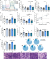
Figure 7. DMF improves mdx muscle histopathology.
TA architecture was assessed using hematoxylin and eosin staining. The (A) healthy and (B) unhealthy tissue as well as (C) the unhealthy to healthy tissue ratio and (D) percentage regenerating centronucleated fibers are shown. Representative images of (E) WT VEH, (F) WT DMF, (G) mdx VEH, (H) mdx DMF, and (I) mdx PRED TA muscles are provided where arrow pointers indicate regenerating centronucleated fibers. Statistical significance was tested by 2-way (genotype and DMF treatment) and 1-way (mdx treatment) ANOVA. Treatment effect: *P < 0.05, **P < 0.01, ***P < 0.001, ****P < 0.0001; genotype effect: ####P < 0.0001. (EI) Scale bar = 50 mm.

References

    1. Ismail HM, et al. The potential and benefits of repurposing existing drugs to treat rare muscular dystrophies. Expert Opin Orphan Drugs. 2018;6(4):259–271. doi: 10.1080/21678707.2018.1452733. - DOI
    1. Duan D, et al. Duchenne muscular dystrophy. Nat Rev Dis Primers. 2021;7(1):13. doi: 10.1038/s41572-021-00248-3. - DOI - PMC - PubMed
    1. Passamano L, et al. Improvement of survival in Duchenne muscular dystrophy: retrospective analysis of 835 patients. Acta Myol. 2012;31(2):121–125. - PMC - PubMed
    1. Wahlgren L, et al. One in five patients with Duchenne muscular dystrophy dies from other causes than cardiac or respiratory failure. Eur J Epidemiol. 2022;37(2):147–156. doi: 10.1007/s10654-021-00819-4. - DOI - PMC - PubMed
    1. Bach JR, et al. Duchenne muscular dystrophy: the effect of glucocorticoids on ventilator use and ambulation. Am J Phys Med Rehabil. 2010;89(8):620–624. doi: 10.1097/PHM.0b013e3181e72207. - DOI - PubMed

Publication types