. 2023 Nov 15;133(22):e169200.
doi: 10.1172/JCI169200.

The catalytic subunit of DNA-PK regulates transcription and splicing of AR in advanced prostate cancer

Affiliations

The catalytic subunit of DNA-PK regulates transcription and splicing of AR in advanced prostate cancer

Beth Adamson et al. J Clin Invest. .

Abstract

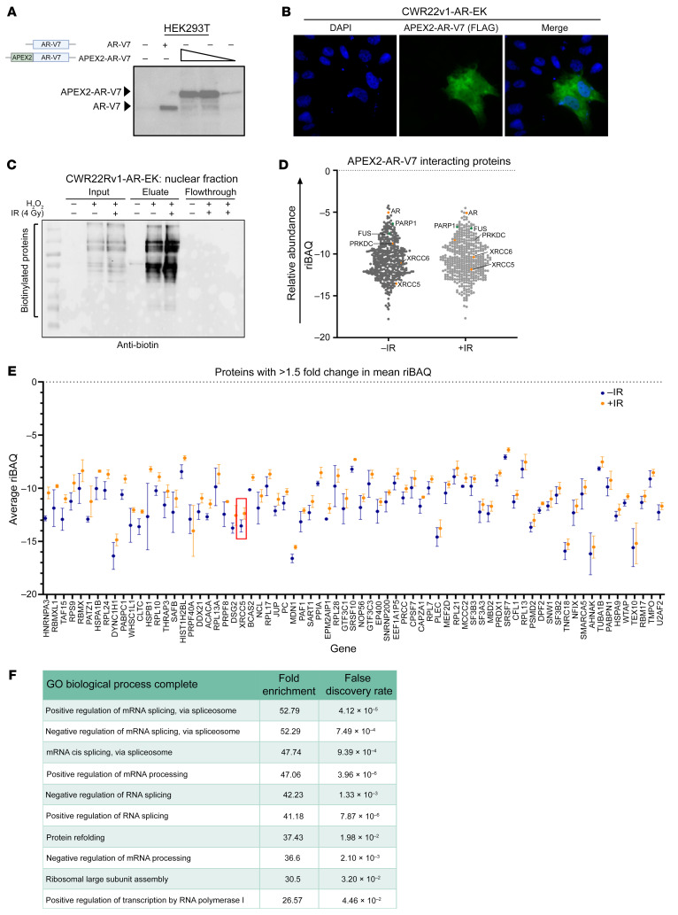
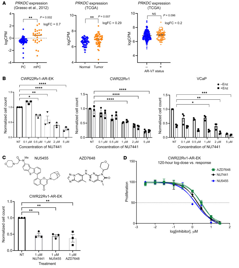
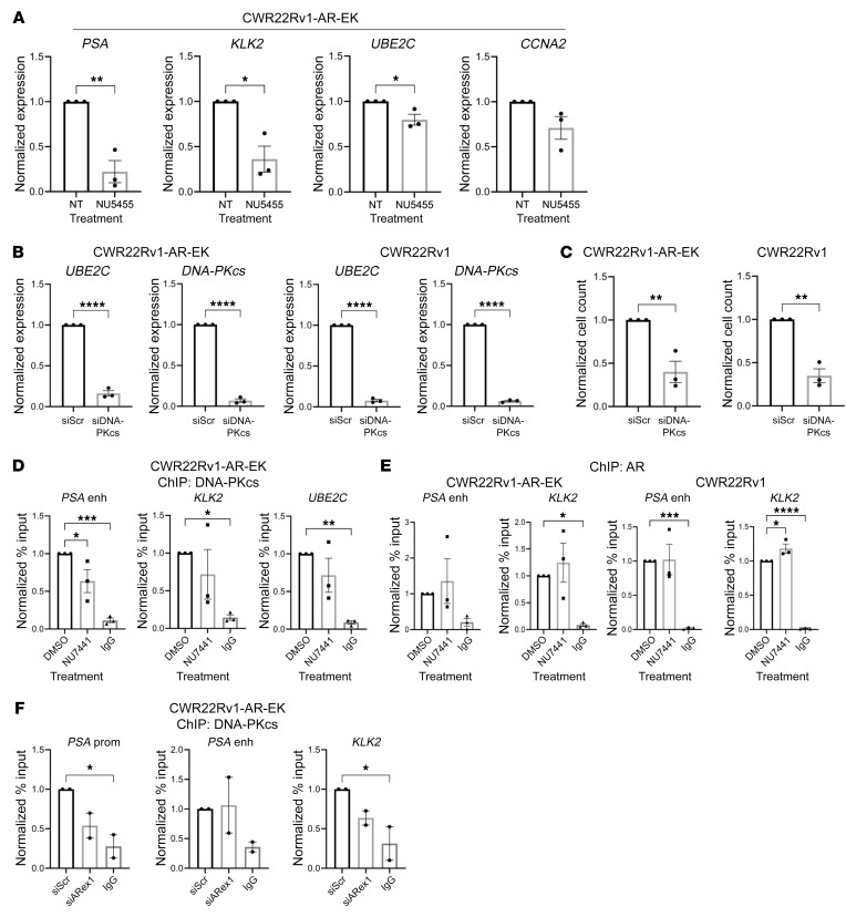
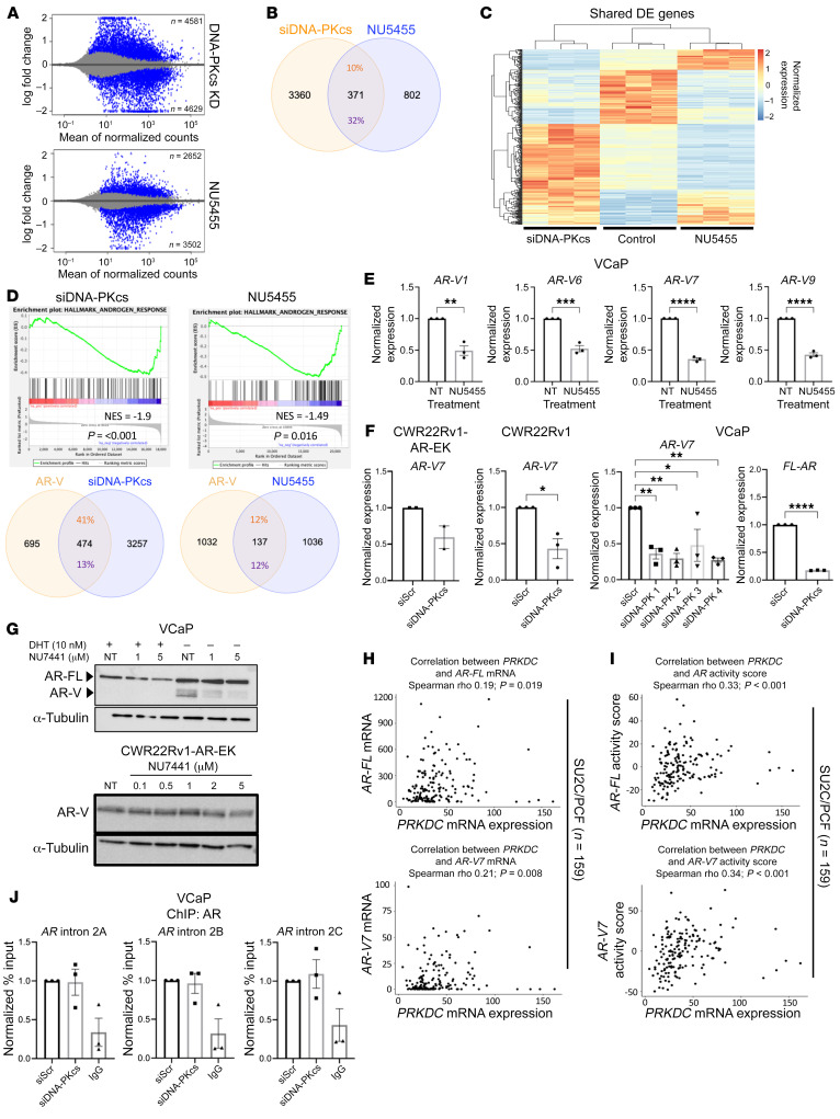
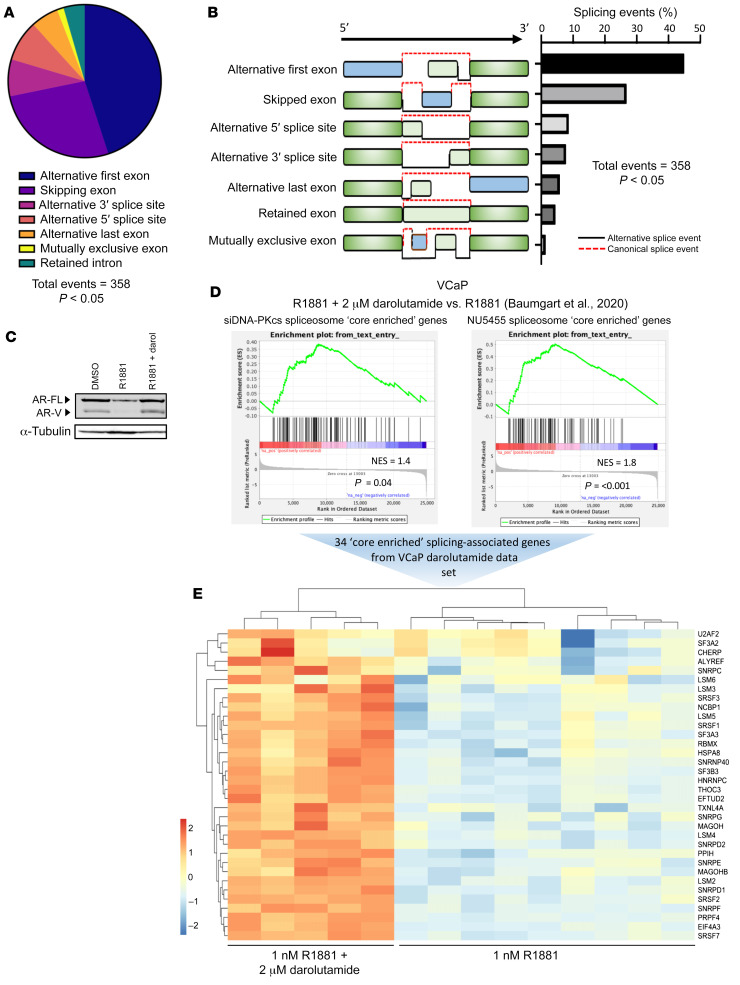
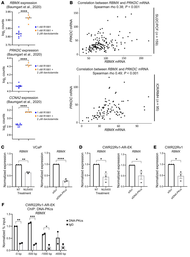
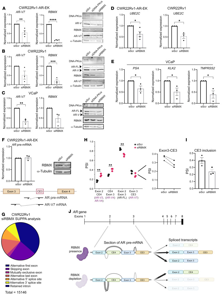
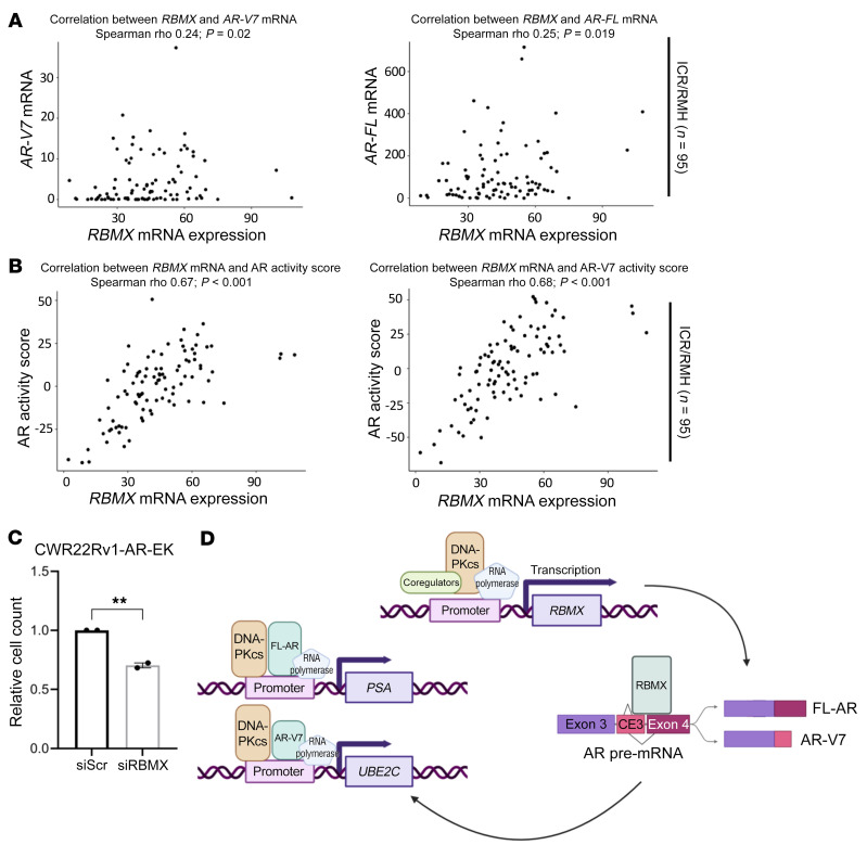
Aberrant androgen receptor (AR) signaling drives prostate cancer (PC), and it is a key therapeutic target. Although initially effective, the generation of alternatively spliced AR variants (AR-Vs) compromises efficacy of treatments. In contrast to full-length AR (AR-FL), AR-Vs constitutively activate androgenic signaling and are refractory to the current repertoire of AR-targeting therapies, which together drive disease progression. There is an unmet clinical need, therefore, to develop more durable PC therapies that can attenuate AR-V function. Exploiting the requirement of coregulatory proteins for AR-V function has the capacity to furnish tractable routes for attenuating persistent oncogenic AR signaling in advanced PC. DNA-PKcs regulates AR-FL transcriptional activity and is upregulated in both early and advanced PC. We hypothesized that DNA-PKcs is critical for AR-V function. Using a proximity biotinylation approach, we demonstrated that the DNA-PK holoenzyme is part of the AR-V7 interactome and is a key regulator of AR-V-mediated transcription and cell growth in models of advanced PC. Crucially, we provide evidence that DNA-PKcs controls global splicing and, via RBMX, regulates the maturation of AR-V and AR-FL transcripts. Ultimately, our data indicate that targeting DNA-PKcs attenuates AR-V signaling and provide evidence that DNA-PKcs blockade is an effective therapeutic option in advanced AR-V-positive patients with PC.

Keywords: Endocrinology; Oncology; Prostate cancer.


Conflict of interest statement

Conflict of interest: AS is an employee of The Institute for Cancer Research (ICR), which has a commercial interest in abiraterone, PARP inhibition in DNA repair–defective cancers, and PI3K/AKT pathway inhibitors (with no financial interest). AS has received travel support from Sanofi, Roche-Genentech, and Nurix and speaker honoraria from Astellas Pharma and Merck Sharp & Dohme. He has served as an advisor to DE Shaw Research and CHARM Therapeutics. AS has been the chief investigator/principal investigator of industry-sponsored clinical trials. JSDB has served on advisory boards and received fees from many companies, including Amgen, AstraZeneca, Astellas, Bayer, Bioxcel Therapeutics, Boehringer Ingelheim, Cellcentric, Daiichi, Eisai, Genentech/Roche, Genmab, GSK, Harpoon, ImCheck Therapeutics, Janssen, Merck Serono, Merck Sharp & Dohme, Menarini/Silicon Biosystems, Orion, Pfizer, Qiagen, Sanofi Aventis, Sierra Oncology, Taiho, Terumo, and Vertex Pharmaceuticals. He is an employee of The ICR, which has received funding or other support for his research work from AstraZeneca, Astellas, Bayer, Cellcentric, Daiichi, Genentech, Genmab, GSK, Janssen, Merck Serono, MSD, Menarini/Silicon Biosystems, Orion, Sanofi Aventis, Sierra Oncology, Taiho, Pfizer, and Vertex; The ICR has a commercial interest in abiraterone, PARP inhibition in DNA repair–defective cancers, and PI3K/AKT pathway inhibitors (with no financial interest). JSDB was named as an inventor, with no financial interest for patent 8,822,438 (Methods and Compositions for Treating Cancer), submitted by Janssen, which covers the use of abiraterone acetate with corticosteroids. He has been the chief investigator/principal investigator of many industry-sponsored clinical trials.

Figures

Figure 1
Figure 1. AR-V7 proximal biotinylation experiments identify known AR-V7 interactors and the DNA-PKcs holoenzyme.
(A) Diagrammatic representation of APEX2-AR-V7 construct and anti-AR Western blot of HEK293T cells transiently transfected with either AR-V7 or increasing quantities of FLAG-APEX2-AR-V7 constructs. (B) 1 × 10⁵ CWR22Rv1-AR-EK cells were transfected with 2 μg of a FLAG-tagged APEX2-AR-V7 construct for 48 hours and again for an additional 24 hours prior to immunofluorescence using an anti-FLAG antibody. Magnification ×40. (C) 5 × 10⁶ CWR22Rv1-AR-EK cells were transfected with 10 μg pLV-FLAG-APEX2-AR-V7 and again 48 hours later prior to treatment with biotin-phenol and with or without IR (4 Gy) for 2 hours. In the –IR and +IR arms, H2O2 was added to cells to induce the labeling reaction. Cells were then quenched and harvested, and the cytoplasmic and nuclear fractions were isolated and quantified. 10 μg resultant nuclear lysate was analyzed by Western blotting using an HRP-linked anti-biotin antibody. Corresponding Ponceau Red stain is shown to indicate equal sample loading. (D) Plot of mean riBAQ scores of all APEX2-AR-V7–interacting proteins identified by mass spectrometry. AR and components of the DNA-PK holoenzyme are highlighted in orange, and two known AR interactors, PARP1 and FUS, are highlighted in green. (E) APEX2-AR-V7–interacting proteins that have a riBAQ score >1.5-fold in response to irradiation. Ku80 (XRCC5) is highlighted in a red box. Data points represent the mean of 2–3 replicates (depending on if the protein is identified in 2 or 3 replicates) ± SEM. (F) Top 10 biological processes that are enriched in the list of proteins that are more abundant AR-V7 interactors in response to irradiation.
Figure 2
Figure 2. DNA-PKcs inhibition represses growth of AR-V–expressing PC cell lines.
(A) The TCGA data set was analyzed to compare PRKDC expression in matched normal and tumor samples (n = 51) and in localized (n = 49) and metastatic (n = 27) PC from a publicly available microarray data set (Grasso et al., ref. 10). **P < 0.01. (B) CWR22Rv1-AR-EK cells grown in serum-containing media and CWR22Rv1 and VCaP cells grown in steroid-depleted media supplemented with 10 M enzalutamide (Enz) were treated with increasing concentrations of NU7441 for 96 hours prior to cell count. Data were normalized to the untreated (NT) control arm (–Enz/–DHT group) and are representative of 3 independent repeats ± SEM. One-way ANOVA using Bonferroni’s post hoc analysis was used to determine the statistical significance for CWR22Rv1-AR-EK and 2-way ANOVA was used for CWR22Rv1, LNCaP, and VCaP cells. *P < 0.05, **P < 0.01, ***P < 0.001. (C) Representative structures of DNA-PKcs inhibitors NU5455 and AZD7648 are shown adjacent to cell count data from CWR22Rv1-AR-EK cells treated with 1 mM NU7441, NU5455, and AZD7648 for 24 hours. Data represent an average of 3 repeats ± SEM (*P < 0.05, **P < 0.01). (D) CWR22Rv1-AR-EK cells were treated with increasing concentrations of AZD7648, NU7441, and NU5455 for 120 hours before harvesting for an SRB proliferation assay. Data are shown as the mean ± SEM across 3 independent repeats that included 3 technical replicates for each experimental arm.
Figure 3
Figure 3. DNA-PKcs is a transcriptional coregulator of AR-Vs.
(A) CWR22Rv1-AR-EK cells were cultured in serum-containing media for 48 hours and then treated with 1 μM NU5455 for 24 hours prior to qRT-PCR. Data were normalized to the DMSO treatment arm for each target gene and are representative of 3 independent repeats ± SEM. One-way ANOVA using Bonferroni’s post hoc analysis was used to determine the statistical significance. *P < 0.05, **P < 0.01. (B) CWR22Rv1-AR-EK and CWR22Rv1 cells were transfected with either siScr or siDNA-PK for 72 hours prior to qRT-PCR. Data represent the mean of 3 repeats ± SEM. An unpaired 2-tailed t test was used to determine statistical significance. ***P < 0.001, ****P < 0.0001. (C) CWR22Rv1-AR-EK cells cultured in serum-containing media and CWR22Rv1 cells cultured in steroid-depleted conditions were transfected with either siScr or siDNA-PKcs for 96 hours prior to cell count. Data are representative of 3 independent repeats ± SEM. An unpaired 2-tailed t test was used to determine the statistical significance. **P < 0.01. (D and E) CWR22Rv1-AR-EK cells grown in serum-containing media were treated with 1 μM NU7441 prior to ChIP using (D) anti-DNA-PKcs, (E) anti-AR, and isotype control (IgG) antibodies. ChIP-qPCR readouts represent the normalized percentage input to the control of 3 independent experiments incorporating 1-way ANOVA using Bonferroni’s post hoc analysis to determine the statistical significance. *P < 0.05, **P < 0.01, ***P < 0.001, ****P < 0.0001. (F) CWR22Rv1-AR-EK cells grown in serum-containing media were transfected with either scrambled control (siScr) or AR exon 1-targeting (siARex1) siRNAs and incubated for 72 hours before ChIP using DNA-PKcs and isotype control (IgG) antibodies. Data shown represent the normalized percentage input to the control and represents 2 independent repeats. One-way ANOVA using Bonferroni’s post hoc analysis was used to determine the statistical significance. *P < 0.05.
Figure 4
Figure 4. DNA-PKcs blockade and knockdown markedly affects the AR-V transcriptome.
(A) MA plot (log fold change [M] versus mean of normalised counts [A]) showing the number of up- and downregulated genes in response to DNA-PKcs knockdown and inhibition with NU5455 (blue represents statistically significant differentially expressed genes [DEGs], P adjusted < 0.05). (B) Venn diagram indicating the percentage overlap of DEGs (P < 0.05, fold change ± 1.5) between DNA-PKcs depletion (siDNA-PKcs) and inhibition (NU5455). (C) Heatmap of overlapping DEGs between DNA-PKcs knockdown and inhibition compared with control. (D) Unfiltered DEG lists from NU5455 and siDNA-PKcs treatment were compared with the “androgen response hallmark” gene lists using GSEA. Venn diagrams show the percentage overlap between AR-V transcriptome (Kounatidou et al., ref. 16) and DNA-PKcs knockdown or inhibition DEGs. (E) VCaP cells were treated for 24 hours with 1 μM NU5455 with and without DHT before RT-qPCR analysis. Data represent the mean of 3 repeats ± SEM. An unpaired 2-tailed t test was used to determine the statistical significance. *P < 0.05, **P < 0.01, ***P < 0.001, ****P < 0.0001. (F) Cells were transfected with oligonucleotides targeting DNA-PKcs or a scrambled (siScr) control for 72 hours prior to RT-QPCR, as in E. (G) AR immunoblotting of VCaP and CWR22Rv1-AR-EK cells grown in increasing doses of NU7441 or vehicle control (NT) for 24 hours. (H and I) Association of DNA-PKcs (PRKDC) mRNA levels with (H) AR and AR-V7 mRNA levels and (I) AR and AR-V7 activity scores in SU2C/PCF (n = 159) CRPC transcriptomes. r and P values were calculated using Spearman’s correlation. (J) VCaP cells grown in steroid-depleted media were transfected with DNA-PKcs–targeting (siDNA-PKcs) or control scrambled siRNA (siScr) for 72 hours prior ChIP using AR or isotype control (IgG) antibodies. Data shown represent the normalized fold enrichment to siScr control and are the mean of 3 independent repeats.
Figure 5
Figure 5. DNA-PKcs regulates a splicing-associated gene signature.
(A) RNA-Seq data derived from CWR22Rv1-AR-EK cells depleted of DNA-PKcs was analyzed for differential splicing activity using SUPPA2 inbuilt statistical test (ref. 65). Events that passed a P value cut off of <0.05 were plotted in the pie chart. (B) Diagrammatic representation and quantification of the statistically significant splicing alterations detected in response to DNA-PKcs depletion, as determined using SUPPA2 inbuilt statistical test (ref 65). (C) Upregulation of AR-V7 in response to darolutamide was validated by Western blotting using an anti-AR-V7 antibody. (D) Differentially expressed splicing-associated genes from DNA-PKcs depleted and NU5455-treated cells were analyzed by GSEA using a darolutamide-responsive gene set (Baumgart et al., ref. 54) to identify splicing factor expression correlating with AR-V7 synthesis. (E) 34 splicing-associated genes were found to be upregulated in response to darolutamide and are shown in the heatmap.
Figure 6
Figure 6. RBMX is a bona fide DNA-PKcs–regulated gene.
(A) Graphical representations of RBMX, DNA-PKcs (PRKDC), and CCNA2 expression from Baumgart et al. (54). Statistics were determined with limma and GEO2R. ****P < 0.0001. (B) Association of DNA-PKcs (PRKDC) mRNA levels with RBMX mRNA levels in SU2C/PCF (n = 159) and ICR/RMH (n = 95) CRPC transcriptomes. r and P values are shown and were calculated using Spearman’s correlation. (C) VCaP, (D) CWR22Rv1-AR-EK, and (E) CWR22Rv1 cells were either reverse transfected with siScr/siDNA-PKcs for 72 hours or treated for 24 hours with 1 μM DNA-PKcs inhibitors (NU5455) prior to RT-qPCR analysis of RBMX expression. An unpaired 2-tailed t test was used to determine the statistical significance from 3 independent experiments. *P < 0.05, **P < 0.01, ****P < 0.0001. (F) CWR22Rv1-AR-EK cells grown in serum-containing media were subject to ChIP using either DNA-PKcs or isotype control (IgG) antibodies prior to qPCR analysis to assess DNA-PKcs enrichment at and upstream of the RBMX transcriptional start site (0, –500, –1,000, and –4,000 bp) Data shown represent the normalized percentage input to the DNA-PKcs ChIP at the –0 bp site and represent 2 independent repeats. Two-way ANOVA using Šídák’s multiple comparisons test was used to determine the statistical significance. *P < 0.05, **P < 0.01, ***P < 0.001.
Figure 7
Figure 7. RBMX regulates AR-V synthesis in prostate cancer.
(A) CWR22Rv1-AR-EK, (B) CWR22Rv1, and (C) VCaP cells grown in serum-containing and steroid-depleted media, respectively, were transfected with RBMX (siRBMX) or scrambled control (siScr) siRNAs for 72 hours prior to AR-V7 and RBMX transcript analysis using RT-qPCR. Data represent the mean of 3 repeats ± SEM. An unpaired 2-tailed t test was used to determine the statistical significance. *P < 0.05, **P < 0.01, ***P < 0.001, ****P < 0.0001. In parallel, AR, AR-V7, DNA-PKcs, and RBMX protein levels were analyzed by Western blot in cells depleted of DNA-PKcs and RBMX for 72 hours. (D) CWR22Rv1-AR-EK and CWR22Rv1 and (E) VCaP cells were depleted of RBMX, as in AC, and canonical AR-V–target gene expression was analyzed by qRT-PCR. Data represent the mean of 3 repeats ± SEM. An unpaired 2-tailed t test was used to determine the statistical significance. *P < 0.05. (F) CWR22Rv1-AR-EK cells depleted of RBMX for 72 hours were subject to qRT-PCR analysis to assess unspliced, pre-mRNA AR transcript abundance compared with scrambled siRNA (siScr) control. Representative Western analysis is shown to demonstrate successful RBMX knockdown. (G) RNA-Seq data derived from CWR22Rv1 cells depleted of RBMX was analyzed for differential splicing activity using SUPPA2. Events that passed a P value cut off of < 0.05 were plotted in the pie chart. (H and I) Altered exon composition of distinct AR transcripts as calculated by investigating relative exon inclusion (PSI) for all junctions measured using (H) hisat2 and (I) SUPPA2. (J) Diagrammatic representation of exon inclusion dynamics across exon 2 to cryptic exon 3 (CE3) in steady-state and in response to RBMX knockdown.
Figure 8
Figure 8. Expression of RBMX correlates with AR isoforms and activity in CRPC.
(A) Association of RBMX mRNA with AR-V7 and AR-FL mRNA levels and (B) activity scores in the ICR/RMH (n = 95) CRPC transcriptomes. r and P values are shown and were calculated using Spearman’s correlation. (C) CWR22Rv1-AR-EK cells cultured in serum-containing media were transfected with either siRBMX or siScr for 72 hours prior to cell counts analysis. Data are representative of 2 independent repeats ± SEM. An unpaired 2-tailed t test was used to determine the statistical significance. **P < 0.01. (D) Proposed mechanism of DNA-PKcs modulated AR/AR-V splicing and transcriptional activity. In normal conditions, DNA-PKcs is involved in the transcription of the RNA binding protein RBMX, which is directly involved in splicing FL-AR and multiple AR-V mRNA transcripts, including AR-V7. Resultant FL-AR and AR-V7 protein interacts with and is subsequently coactivated by DNA-PKcs to facilitate expression of canonical AR target genes.

References

    1. Westaby D, et al. Beyond the androgen receptor: the sequence, the mutants, and new avengers in the treatment of castrate-resistant metastatic prostate cancer. Am Soc Clin Oncol Educ Book. 2021;41:e190–e202. doi: 10.1200/EDBK_321209. - DOI - PubMed
    1. Obinata D, et al. Recent discoveries in the androgen receptor pathway in castration-resistant prostate cancer. Front Oncol. 2020;10:581515. doi: 10.3389/fonc.2020.581515. - DOI - PMC - PubMed
    1. Messner EA, et al. The androgen receptor in prostate cancer: effect of structure, ligands and spliced variants on therapy. Biomedicines. 2020;8(10):422. doi: 10.3390/biomedicines8100422. - DOI - PMC - PubMed
    1. Nadal M, et al. Structure of the homodimeric androgen receptor ligand-binding domain. Nat Commun. 2017;8:14388. doi: 10.1038/ncomms14388. - DOI - PMC - PubMed
    1. Einstein DJ, et al. Targeting the androgen receptor and overcoming resistance in prostate cancer. Curr Opin Oncol. 2019;31(3):175–182. doi: 10.1097/CCO.0000000000000520. - DOI - PMC - PubMed

Publication types