

Plasminogen Activator Inhibitor 1 Is a Novel Faecal Biomarker for Monitoring Disease Activity and Therapeutic Response in Inflammatory Bowel Diseases
- PMID: 37751311
- PMCID: PMC10906952
- DOI: 10.1093/ecco-jcc/jjad160
Plasminogen Activator Inhibitor 1 Is a Novel Faecal Biomarker for Monitoring Disease Activity and Therapeutic Response in Inflammatory Bowel Diseases
Abstract
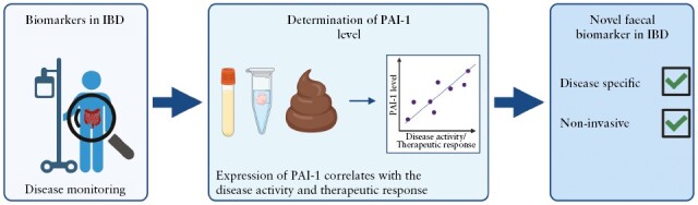
Background and aims: Crohn's disease [CD] and ulcerative colitis [UC] require lifelong treatment and patient monitoring. Current biomarkers have several limitations; therefore, there is an unmet need to identify novel biomarkers in inflammatory bowel disease [IBD]. Previously, the role of plasminogen activator inhibitor 1 [PAI-1] was established in the pathogenesis of IBD and suggested as a potential biomarker. Therefore, we aimed to comprehensively analyse the selectivity of PAI-1 in IBD, its correlation with disease activity, and its potential to predict therapeutic response.
Methods: Blood, colon biopsy, organoid cultures [OC], and faecal samples were used from active and inactive IBD patients and control subjects. Serpin E1 gene expressions and PAI-1 protein levels and localisation in serum, biopsy, and faecal samples were evaluated by qRT-PCR, ELISA, and immunostaining, respectively.
Results: The study population comprised 132 IBD patients [56 CD and 76 UC] and 40 non-IBD patients. We demonstrated that the serum, mucosal, and faecal PAI-1 concentrations are elevated in IBD patients, showing clinical and endoscopic activity. In responders [decrease of eMayo ≥3 in UC; or SES-CD 50% in CD], the initial PAI-1 level decreased significantly upon successful therapy. OCs derived from active IBD patients produced higher concentrations of PAI-1 than the controls, suggesting that epithelial cells could be a source of PAI-1. Moreover, faecal PAI-1 selectively increases in active IBD but not in other organic gastrointestinal diseases.
Conclusions: The serum, mucosal, and faecal PAI-1 concentration correlates with disease activity and therapeutic response in IBD, suggesting that PAI-1 could be used as a novel, non-invasive, disease-specific, faecal biomarker in patient follow-up.
Keywords: Inflammatory bowel disease; PAI-1; faecal marker.
© The Author(s) 2023. Published by Oxford University Press on behalf of European Crohn’s and Colitis Organisation.
Conflict of interest statement
BJ is a scientist at Ladon Therapeutics. JM is a chief scientific officer of Ladon Therapeutics and external consultant of Tavanta Therapeutics. KF has received speaker’s honoraria from AbbVie, Janssen, Ferring, Takeda, and Goodwill Pharma. TM has received speaker’s honoraria from MSD, AbbVie, Egis, Goodwill Pharma, Takeda, Pfizer, Teva, Janssen, and Swixx.
Figures


Similar articles
-
Faecal volatile organic compounds differ according to inflammatory bowel disease sub-type, severity, and response to treatment in paediatric patients.United European Gastroenterol J. 2024 Jul;12(6):780-792. doi: 10.1002/ueg2.12603. Epub 2024 Jun 22. United European Gastroenterol J. 2024. PMID: 38922802 Free PMC article.
-
Faecal Myeloperoxidase as a Biomarker of Endoscopic Activity in Inflammatory Bowel Disease.J Crohns Colitis. 2022 Dec 5;16(12):1862-1873. doi: 10.1093/ecco-jcc/jjac098. J Crohns Colitis. 2022. PMID: 35803583 Free PMC article.
-
Are faecal markers good indicators of mucosal healing in inflammatory bowel disease?World J Gastroenterol. 2015 Oct 28;21(40):11469-80. doi: 10.3748/wjg.v21.i40.11469. World J Gastroenterol. 2015. PMID: 26523111 Free PMC article. Review.
-
Investigation of faecal volatile organic metabolites as novel diagnostic biomarkers in inflammatory bowel disease.Aliment Pharmacol Ther. 2016 Mar;43(5):596-611. doi: 10.1111/apt.13522. Epub 2016 Jan 25. Aliment Pharmacol Ther. 2016. PMID: 26806034
-
Conventional therapy for moderate to severe inflammatory bowel disease: A systematic literature review.World J Gastroenterol. 2019 Mar 7;25(9):1142-1157. doi: 10.3748/wjg.v25.i9.1142. World J Gastroenterol. 2019. PMID: 30863001 Free PMC article.
Cited by
-
In vitro neutralization of IL-6 receptor exacerbates damage to intestinal epithelial cells during Mycobacterium avium paratuberculosis infection.Front Immunol. 2024 Aug 7;15:1412800. doi: 10.3389/fimmu.2024.1412800. eCollection 2024. Front Immunol. 2024. PMID: 39170608 Free PMC article.
-
Pathophysiological Links Between Inflammatory Bowel Disease and Cardiovascular Disease: The Role of Dysbiosis and Emerging Biomarkers.Biomedicines. 2025 Jul 31;13(8):1864. doi: 10.3390/biomedicines13081864. Biomedicines. 2025. PMID: 40868118 Free PMC article. Review.
-
Serum regenerative islet-derived protein 1α: a novel and sensitive biomarker for endoscopic disease activity in ulcerative colitis.Ann Med. 2025 Dec;57(1):2496404. doi: 10.1080/07853890.2025.2496404. Epub 2025 Apr 28. Ann Med. 2025. PMID: 40289639 Free PMC article.
-
MCT4 inhibition attenuates inflammatory response to Mycobacterium avium paratuberculosis infection and restores intestinal epithelial integrity in vitro.Front Immunol. 2025 Apr 14;16:1562100. doi: 10.3389/fimmu.2025.1562100. eCollection 2025. Front Immunol. 2025. PMID: 40297589 Free PMC article.
References
MeSH terms
Substances
Grants and funding
LinkOut - more resources
Full Text Sources
Medical
Miscellaneous
