. 2023 Sep 26;18(9):e0291977.
doi: 10.1371/journal.pone.0291977. eCollection 2023.

Identification of the functional domain of the dense core vesicle biogenesis factor HID-1

Affiliations

Identification of the functional domain of the dense core vesicle biogenesis factor HID-1

Blake H Hummer et al. PLoS One. .

Abstract

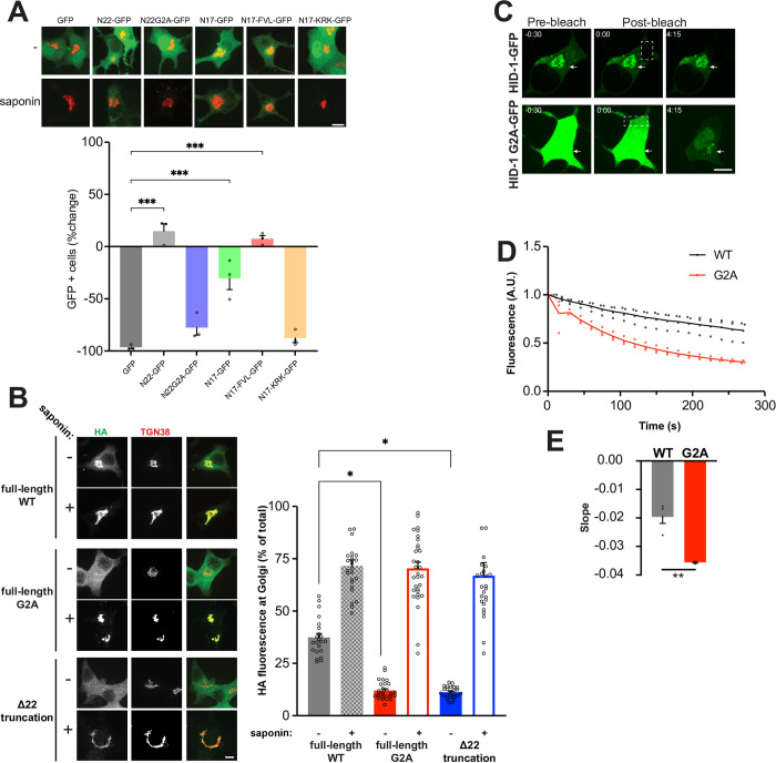
Large dense core vesicles (LDCVs) mediate the regulated release of neuropeptides and peptide hormones. HID-1 is a trans-Golgi network (TGN) localized peripheral membrane protein contributing to LDCV formation. There is no information about HID-1 structure or domain architecture, and thus it remains unknown how HID-1 binds to the TGN and performs its function. We report that the N-terminus of HID-1 mediates membrane binding through a myristoyl group with a polybasic amino acid patch but lacks specificity for the TGN. In addition, we show that the C-terminus serves as the functional domain. Indeed, this isolated domain, when tethered to the TGN, can rescue the neuroendocrine secretion and sorting defects observed in HID-1 KO cells. Finally, we report that a point mutation within that domain, identified in patients with endocrine and neurological deficits, leads to loss of function.


Conflict of interest statement

The authors declare that the research was conducted in the absence of any commercial or financial relationships that could be construed as a potential conflict of interest. This does not alter our adherence to PLOS ONE policies on sharing data and materials.

Figures

Fig 1
Fig 1. HID-1 N-terminus mediates membrane binding but is not strictly required for TGN localization.
(A) INS-1 cells were co-transfected with sialyltransferase-RFP657 and indicated GFP constructs. Cells treated or not with saponin were fixed and imaged. Quantification of the number of GFP positive cells was performed using flow cytometry and data represent the percentage changes in the number of GFP positive cells after saponin treatment compared to untreated cells (>3000 cells per experiment). ***, p<0.001 relative to GFP by one-way ANOVA (n = 3 independent transfections). The bar graphs indicate mean ± standard error (s.e.) (B) INS-1 cells were transfected with WT, G2A or Δ22 HID-1-HA and treated or not with saponin before fixation and immunostained for HA and TGN38. TGN-localized and total HA immunoreactivity was quantified and expressed as a ratio. *, p < 0.05 relative to WT by one-way ANOVA (n = 22 cells for WT and n = 25 cells for WT + saponin; 27 cells for G2A and n = 31 cells for G2A + saponin; n = 25 cells for Δ22 and n = 25 cells for Δ22 + saponin). (C-E) INS-1 HID-1 KO cells stably expressing WT or G2A HID-1-GFP were transiently transfected with sialyltransferase-RFP657 and analyzed by fluorescence loss in photobleaching. The white boxes represent the defined photobleaching region of interest (ROI). Arrows designate the TGN (determined by co-transfected sialyltransferase-RFP657—not shown). Time (0:00) indicates the timeframe immediately after the first photobleaching pulse. Quantification of the change in fluorescence at the TGN is shown in (D) and of the initial decay slope in (E). **, p<0.01; WT n = 4, G2A n = 3 independent sialyltransferase-RFP657 transfections. Scale bar indicates 10μm. The bar graphs indicate mean ± s.e.
Fig 2
Fig 2. HID-1 myristoylation is not required for function.
(A-C) HID-1 KO PC12 cells transduced with indicated HID-1-HA lentivirus were washed and incubated for 30 minutes in Tyrode’s solution containing 2.5 mM K+ (basal) or 90 mM K+ (stimulated). Cellular and secreted SgII were measured by quantitative fluorescent immunoblotting (A), with the secreted SgII normalized to tubulin and expressed as percent of basal secretion in the KO (B), and the cellular SgII normalized to tubulin (C). *, p < 0.05 relative to HID-1 KO by one-way ANOVA (n = 4). The bar graphs indicate mean ± s.e.
Fig 3
Fig 3. The highly conserved C-terminus of HID-1 serves as the functional domain.
(A) Secretion assays were performed using HID-1 KO PC12 cells stably expressing WT HID-1 or indicated HID-1 truncations. Cellular and secreted SgII were measured by quantitative fluorescent immunoblotting as described in Fig 2, with the secreted SgII normalized to actin and expressed as percent of basal secretion in the KO (B), and the cellular SgII normalized to actin (C). *, p < 0.05; **, p < 0.01 relative to KO (n = 3) by one-way ANOVA. The bar graphs indicate mean ± s.e. (D) HID-1 KO INS-1 cells were stably transfected with indicated constructs. Cells were fixed, immunostained for HA and TGN38, and imaged using a spinning disk confocal. Scale bars indicate 10μm.
Fig 4
Fig 4. Artificial tethering of the HID-1 C-terminus domain to the TGN is sufficient to rescue the HID-1 KO phenotype.
(A-C) HID-1 KO PC12 cells were transiently co-transfected with GFP-TGN38, GFP-TGN38-HID-1-C-terminus or HID-1-GFP and NPY-sfCherry3. (A) Anti-GFP immunoblot showing expression level of the fusion proteins. (B-C) Cells were washed and incubated in Tyrode’s buffer containing 2.5 mM K+ (basal) or 90 mM K+ (stimulated) for 30 min at 37°C. The amount of cellular and secreted NPY-sfCherry3 was determined using a plate reader (Tecan). *, p < 0.05, **, p < 0.01 relative to GFP-TGN38 by one-way ANOVA (n = 6). The bar graphs indicate mean ± s.e. (D) HID-1 KO PC12 cells were transiently co-transfected with GFP, GFP-TGN38-HID-1-C-terminus, immunostained for SgII and the amount of fluorescence in GFP positive cells was quantified and normalized to WT PC12 cells. ***, p<0.001 relative to HID-1-KO by one-way ANOVA (n = 226 cells for WT, n = 217 cells for KO, n = 186 cells for GFP, n = 185 cells for GFP-TGN38-Cterm from three independent experiments). The bar graphs indicate mean ± s.e.
Fig 5
Fig 5. Homozygous mutation within HID-1 C-terminus identified in human patients fail to rescue regulated secretion.
(A) HID-1 KO PC12 cells stably expressing WT HID-1 or the human mutant were immunostained for HA and TGN38. (B-D) Secretion assays were performed and quantified as described in Fig 2 using HID-1 KO PC12 cells stably expressing WT or HID-1 human mutant. *, p < 0.05 relative to WT by one-way ANOVA (n = 4). The bar graphs indicate mean ± s.e. Scale bar indicates 10μm.

Similar articles

References

    1. Argiolas A, Melis MR. Neuropeptides and central control of sexual behaviour from the past to the present: a review. Prog Neurobiol. 2013;108:80–107. doi: 10.1016/j.pneurobio.2013.06.006 - DOI - PubMed
    1. Richter C, Woods IG, Schier AF. Neuropeptidergic control of sleep and wakefulness. Annu Rev Neurosci. 2014;37:503–31. doi: 10.1146/annurev-neuro-062111-150447 - DOI - PubMed
    1. Sohn JW, Elmquist JK, Williams KW. Neuronal circuits that regulate feeding behavior and metabolism. Trends Neurosci. 2013;36(9):504–12. doi: 10.1016/j.tins.2013.05.003 - DOI - PMC - PubMed
    1. Ahras M, Otto GP, Tooze SA. Synaptotagmin IV is necessary for the maturation of secretory granules in PC12 cells. J Cell Biol. 2006;173(2):241–51. doi: 10.1083/jcb.200506163 - DOI - PMC - PubMed
    1. Borgonovo B, Ouwendijk J, Solimena M. Biogenesis of secretory granules. Curr Opin Cell Biol. 2006;18(4):365–70. doi: 10.1016/j.ceb.2006.06.010 - DOI - PubMed

Publication types