. 2023 Sep 26;13(1):16130.
doi: 10.1038/s41598-023-37850-2.

Preventing inadvertent drain removal using a novel catheter securement device

Affiliations

Preventing inadvertent drain removal using a novel catheter securement device

Mario Russo et al. Sci Rep. .

Abstract

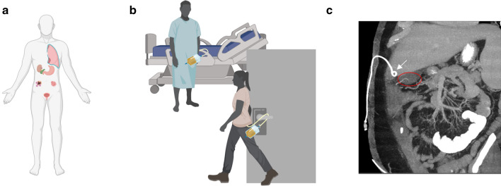
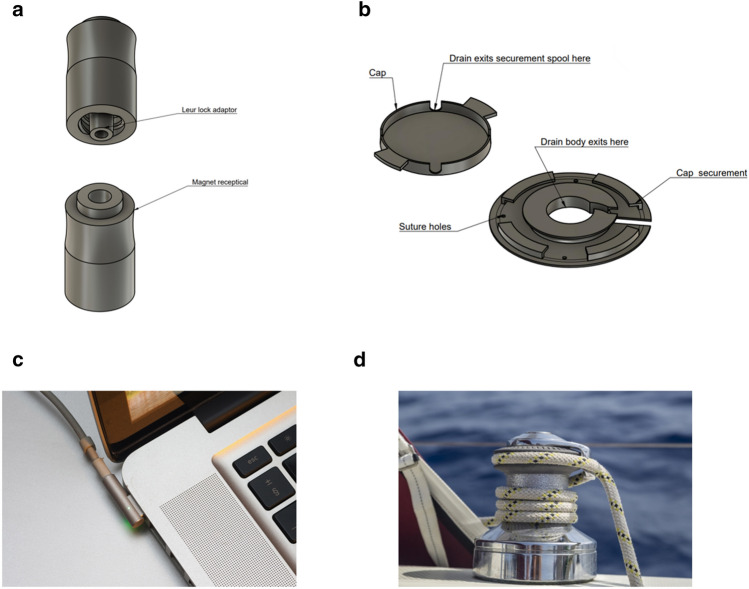
Percutaneous drains have provided a minimally invasive way to treat a wide range of disorders from abscess drainage to enteral feeding solutions to treating hydronephrosis. These drains suffer from a high rate of dislodgement of up to 30% resulting in emergency room visits, repeat hospitalizations, and catheter repositioning/replacement procedures, which incur significant morbidity and mortality. Using ex vivo and in vivo models, a force body diagram was utilized to determine the forces experienced by a drainage catheter during dislodgement events, and the individual components which contribute to drainage catheter securement were empirically collected. Prototypes of a skin level catheter securement and valved quick release system were then developed. The system was inspired by capstans used in boating for increasing friction of a line around a central spool and quick release mechanisms used in electronics such as the Apple MagSafe computer charger. The device was tested in a porcine suprapubic model, which demonstrated the effectiveness of the device to prevent drain dislodgement. The prototype demonstrated that the miniaturized versions of technologies used in boating and electronics industries were able to meet the needs of preventing dislodgement of patient drainage catheters.


Conflict of interest statement

Dr. Di Capua and Dr. Som are listed inventors on a patent pending application (#63306932) which describes the securement device evaluated by this manuscript. As the intellectual property was created as part of an employment role, the employing institution, Massachusetts General Hospital, holds full ownership and rights to the intellectual property, in accordance with local legislation. The authors disclose no other financial or non-financial competing interests.

Figures

Figure 1
Figure 1
Clinical Scenario—(A) Illustration of the variety of locations drainage catheters are used, including pleura, intrahepatic, gastric, bladder, malignant and infectious foci; (B) demonstration of common dislodgement situations; (C) computed tomography (CT) imaging demonstrating a pigtail catheter (white arrow) after it has been unintentionally retracted from its intended drainage site, the gallbladder (red oval). Figure 1a-b produced using www.BioRender.com Individual License and Fig. 1c is exported from Visage 7.
Figure 2
Figure 2
A free body diagram describing the forces that resist dislodgement of a drainage catheter. Force F represents the pulling force imparted on the drainage catheter externally. Resisting this, the adhesive forces (N), drain suture tension (Ts), and the pigtail tension (Tp) all act in opposition to the pulling force, and these are overcome when a dislodgement event occurs. Illustration produced using www.BioRender.com Individual License.
Figure 3
Figure 3
Inspiration of a proposed securement device from sailing and tech industries—pictured are the quick-release unit (QR, a) and securement component (SC, b). The QR is placed in-line with the catheter and does not allow internal transmission of forces great enough for dislodgement. A percutaneous catheter is wound around the SC, which is adhered to the body and prevents migration of the catheter. See supplemental video 1 which demonstrates an animated model of the device in action. The inspiration for the QR draws from current technology solutions for a safe power supply connection (c), and the inspiration for the SC is inspired from sailing winches, which also employ the Capstan principle (d). Figure 3a-b were produced using Fusion360 V.2.0.15299. Figure 3c was produced by Ashley Pomeroy (see acknowledgment)—Own work, CC BY-SA 4.0, Fig. 3d was produced by George Hodan (see acknowledgement)–—wn work, CC0.
Figure 4
Figure 4
Conceptual demonstration of the novel, 3D-printed securement device—pictured are a kidney, percutaneous nephrostomy tube, and the securement device. The device noninvasively and safely prevents migration or dislodgement of the drain by reducing the profile of external drain tubing and eliminating the transmission of dislodgement-level pull forces. Figure was produced using www.BioRender.com Individual License and Microsoft PowerPoint V16.72.
Figure 5
Figure 5
Ex vivo force measurements for dislodgements: the force to break a drain stitch (a), the force to break the inner securement of the drainage catheter pigtail (b), the force to disengage the quick-release component (c), the force to remove an ostomy adhesive (d), the force resisted by a linear fixation device (e), and the force resisted by the proposed device which implements a spooled design (f). The black dotted line refers to the average force for a 1 L amount of fluid at 1 g (acceleration ~ 9.8 m/s2), about two times the average volume held by drainage bags, and the clinical utility force required to be kept with accidental dislodgement. Illustrations produced using www.BioRender.com Individual License.
Figure 6
Figure 6
In vivo dislodgement and fluoroscopic evaluation—Following fluoroscopic drain placement, the impacts of pull forces on internal drain placement were compared between our novel securement component (SC), with a break away mechanism, and a drain stitch, the current standard of care. Fluoroscopy demonstrates proper drain positioning (a) with our novel SC attached and no pull force applied (green circle). A pull force is applied (blue circle) with maintenance of the drain pigtail position (b). The pull force was increased until the SC break away mechanism reached its detachment threshold (red circle) throughout which time the drain pigtail remains in an identical location (c). In contrast, a drain was placed under fluoroscopy and secured using a drain stitch (d). A pull force was applied with partial internal migration of the drain tip (e). The pull force was increased until failure of the drain stitch resulting in complete dislodgement of the drain (f). Figure was produced using Microsoft PowerPoint V16.72.
Figure 7
Figure 7
In vivo forces resisted by the standard of care and proposed device. Under the standard of care (a), forces peak at the drain stitch breaking (47.7 ± 4.6 N), resulting in complete dislodgement of the drain. Conversely, when implementing the proposed device (b), the drain first coils around the spool, removing slack. Then, once forces reach a sufficient level, the quick-release detached, keeping the drain in place. The maximum force resisted by the quick-release device was 15.1 ± 0.7 N. Illustrations produced using www.BioRender.com Individual License.

References

    1. Moureau N. Impact and safety associated with accidental dislodgement of vascular access devices: A survey of professions, settings, and devices. J. Assoc. Vasc. Access. 2018;23:203–215. doi: 10.1016/j.java.2018.07.002. - DOI
    1. Feil M. Dislodged gastrostomy tubes: Preventing a potentially. Fatal Comp. 2017;14:9.
    1. Lorente L, Huidobro MS, Martín MM, Jiménez A, Mora ML. Accidental catheter removal in critically ill patients: A prospective and observational study. Crit. Care. 2004;8:R229. doi: 10.1186/cc2874. - DOI - PMC - PubMed
    1. Huang S, Engstrom B, Lungren M, Kim C. Management of dysfunctional catheters and tubes inserted by interventional radiology. Semin. Interv. Radiol. 2015;32:067–077. doi: 10.1055/s-0035-1549371. - DOI - PMC - PubMed
    1. Collares FB, Faintuch S, Kim SK, Rabkin DJ. Reinsertion of accidentally dislodged catheters through the original track: What is the likelihood of success? J. Vasc. Interv. Radiol. 2010;21:861–864. doi: 10.1016/j.jvir.2010.02.022. - DOI - PubMed

Publication types