. 2023 Sep 26;23(1):906.
doi: 10.1186/s12885-023-11393-1.

Hepatocyte-specific HDAC3 ablation promotes hepatocellular carcinoma in females by suppressing Foxa1/2

Affiliations

Hepatocyte-specific HDAC3 ablation promotes hepatocellular carcinoma in females by suppressing Foxa1/2

Yahong Xu et al. BMC Cancer. .

Abstract

Background: Hepatocellular carcinoma (HCC), the most common primary liver cancer, prevails mainly in males and has long been attributed to androgens and higher circumstantial levels of interleukin-6 (IL-6) produced by resident hepatic macrophages.

Methods: Constitutively hepatocyte-specific histone deacetylase 3 (HDAC3)-deficient (HDAC3LCKO) mice and constitutively hepatocyte-specific HDAC3 knockout and systemic IL-6 simultaneously ablated (HDAC3LCKO& IL-6-/-) mice were used in our study to explore the causes of sex differences in HCC. Additionally, we performed human HCC tissues with an IHC score. Correlation analysis and linear regression plots were constructed to reveal the association between HDAC3 and its candidate genes. To further elucidate that HDAC3 controls the expression of Foxa1/2, we knocked down HDAC3 in HUH7 liver cancer cells.

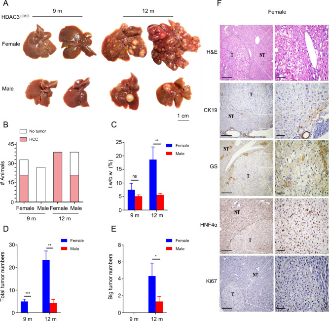
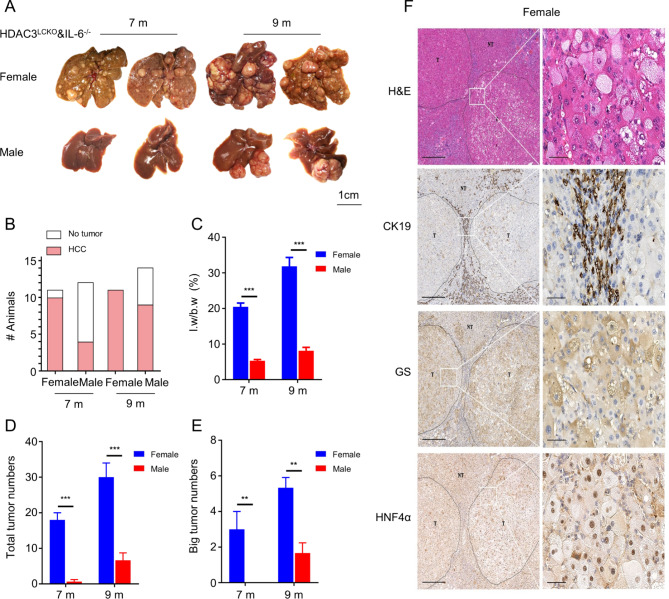
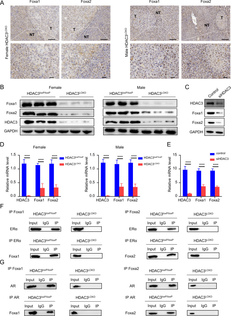
Results: We observed a contrary sex disparity, with an earlier onset and higher incidence of HCC in female mice when HDAC3 was selectively ablated in the liver. Loss of HDAC3 led to constant liver injury and the spontaneous development of HCC. Unlike the significant elevation of IL-6 in male mice at a very early age, female mice exhibit stable IL-6 levels, and IL-6 ablation did not eliminate the sex disparity in hepatocarcinogenesis in HDAC3-deficient mice. Oestrogen often protects the liver when combined with oestrogen receptor alpha (ERα); however, ovariectomy in HDAC3-ablated female mice significantly delayed tumourigenesis. The oestrogen-ERα axis can also play a role in tumour promotion in the absence of Foxa1 and Foxa2 in the receptor complex. Loss of HDAC3 profoundly reduced the expression of both Foxa1 and Foxa2 and impaired the binding between Foxa1/2 and ERα. Furthermore, a more frequent HDAC3 decrease accompanied by the simultaneous Foxa1/2 decline was found in female HCC compared to that in male HCC.

Conclusion: In summary, we reported that loss of HDAC3 reduces Foxa1/2 and thus promotes HCC development in females in an oestrogen-dependent manner.

Keywords: Foxa1; Foxa2; HCC; HDAC3; IL-6; Oestrogen receptor; Sex difference.


Conflict of interest statement

The authors declare no conflict of Interests.

Figures

Fig. 1
Fig. 1
Significant hepatocyte damage in 2-month-old HDAC3LCKO mice. (A) Western blot detection of HDAC3 protein in livers from HDAC3LCKO mice. GAPDH and histone H3 were used as the loading controls. (B) Immunohistochemistry staining shows the negative expression of HDAC3 in HDAC3LCKO hepatocytes. Scale bar, 100 μm. (C) Gross morphology of HDAC3loxP/loxP and HDAC3LCKO livers. (D) The ratio of liver weight (l.w) to body weight (b.w) between HDAC3loxP/loxP mice and HDAC3LCKO mice. (E-F) Serum levels of ALT and AST. (G) H&E staining and immunohistochemistry staining of glutamine synthetase (GS), cytokeratin 19 (CK19), and Ki67 in livers from 2-month-old HDAC3LCKO mice. Scale bar, 100 μm. * p < 0.05, **** p < 0.0001, and ns, not significant
Fig. 2
Fig. 2
HDAC3 deletion results in earlier HCC onset in female mice. (A) Gross morphology of HDAC3LCKO livers at 9 and 12 months old. (B) The number of female HDAC3LCKO mice and male HDAC3LCKO mice with liver nodules. (C) The ratio of liver weight (l.w) to body weight (b.w) in HDAC3LCKO mice at 9 and 12 months old. (D) The number of tumours in female and male HDAC3LCKO mice at the indicated times. (E) The number of tumours larger than 5 mm in diameter in female and male HDAC3LCKO mice. (F) Histological detection of the livers of 9-month-old female HDAC3LCKO mice. T, tumour; NT, not tumour. Scale bar, 100 μm. * p < 0.05, ** p < 0.01, *** p < 0.001, and ns, not significant
Fig. 3
Fig. 3
HDAC3 deletion resulted in higher IL-6 levels in female mice and more severe liver damage was observed in HDAC3LCKO&IL-6−/− mice at 2 months of age. (A) Serum IL-6 levels in mice of different genotypes and sexes at 2, 6, and 9 months. (B) Western blot detection of HDAC3 protein in the liver. (C) Serum IL-6 levels in HDAC3LCKO and HDAC3LCKO&IL-6−/− mice. (D) Gross morphology of HDAC3LCKO and HDAC3LCKO&IL-6−/− livers. (E) The ratio of liver weight (l.w) to body weight (b.w) between HDAC3LCKO and HDAC3LCKO&IL-6−/− mice. (F-G) Serum AST and ALT levels. (H) H&E staining and immunohistochemistry staining of GS, CK19, and Ki67 in livers from 2-month-old HDAC3LCKO mice. Scale bar, 100 μm. * p < 0.05, ** p < 0.01, *** p < 0.001, **** p< 0.0001, and ns, not significant
Fig. 4
Fig. 4
HDAC3LCKO&IL-6-/- female mice developed HCC earlier than males. (A) Gross liver morphology in HDAC3LCKO&IL-6−/− mice at 7 and 9 months old. (B) The number of female HDAC3LCKO&IL-6−/− mice and male HDAC3LCKO&IL-6−/− mice with liver nodules. (C) The ratio of liver weight to body weight in HDAC3LCKO&IL-6−/− mice at 7 and 9 months old. (D) The number of tumours in female HDAC3LCKO&IL-6−/− mice and male HDAC3LCKO&IL-6−/− mice. (E) The number of tumours larger than 5 mm in diameter in female HDAC3LCKO&IL-6−/− mice and male HDAC3LCKO&IL-6−/− mice. (F) Histological detection of the liver in 7-month-old female HDAC3LCKO&IL-6−/− mice. T, tumour; NT, not tumour. Scale bar, 100 μm. * p < 0.05, ** p < 0.01, and *** p < 0.001
Fig. 5
Fig. 5
Ovariectomy significantly delayed tumorigenesis. (A) Gross morphology of HDAC3LCKO-OVX livers at 9 and 12 months old. (B) The number of HDAC3LCKO and HDAC3LCKO-OVX mice with liver nodules at the indicated times. (C) The ratio of liver weight (l.w) to body weight (b.w) in HDAC3LCKO and HDAC3LCKO-OVX mice. ** p < 0.01, *** p < 0.001, and **** p < 0.0001
Fig. 6
Fig. 6
HDAC3 ablation suppresses Foxa1 and Foxa2 expression and their binding to ERα and AR. (A) Immunohistochemical detection of Foxa1 and Foxa2 in the livers of HDAC3LCKO mice. Scale bar, 100 μm. (B) Western blot detection of Foxa1 and Foxa2 protein expression in the livers of HDAC3LCKO mice. (C) Western blotting indicating that knockdown of HDAC3 reduced Foxa1 and Foxa2 in HUH7 cells. (D) The mRNA level of HDAC3, Foxa1, and Foxa2 in HDCA3loxP/loxP and HDAC3LCKO mice. (E) The mRNA level of Foxa1 and Foxa2 in HUH7 cells. (F) The interactions between ERα and Foxa1/2 were analyzed by co-immunoprecipitation (co-IP). (G) The interactions between AR and Foxa1/2 were analyzed by co-immunoprecipitation (co-IP).
Fig. 7
Fig. 7
Human HCCs display sex differences in HDAC3 levels. (A) staining intensity of HDAC3 in HCC. Scale bar, 100 μm. (B-C) Staining intensity of Foxa1 and Foxa2 in HCC. Scale bar, 100 μm. (D) The chi-square test revealed HDAC3 expression was presumably lower in female HCC. (E-F) Correlation analysis revealed that Foxa1 (r = 0.5265; p < 0.0001) and Foxa2 (r = 0.5550; p < 0.0001) were positively associated with HDAC3 expression in female HCC patients. (G) Diagram of the hypothetical roles of HDAC3 in female HCC development by regulating the Foxa1/2 signalling pathway. * p < 0.05 and **** p < 0.0001

Similar articles

Cited by

References

    1. Sung H, Ferlay J, Siegel RL, Laversanne M, Soerjomataram I, Jemal A, Bray F. Global Cancer Statistics 2020: GLOBOCAN estimates of incidence and Mortality Worldwide for 36 cancers in 185 countries. CA Cancer J Clin. 2021;71(3):209–49. doi: 10.3322/caac.21660. - DOI - PubMed
    1. Thomas L, Petrick J, McGlynn K. Liver cancer. In: Thun M, Linet M, Cerhan J, Haiman C, Schottenfeld D, editors. Cancer Epidemiology and Prevention. 4th ed. Oxford University Press; 2018. pp. 635–60.
    1. Cook MB, McGlynn KA, Devesa SS, Freedman ND, Anderson WF. Sex disparities in Cancer Mortality and Survival. Cancer Epidemiol Biomarkers Prev. 2011;20(8):1629–37. doi: 10.1158/1055-9965.EPI-11-0246. - DOI - PMC - PubMed
    1. Dorak M, Karpuzoglu E. Gender differences in cancer susceptibility: an inadequately addressed issue. Front Genet. 2012;28:268. - PMC - PubMed
    1. El-Serag H, Rudolph K. Hepatocellular Carcinoma: Epidemiology and Molecular Carcinogenesis. Gastroenterology. 2007;132(7):2557–76. doi: 10.1053/j.gastro.2007.04.061. - DOI - PubMed