. 2023 Sep 27;23(1):340.
doi: 10.1186/s12883-023-03393-2.

Obstructive sleep apnea: a major risk factor for COVID-19 encephalopathy?

Affiliations

Obstructive sleep apnea: a major risk factor for COVID-19 encephalopathy?

Gautier Breville et al. BMC Neurol. .

Abstract

Background: This study evaluates the impact of high risk of obstructive sleep apnea (OSA) on coronavirus disease 2019 (COVID-19) acute encephalopathy (AE).

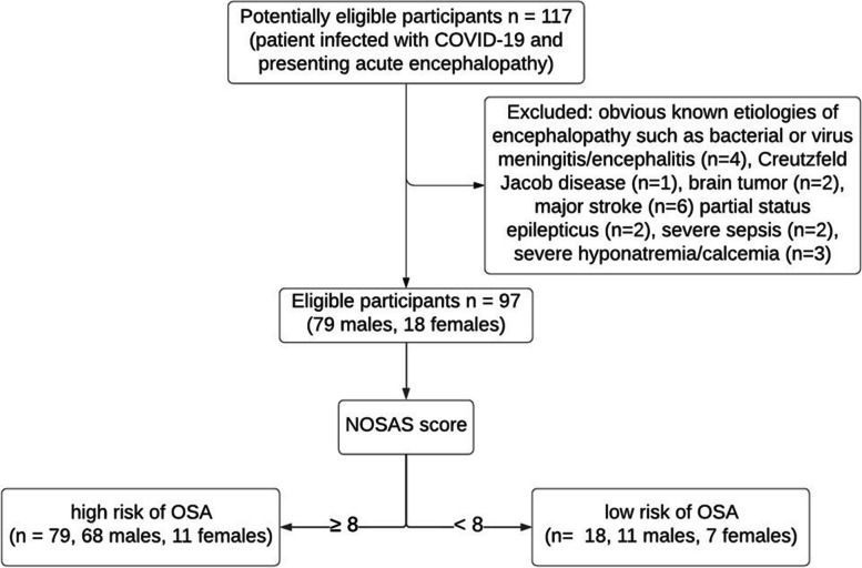
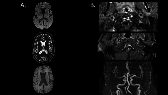
Methods: Between 3/1/2020 and 11/1/2021, 97 consecutive patients were evaluated at the Geneva University Hospitals with a neurological diagnosis of COVID-19 AE. They were divided in two groups depending on the presence or absence of high risk for OSA based on the modified NOSAS score (mNOSAS, respectively ≥ 8 and < 8). We compared patients' characteristics (clinical, biological, brain MRI, EEG, pulmonary CT). The severity of COVID-19 AE relied on the RASS and CAM scores.

Results: Most COVID-19 AE patients presented with a high mNOSAS, suggesting high risk of OSA (> 80%). Patients with a high mNOSAS had a more severe form of COVID-19 AE (84.8% versus 27.8%), longer mean duration of COVID-19 AE (27.9 versus 16.9 days), higher mRS at discharge (≥ 3 in 58.2% versus 16.7%), and increased prevalence of brain vessels enhancement (98.1% versus 20.0%). High risk of OSA was associated with a 14 fold increased risk of developing a severe COVID-19 AE (OR = 14.52).

Discussion: These observations suggest an association between high risk of OSA and COVID-19 AE severity. High risk of OSA could be a predisposing factor leading to severe COVID-19 AE and consecutive long-term sequalae.

Keywords: COVID-19 encephalopathy; Obstructive sleep apnea; SARS-CoV-2.


Conflict of interest statement

The authors declare no competing interests.

Figures

Fig. 1
Fig. 1
Flow chart
Fig. 2
Fig. 2
Brain MRI of one patient from the OSA group with evidence of intracranial vessel gadolinium enhancement without vascular stenosis nor any parenchymal lesion. Legend: A from the top to the bottom, we show brain axial slides of diffusion-weighted imaging (DWI, top), T2-weighted (middle) and susceptibility-weighted images (SWI, bottom) brain MRI sequences. They do not find any brain parenchymal damage. B from the top to the bottom, we show axial slide of dynamic 3D contrast-enhanced MR angiography (MRA) of the neck vessels at the vertebral arteries levels (top) and at the basilar artery level (middle) as well as 3D time-of-flight (TOF) MRA of the intracranial vessels (bottom). The first two images show gadolinium contrast enhancement of both vertebral arteries and basilar artery, without any vascular stenosis (TOF, third image)

Similar articles

Cited by

References

    1. Gupta A, Madhavan MV, Sehgal K, Nair N, Mahajan S, Sehrawat TS, et al. Extrapulmonary manifestations of COVID-19. Nat Med. 2020;26(7):1017–1032. doi: 10.1038/s41591-020-0968-3. - DOI - PMC - PubMed
    1. Koralnik IJ, Tyler KL. COVID -19: a global threat to the nervous system. Ann Neurol. 2020;88(1):1–11. doi: 10.1002/ana.25807. - DOI - PMC - PubMed
    1. Hernández-Fernández F, Valencia HS, Barbella-Aponte RA, Collado-Jiménez R, Ayo-Martín Ó, Barrena C, et al. Cerebrovascular disease in patients with COVID-19: neuroimaging, histological and clinical description. Brain. 2020;143(10):3089–3103. doi: 10.1093/brain/awaa239. - DOI - PMC - PubMed
    1. Helms J, Kremer S, Merdji H, Schenck M, Severac F, Clere-Jehl R, et al. Delirium and encephalopathy in severe COVID-19: a cohort analysis of ICU patients. Crit Care. 2020;24(1):491. doi: 10.1186/s13054-020-03200-1. - DOI - PMC - PubMed
    1. Uhlen M, Fagerberg L, Hallstrom BM, Lindskog C, Oksvold P, Mardinoglu A, et al. Tissue-based map of the human proteome. Science. 2015;347(6220):1260419. doi: 10.1126/science.1260419. - DOI - PubMed

MeSH terms