. 2023 Sep 26;24(1):33.
doi: 10.1186/s12865-023-00569-w.

Immune cell profiles of idiopathic inflammatory myopathy patients expressed anti-aminoacyl tRNA synthetase or anti-melanoma differentiation-associated gene 5 autoantibodies

Affiliations

Immune cell profiles of idiopathic inflammatory myopathy patients expressed anti-aminoacyl tRNA synthetase or anti-melanoma differentiation-associated gene 5 autoantibodies

Joung-Liang Lan et al. BMC Immunol. .

Abstract

Background: Patients with idiopathic inflammatory myopathy (IIM) often express a different type of myositis-specific autoantibodies (MSAs), each associated with different clinical symptoms. Understanding the immunopathogenesis of various IIM subgroups can help improve the diagnosis and prognosis of IIM patients with different MSAs. However, the immune cell profiles of these IIM patients with anti-aminoacyl tRNA synthetase (ARS) or anti-melanoma differentiation-associated gene 5 (MDA5) autoantibodies remain unclear. We focused on the immune cell profiles of IIM patients with anti-ARS or anti-MDA5 autoantibodies.

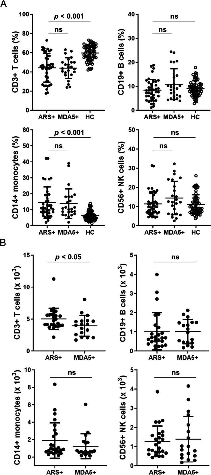
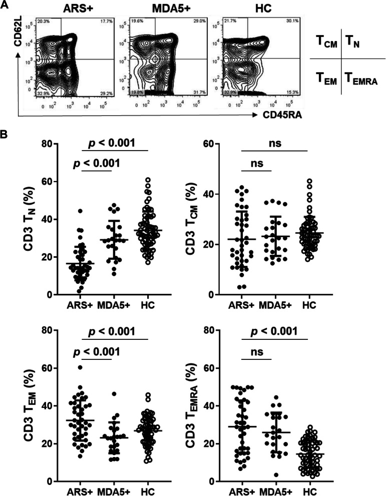
Results: The peripheral blood from IIM patients with anti-MDA5 autoantibody (MDA5 + group, n = 24) or one of the anti-ARS autoantibodies (ARS + group, n = 40) autoantibodies, and healthy controls (HC group, n = 60) were collected and examined. We found that IIM patients had a lower CD3 T cell population compared to the HC group. IIM patients showed a significantly lower TN cell population and a higher TEMRA cell population. Higher Th17 and Treg cell populations were found in these IIM patients than in the HC group. In these IIM patients, the MDA5 + group exhibited the higher percentages of Th17 and Treg cells than the ARS + group. It is noteworthy that the percentage of Th1 cells in the survival subgroup was higher than in the death subgroup in IIM patients with ARS + or MDA5 + . Furthermore, in the MDA5 + group, the percentage of Treg cells was higher in the survival subgroup compared to the death subgroup.

Conclusions: Our study demonstrated that elevated Th1 may be a good prognostic indicator in IIM patients with ARS + or MDA5 + . Elevated Treg may also help predict a good prognosis in MDA5 + IIM patients. However, more large-scale studies and clinical samples are needed to verify the significance of Th1 and Treg cell subsets in clinical outcomes for these IIM patients with ARS + or MDA5 + . These data may help design a therapeutic approach that specifically targets the pathogenic immune molecular responsible for autoimmune attacks in IIM.

Keywords: Aminoacyl-tRNA synthetase (ARS); Idiopathic inflammatory myositis (IIM); Immune cell profiles; Melanoma differentiation-associated gene 5 (MDA5); Myositis-specific autoantibody (MSA).


Conflict of interest statement

The authors declare no competing interests.

Figures

Fig. 1
Fig. 1
IIM patients have a lower CD3 + cell population and a higher CD14 + cell population in the pheripheral blood. A Flow cytometry analyses of CD3, CD14, CD19, and CD56 surface markers in peripheral blood mononuclear cells (PBMCs) of IIM patients with anti-aminoacyl-tRNA synthetase (ARS + , n = 40), or with anti-MDA5 antibody (MDA5 + , n = 24), and healthy controls (HC, n = 60). B Absolute cell numbers of CD3 + , CD14 + , CD19 + , and CD56 + cells of IIM patients with anti-aminoacyl-tRNA synthetase (ARS + , n = 25), or with anti-MDA5 antibody (MDA5 + , n = 18). Each dot was displayed for each data from the enrolled subjects. Data are presented as means ± SD. The p values were calculated using Kruskal–Wallis test between the ARS + , MDA5 + , and HC groups. ns, not significant
Fig. 2
Fig. 2
IIM patients exhibit a lower CD3 TN cell population and a higher CD3 TEMRA cell population in the pheripheral blood. Flow cytometry analyses of surface markers CD3, CD62L and CD45RA surface markers in PBMCs of IIM patients with anti-aminoacyl-tRNA synthetase (ARS + , n = 40), or with anti-MDA5 antibody (MDA5 + , n = 24), and healthy controls (HC, n = 60). A A representative data was shown. Naïve T (TN, CD45RA + CD62L +), central memory T (TCM, CD45RA-CD62L +), effector memory T (TEM, CD45RA-CD62L-), and terminally differentiated effector memory T cells (TEMRA, CD45R + CD62L-) (B) Each dot was displayed for each data from the enrolled subjects. Data are presented as means ± SD. The p values were calculated using Kruskal–Wallis test between ARS + , MDA5 + , and HC groups. ns, not significant
Fig. 3
Fig. 3
IIM patients with anti-MDA5 autoantibody have a higher Th17 cell population in the pheripheral blood. Flow cytometry analyses of CD3, CD4, CXCR3, CCR6, and CCR4 surface markers in PBMCs of IIM patients with anti-aminoacyl-tRNA synthetase (ARS + , n = 36), or with anti-MDA5 antibody (MDA5 + , n = 22), and healthy controls (HC, n = 60). A One represented data was shown. Th1, CXCR3 + CD4 + ; Th2, CXCR3-CCR4 + CCR6-CD4 + ; Th9, CXCR3-CCR4-CCR6 + CD4 + ; and Th17 cells, CXCR3-CCR4 + CCR6 + CD4 + . B Each dot was displayed for each data from the enrolled subjects. Data are presented as means ± SD. The p values were calculated using Kruskal–Wallis test between the MDA5, ARS, and HC groups. ns, not significant
Fig. 4
Fig. 4
IIM patients with anti-MDA5 autoantibody have a higher Treg cell population in the pheripheral blood. Flow cytometry analyses of CD3, CD4, and Foxp3 markers in PBMCs of IIM patients with anti-aminoacyl-tRNA synthetase (ARS + , n = 40), or with anti-MDA5 antibody (MDA5 + , n = 22), and healthy controls (HC, n = 60). A A representative data was shown. Treg cells were defined as CD3 + CD4 + Foxp3 + . B Each dot was displayed for each data from the enrolled subjects. Data are presented as means ± SD. The p values were calculated using Mann–Whitney U test between the MDA5 and ARS groups, or using Kruskal–Wallis test between the MDA5, ARS, and HC groups
Fig. 5
Fig. 5
A higher Th1 cell population is associated with survival in IIM patients. A Differences in Th1, Th17, and Treg cell populations between death and survival in the IIM patients with ARS + and MDA5 + were compared. B Differences in Th1, Th17, and Treg cell populations were compared between death (n = 5) and survival (n = 17) in the MDA5 + group. Each dot was displayed for each data from the enrolled subjects. Data are presented as means ± SD. The p values were calculated using the Mann–Whitney U test. ns, not significant. C The cumulative incidence of overall survival in the MDA5 + group according to their Treg levels (n = 22). Treg-High, the Treg level was higher than average Treg level. Treg-Low, lower than average Treg level. The p values were calculated using log-rank test

Similar articles

Cited by

References

    1. Cheeti A, Brent LH, Panginikkod S: Autoimmune Myopathies. In: StatPearls. edn. Treasure Island (FL): StatPearls Publishing Copyright © 2023, StatPearls Publishing LLC.; 2023. - PubMed
    1. Sarwar A, Dydyk AM, Jatwani S. Polymyositis. In: StatPearls. edn. Treasure Island (FL): StatPearls Publishing LLC; 2023. StatPearls Publishing Copyright © 2023. - PubMed
    1. Lu X, Peng Q, Wang G. Discovery of new biomarkers of idiopathic inflammatory myopathy. Clin Chim Acta. 2015;444:117–125. doi: 10.1016/j.cca.2015.02.007. - DOI - PubMed
    1. Damoiseaux J, Andrade LE, Fritzler MJ, Shoenfeld Y. Autoantibodies 2015: From diagnostic biomarkers toward prediction, prognosis and prevention. Autoimmun Rev. 2015;14(6):555–563. doi: 10.1016/j.autrev.2015.01.017. - DOI - PubMed
    1. Stuhlmüller B, Schneider U, González-González J-B, Feist E. Disease Specific Autoantibodies in Idiopathic Inflammatory Myopathies. Front Neurol. 2019;10:438. doi: 10.3389/fneur.2019.00438. - DOI - PMC - PubMed

Publication types