. 2023 Sep 27;20(1):216.
doi: 10.1186/s12974-023-02891-x.

Type I/type III IFN and related factors regulate JEV infection and BBB endothelial integrity

Affiliations

Type I/type III IFN and related factors regulate JEV infection and BBB endothelial integrity

Ya-Ge Zhang et al. J Neuroinflammation. .

Abstract

Background: Japanese encephalitis virus (JEV) remains a predominant cause of Japanese encephalitis (JE) globally. Its infection is usually accompanied by disrupted blood‒brain barrier (BBB) integrity and central nervous system (CNS) inflammation in a poorly understood pathogenesis. Productive JEV infection in brain microvascular endothelial cells (BMECs) is considered the initial event of the virus in penetrating the BBB. Type I/III IFN and related factors have been described as negative regulators in CNS inflammation, whereas their role in JE remains ambiguous.

Methods: RNA-sequencing profiling (RNA-seq), real-time quantitative PCR, enzyme-linked immunosorbent assay, and Western blotting analysis were performed to analyze the gene and protein expression changes between mock- and JEV-infected hBMECs. Bioinformatic tools were used to cluster altered signaling pathway members during JEV infection. The shRNA-mediated immune factor-knockdown hBMECs and the in vitro transwell BBB model were utilized to explore the interrelation between immune factors, as well as between immune factors and BBB endothelial integrity.

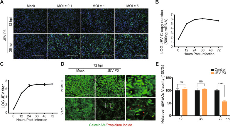
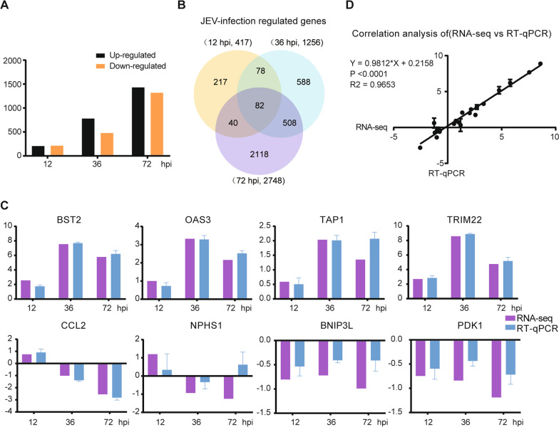
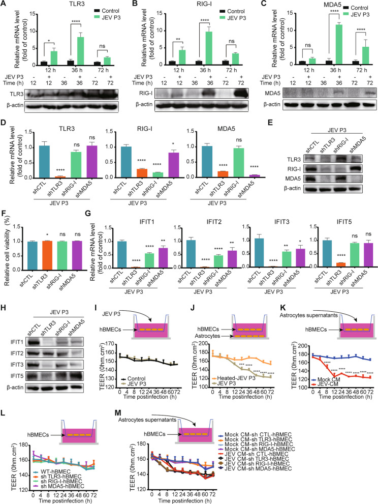
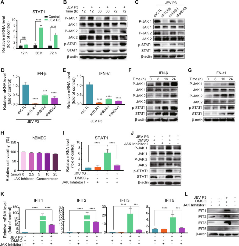
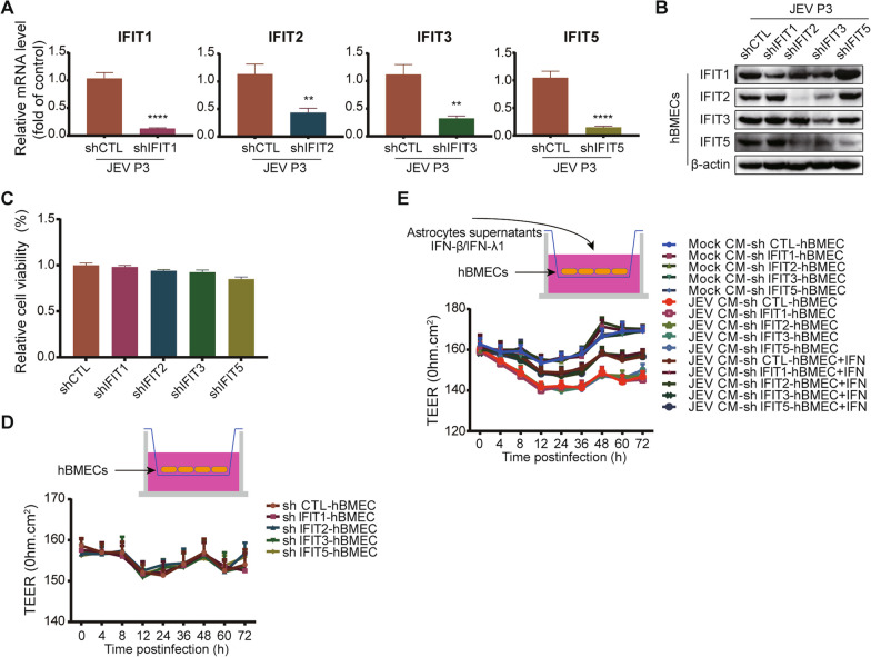
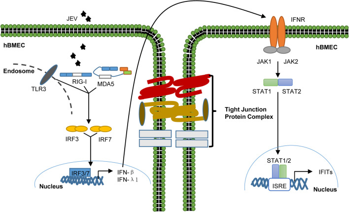
Results: RNA-Seq data of JEV-infected hBMECs identified 417, 1256, and 2748 differentially expressed genes (DEGs) at 12, 36, and 72 h post-infection (hpi), respectively. The altered genes clustered into distinct pathways in gene ontology (GO) terms and KEGG pathway enrichment analysis, including host antiviral immune defense and endothelial cell leakage. Further investigation revealed that pattern-recognition receptors (PRRs, including TLR3, RIG-I, and MDA5) sensed JEV and initiated IRF/IFN signaling. IFNs triggered the expression of interferon-induced proteins with tetratricopeptide repeats (IFITs) via the JAK/STAT pathway. Distinct PRRs exert different functions in barrier homeostasis, while treatment with IFN (IFN-β and IFN-λ1) in hBMECs stabilizes the endothelial barrier by alleviating exogenous destruction. Despite the complex interrelationship, IFITs are considered nonessential in the IFN-mediated maintenance of hBMEC barrier integrity.

Conclusions: This research provided the first comprehensive description of the molecular mechanisms of host‒pathogen interplay in hBMECs responding to JEV invasion, in which type I/III IFN and related factors strongly correlated with regulating the hBMEC barrier and restricting JEV infection. This might help with developing an attractive therapeutic strategy in JE.

Keywords: Human brain microvascular endothelial cells; IFITs; IFNs; Japanese encephalitis virus; PRRs; RNA-Seq.


Conflict of interest statement

The authors declare no competing financial interests.

Figures

Fig. 1
Fig. 1
JEV infection of hBMECs. A hBMECs were infected with JEV P3 at MOIs of 0.1, 1, and 5. At 12 and 36 h post-infection (hpi), JEV-E was detected by indirect immunofluorescence (green), and nuclei are shown by Hoechst (blue) staining. B, C JEV RNA levels were assayed by quantitative real-time PCR (RT-qPCR) (B), and the titers were determined by PFU assay (C). D Vero or hBMECs were infected with JEV P3 for 72 h and then costained with calcein-AM (green [live cells])/propidium iodide (red [dead cells]) for another 45 min. Images were captured by fluorescence microscopy. E hBMECs were infected with JEV P3 for 72 h, and then cell viability was analyzed by CCK8 assay. Data are represented as the mean values ± SEMs from three independent experiments. ****p < 0.0001; ns: not significant
Fig. 2
Fig. 2
Schematic view and validation of transcriptome data in JEV-infected hBMECs. A The number of DEGs at 12, 36, and 72 hpi in hBMECs infected with JEV P3; B Venn diagram showing the distributions of unique and codifferentially expressed DEGs; C RT-qPCR was performed to validate the expression of eight representative genes. D GraphPad software 6.0 (San Diego, CA) was used to perform correlation analysis between RT-qPCR (y-axis) and the RNA-Seq platform (x-axis). Data are represented as the mean values ± SEMs from three independent experiments
Fig. 3
Fig. 3
Induction and antiviral activity of IFITs. A hBMECs were treated with poly I:C at different concentrations, and IFIT mRNA expression was measured. BD hBMECs were infected with JEV P3 or heated-inactivated JEV P3 (heated-JEV P3, 94 °C, 15 min) or treated with poly I: C for different time points, and IFIT expression was measured by performing RT-qPCR (B) and Western blotting (C, D). E, F HEK-293T cells were transfected with IFIT1, IFIT2, IFIT3, and IFIT5 expression plasmids or empty vector plasmids at the indicated doses following infection with JEV P3. Then, the JEV-C gene was analyzed by RT-qPCR (E), and the JEV-E protein was measured by performing an immunofluorescence assay (F). Data are represented as the mean values ± SEMs from three independent experiments. **p < 0.01; ****p < 0.0001; ns: not significant
Fig. 4
Fig. 4
The role of PRRs in IFIT induction and hBMEC barrier maintenance. AC hBMECs were infected with JEV P3 for different time points, and the mRNA and protein levels of TLR3 (A), RIG-I (B), and MDA5 (C) were measured. D, E PRR-knockdown hBMECs were infected with JEV P3, and the expression of TLR3, RIG-I, and MDA5 was assessed by RT-qPCR (D) and Western blotting (E). F PRR-knockdown hBMECs were serum starved after reaching confluence, and cell viability was measured. G, H Expression of IFITs was assessed by RT-qPCR (G) and Western blotting (H) in PRR-knockdown hBMECs. I Confluent monolayers of hBMECs in the upper chamber of the transwell plate were infected with JEV P3, and the TEER (upper graph) was measured at the indicated times. J hBMECs were seeded onto the upper chamber, cultured astrocytes infected with JEV P3 or heated-JEV P3 were placed in the lower compartment of the transwell plate, and the TEER (upper graph) was measured at the indicated times. K The culture supernatants from either mock-infected or JEV-infected astrocytes were collected and mixed with an equal volume of fresh medium (Mock-CM/JEV-CM) and added to confluent hBMECs in the upper chamber of the Transwell plate. Then, the TEER was measured at the indicated times. L, M PRR-knockdown hBMECs were seeded onto the upper chamber of the Transwell plate until confluent. The TEER (upper graph) was measured without treatment (L) or with mock-CM/JEV-CM treatment (M) at the indicated times. Data are represented as the mean values ± SEMs from three independent experiments. *p < 0.05; **p < 0.01; ****p < 0.0001; ns, not significant
Fig. 5
Fig. 5
The role of IFNs in IFIT induction and hBMEC barrier maintenance. hBMECs were infected with JEV P3, the cells/cell culture supernatants were harvested at 0, 12, 36, and 72 hpi, and the expression of IFN-β/IFN-λ1 at the mRNA and protein levels was determined by utilizing RT-qPCR (A, B) and ELISA (C, D). E, F hBMECs were treated with 10 ng/ml rhIFN-β (E) or 100 ng/ml rhIFN-λ1 protein (F) for different time points, and IFIT expression was measured by Western blotting. G, H HEK-293T cells were transfected with either empty vector plasmid (vector) or IFN-β (Vector-IFN-β)/IFN-λ1 (Vector-IFN-λ1) expression plasmid as indicated, and IFIT expression was measured by performing RT-qPCR (G) and Western blotting (H). I, J Vero cells were treated with poly I: C or rhIFN-β/rhIFN-λ1 protein as indicated, and IFIT expression was measured by performing RT-qPCR (I) and Western blotting (J). K HEK-293T cells were transfected with either empty vector plasmid (vector) or plasmids expressing IFIT1 (Vector-IFIT1), IFIT2 (Vector-IFIT2), IFIT3 (Vector-IFIT3), and IFIT5 (Vector-IFIT5) as indicated, and IFN-β and IFN-λ1 mRNA expression was measured by performing RT-qPCR. (L) Dual-luciferase reporter assays detected IRF1 binding to the IFN-β promotor in IFIT-overexpressing HEK-293T cells. HEK-293T cells were transfected with vector-IFIT1, vector-IFIT2, vector-IFIT3, vector-IFIT5, or empty vector plasmid to generate overexpressed cell lines. Simultaneously, empty vector plasmid or vector-IRF1 and IFN-β-Luc alone with pRL-TK plasmids were cotransfected into corresponding overexpressed cell lines. M hBMECs were cultured in the upper chamber of the transwell plate and pretreated with rhIFN-β and rhIFN-λ1 protein, followed by treatment with mock-CM or JEV-CM, and then the TEER (upper graph) was determined at the indicated times. Data are represented as the mean values ± SEMs from three independent experiments. *p < 0.05; **p < 0.01; ***p < 0.001; ****p < 0.0001; ns: not significant
Fig. 6
Fig. 6
The role of the JAK/STAT pathway in IFIT induction. A, B hBMECs were infected with JEV P3 for different time points, and the mRNA of STAT1 (A) and the protein of JAK1/p-JAK1, JAK2/p-JAK2, and STAT1/p-STAT1 (B) were measured. C The protein expression of JAK1/p-JAK1, JAK2/p-JAK2, and STAT1/p-STAT1 was measured in PRR-knockdown hBMECs under JEV infection. D, E IFN-β and IFN-λ1 mRNA levels were measured in PRR-knockdown hBMECs under JEV infection. F, G hBMECs were treated with rhIFN-β or rhIFN-λ1 protein for different time points, and JAK1/p-JAK1, JAK2/p-JAK2, and STAT1/p-STAT1 protein expression was measured. H hBMECs were treated with the carrier control DMSO or JAK inhibitor I at various concentrations for 72 h, and cell cytotoxicity was analyzed. I, J hBMECs were pretreated with carrier control DMSO or 5 μM JAK inhibitor I, followed by JEV P3 infection, and the mRNA of STAT1 (I) and the protein of JAK1/p-JAK1, JAK2/p-JAK2, and STAT1/p-STAT1 (J) were determined. K, L hBMECs were pretreated with the carrier control DMSO or 5 μM JAK inhibitor I, followed by JEV P3 infection, and IFIT mRNA (K) and protein (L) expression was measured. Data are represented as the mean values ± SEMs from three independent experiments. ***p < 0.001; ****p < 0.0001; ns: not significant
Fig. 7
Fig. 7
IFITs are nonessential in hBMEC barrier integrity. A, B The expression of IFITs was assessed by RT-qPCR (A) and Western blotting (B) in IFIT-knockdown hBMECs under JEV infection. C After serum-free starvation, cell viability was determined in IFIT-knockdown hBMECs. D, E IFIT-knockdown hBMECs were seeded onto the upper chamber of the Transwell plate until confluent. At the indicated times, the TEER (upper graph) was measured without treatment (D) or with rhIFN-β and rhIFN-λ1 protein treatment prior to the addition of JEV-CM (E). Data are represented as the mean values ± SEMs from three independent experiments. **p < 0.01; ****p < 0.0001
Fig. 8
Fig. 8
Schematic presentation of host innate immune factor-mediated intracellular communication for inducing IFIT expression, restricting JEV infection, and modulating hBMEC barrier integrity. During infection, hBMECs employed PRRs (TLR3, RIG-I, and MDA5) to sense JEV and subsequently triggered IRF phosphorylation and translocation to the nucleus, leading to the synthesis and secretion of IFNs, which induced IFIT expression in a JAK/STAT-dependent manner. Additionally, these upregulated immune factors differentially regulate hBMEC barrier integrity

Similar articles

Cited by

References

    1. Ghosh D, Basu A. Japanese encephalitis-a pathological and clinical perspective. PLoS Negl Trop Dis. 2009;3:e437. - PMC - PubMed
    1. Wang H, Liang G. Epidemiology of Japanese encephalitis: past, present, and future prospects. Ther Clin Risk Manag. 2015;11:435–448. - PMC - PubMed
    1. Kaur R, Vrati S. Development of a recombinant vaccine against Japanese encephalitis. J Neurovirol. 2003;9:421–431. - PubMed
    1. Deberardinis RJ, Sayed N, Ditsworth D, Thompson CB. Brick by brick: metabolism and tumor cell growth. Curr Opin Genet Dev. 2008;18:54–61. - PMC - PubMed
    1. Eigenmann DE, Xue G, Kim KS, Moses AV, Hamburger M, Oufir M. Comparative study of four immortalized human brain capillary endothelial cell lines, hCMEC/D3, hBMEC, TY10, and BB19, and optimization of culture conditions, for an in vitro blood-brain barrier model for drug permeability studies. Fluids Barriers CNS. 2013;10:33. - PMC - PubMed

LinkOut - more resources