

Spontaneous Rectus Sheath Hematoma
- PMID: 37753012
- PMCID: PMC10519645
- DOI: 10.7759/cureus.44138
Spontaneous Rectus Sheath Hematoma
Abstract
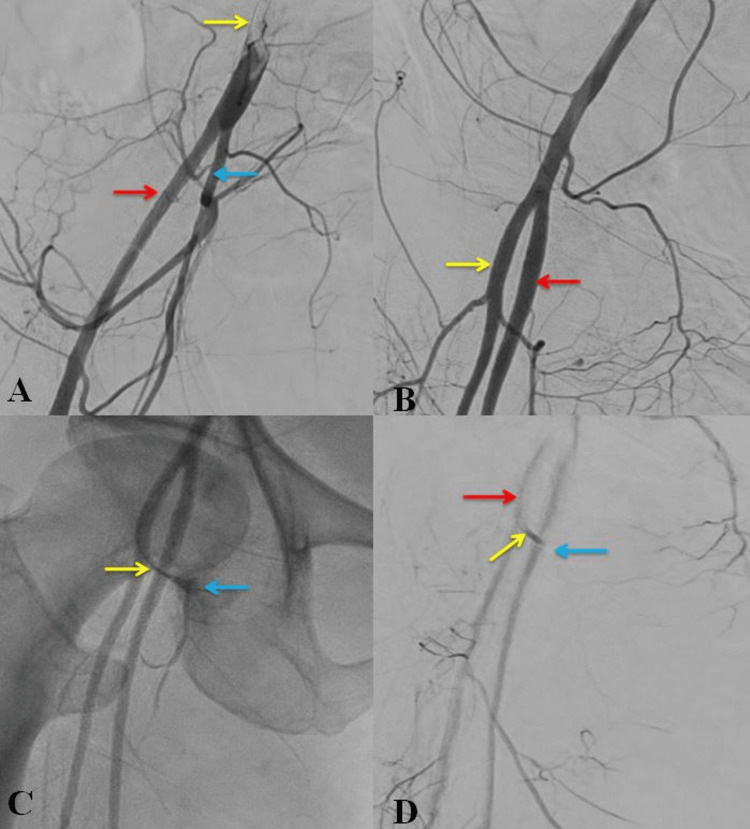
Rectus sheath hematoma (RSH) is one of the surgical emergencies that mimics peritonitis or other causes of acute abdominal pain. It is usually seen in old age, post-trauma, anticoagulation therapy pregnancy, chronic cough, and liver disease. Nevertheless, RSHs can be spontaneous without any underlying predisposing factors. Here, we present a 51-year-old female with sudden onset abdominal pain, abdominal distention, hypotension, and severe pallor. After initial resuscitation, the patient underwent radiological imaging. This suggested an RSH with active bleeding from the inferior epigastric artery or profunda femoris artery. The patient underwent digital subtraction angiography and angioembolization of the profunda femoris branch. After a few days, the patient continued deteriorating and succumbed to acute respiratory distress syndrome (ARDS).
Keywords: angioembolization; digital subtraction angiography; inferior epigastric artery; profundal femoris artery; rectus sheath hematoma.
Copyright © 2023, Das et al.
Conflict of interest statement
The authors have declared that no competing interests exist.
Figures


References
-
- Spontaneous rectus sheath hematoma: a rare clinical entity. Ranga HR, Dev K, Singla S, Marwah S. IJRRMS. 2012;2:39–41.
-
- Rectus sheath hematoma: a review of the literature. Hatjipetrou A, Anyfantakis D, Kastanakis M. Int J Surg. 2015;13:267–271. - PubMed
-
- Rectus sheath hematoma: review of 126 cases at a single institution. Cherry WB, Mueller PS. Medicine (Baltimore) 2006;85:105–110. - PubMed
-
- Rectus sheath hematoma. Osinbowale O, Bartholomew JR. Vasc Med. 2008;13:275–279. - PubMed
Publication types
LinkOut - more resources
Full Text Sources