

Large Water-Clear-Cell Parathyroid Adenoma: A Report of a Rare Case
- PMID: 37753019
- PMCID: PMC10519185
- DOI: 10.7759/cureus.44158
Large Water-Clear-Cell Parathyroid Adenoma: A Report of a Rare Case
Abstract
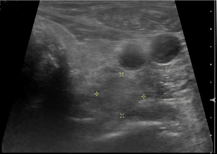
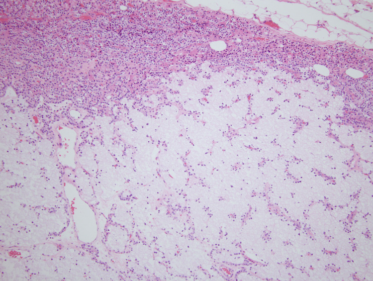
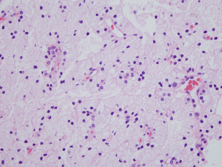
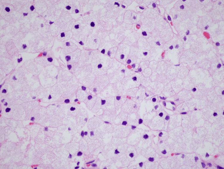
Water-clear-cell parathyroid adenomas are extremely rare tumors characterized by cells that contain clear, foamy cytoplasm. Here we report a case of a large water-clear-cell parathyroid adenoma in a 70-year-old male. The patient was presented to an outside hospital with severe abdominal pain and supporting CT imaging confirming a small bowel obstruction. Initial laboratory studies revealed hypercalcemia and elevated parathyroid hormone levels. Subsequent ultrasound imaging revealed a 2.7 × 2.1 cm neck mass suspicious for a parathyroid adenoma. A parathyroidectomy was performed, and microscopic evaluation revealed an expansile proliferation of cells with characteristic water-clear cell features. Although rare, water-clear-cell parathyroid adenomas are clinically indistinguishable from more common subtypes and should be considered in the differential diagnosis of an anterior neck mass.
Keywords: parathyroid disorder; parathyroid gland adenoma; parathyroid neoplasms; parathyroid pathology; water clear cell parathyroid adenoma.
Copyright © 2023, Durant et al.
Conflict of interest statement
The authors have declared that no competing interests exist.
Figures
References
-
- Clinical practice. Primary hyperparathyroidism. Marcocci C, Cetani F. N Engl J Med. 2011;365:2389–2397. - PubMed
-
- Pathology of the parathyroid glands. Van der Walt J. Diagn Histopathol. 2012;18:221–233.
-
- Oxyphil cell parathyroid adenomas causing primary hyperparathyroidism: a clinico-pathological correlation. Howson P, Kruijff S, Aniss A, et al. Endocr Pathol. 2015;26:250–254. - PubMed
Publication types
LinkOut - more resources
Full Text Sources