Case Reports
. 2023 Aug 26;15(8):e44141.
doi: 10.7759/cureus.44141. eCollection 2023 Aug.

Basilar Invagination With Chiari Type I Malformation and Atlanto-Axial Instability: A Rare Case Report

Affiliations
Case Reports

Basilar Invagination With Chiari Type I Malformation and Atlanto-Axial Instability: A Rare Case Report

Nandini Sanjay et al. Cureus. .

Abstract

Basilar invagination (BI) and Chiari malformation type I (CM-I) are important anomalies involving the craniovertebral junction (CVJ) involving the skull base and occipitocervical region. The incidence of BI is rare involving < 1% of the general population worldwide. They present with varied and complex clinical-radiological features. We present a 36-year-old male who displayed complaints of persistent reeling sensation at our center. Clinical examination revealed bilateral cerebellar signs along with nystagmus and restricted neck movements. Imaging revealed evidence of BI with cerebellar tonsil herniation of ~14.7 mm. Atlantodens interval of 6 mm was noted. The unexpected findings of C1-C2 fusion and instability were also noted. We describe a rare case of BI with C1 prolapse into the foramen magnum along with CM-1 malformation and congenital fusion of C1-C2. We conclude that the treatment algorithm for these rare cases is not very well established and is individually dependent.

Keywords: atlanto-axial fusion; atlantoaxial instability; basilar invagination; chiari malformation; cranio-vertebral junction.


Conflict of interest statement

The authors have declared that no competing interests exist.

Figures

Figure 1
Figure 1. Coronal CT images show that the tip of the odontoid process is 14.1 mm above the bimastoid line (A). The tip of the odontoid process is at a distance of 3.9 mm from the digastric line (B).
Figure 2
Figure 2. Sagittal CT images show Welcher's basal angle measuring 136.2 degrees (within normal limits) (A) and Boogard’s angle measuring 131.4 degrees (B)
Figure 3
Figure 3. Sagittal CT images show that the Chamberlain line, tip of the odontoid process, is ~14.6 mm above this line (A) and the McGregor’s line, tip of the odontoid process is ~14.8 mm above this line (B)
Figure 4
Figure 4. MRI showing caudal herniation of bilateral cerebellar tonsils noted for a distance of ~14.7 mm below the foramen magnum (A). Sagittal CT images show that Wackenheim’s line is intersecting the odontoid process (B). The height index of Klaus, which is the distance between the tip of the dens and the tuberculum torcula line, is 27.1 mm (C), McRae line, which is the tip of the odontoid process is ~7.5 mm above the line (D).
Figure 5
Figure 5. Cranial-caudal, intraoperative image: The left image shows the congenital band (white arrow), and the right image shows the foramen magnum decompression (yellow arrow)
Figure 6
Figure 6. Cranial-caudal, intraoperative image: The left image shows the cisterna magna after opening the dura, and the right image shows the herniation of cerebellar tonsil (yellow arrow) compressing on the spinal cord (black arrow).
Figure 7
Figure 7. The left image shows the left herniated cerebellar tonsil after cauterization, and the right image depicts the following duroplasty (blue arrow) showing the fusion of C1-C2 spinous process (black arrow).
Figure 8
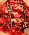
Figure 8. Intraoperative image showing the occipitocervical fusion with occipital screws, C2-C3 screws, and rods

References

    1. Hindbrain herniation syndromes: the Chiari malformations (I and II) Cai C, Oakes WJ. Semin Pediatr Neurol. 1997;4:179–191. - PubMed
    1. The odontoid process invagination in normal subjects, Chiari malformation and Basilar invagination patients: pathophysiologic correlations with angular craniometry. Ferreira JA, Botelho RV. Surg Neurol Int. 2015;6:118. - PMC - PubMed
    1. Donnally III CJ, Munakomi S, Varacallo M. StatPearls. Treasure Island (FL. Vol. 6. Treasure Island, FL: StatPearls Publishing; 2023. Basilar Invagination. - PubMed
    1. Craniovertebral junction: normal anatomy, craniometry, and congenital anomalies. Smoker WR. Radiographics. 1994;14:255–277. - PubMed
    1. Stereological and morphometric analysis of MRI chiari malformation type-1. Alkoç OA, Songur A, Eser O, Toktas M, Gönül Y, Esi E, Haktanir A. J Korean Neurosurg Soc. 2015;58:454–461. - PMC - PubMed

Publication types

LinkOut - more resources