Review
. 2023 Sep 11:13:1229098.
doi: 10.3389/fcimb.2023.1229098. eCollection 2023.

Contribution of viral and bacterial infections to senescence and immunosenescence

Affiliations
Review

Contribution of viral and bacterial infections to senescence and immunosenescence

Antonia Reyes et al. Front Cell Infect Microbiol. .

Abstract

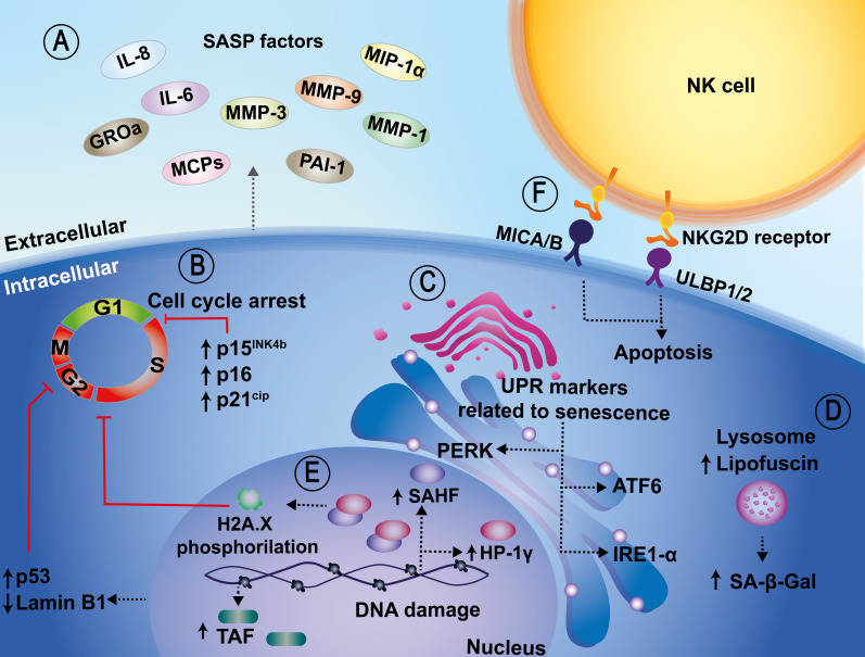
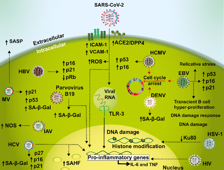
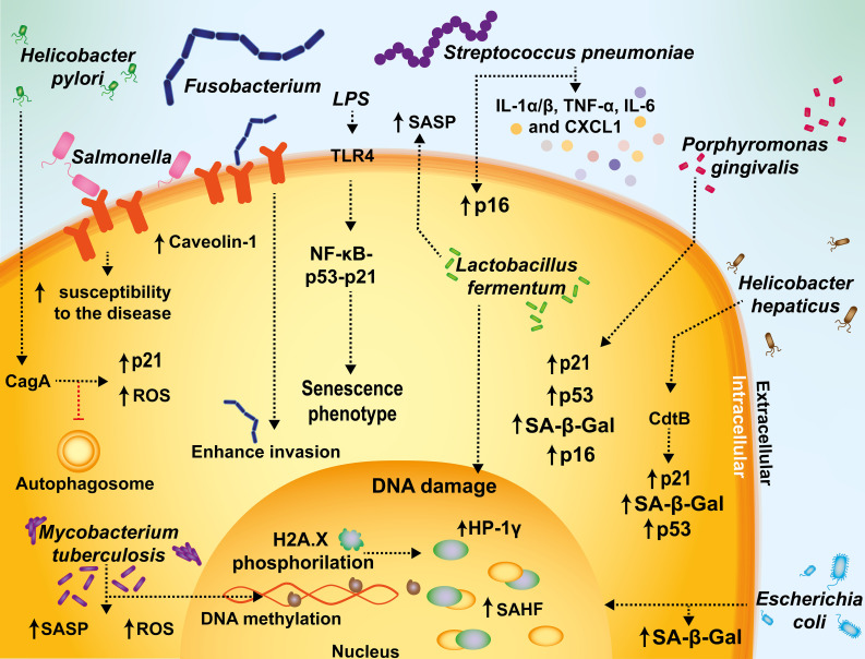
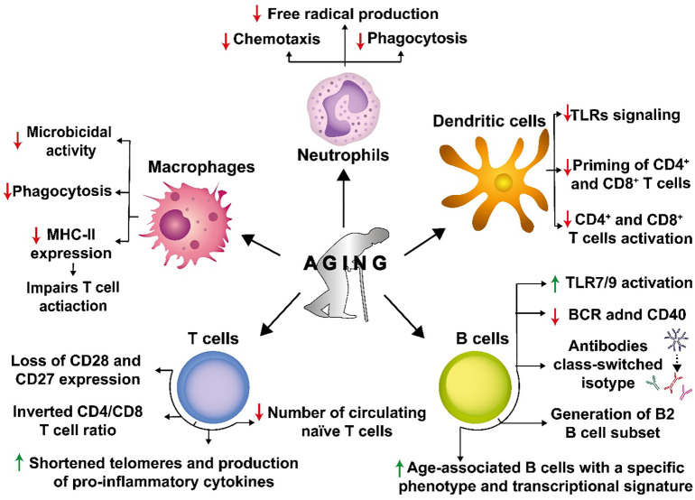
Cellular senescence is a key biological process characterized by irreversible cell cycle arrest. The accumulation of senescent cells creates a pro-inflammatory environment that can negatively affect tissue functions and may promote the development of aging-related diseases. Typical biomarkers related to senescence include senescence-associated β-galactosidase activity, histone H2A.X phosphorylation at serine139 (γH2A.X), and senescence-associated heterochromatin foci (SAHF) with heterochromatin protein 1γ (HP-1γ protein) Moreover, immune cells undergoing senescence, which is known as immunosenescence, can affect innate and adaptative immune functions and may elicit detrimental effects over the host's susceptibility to infectious diseases. Although associations between senescence and pathogens have been reported, clear links between both, and the related molecular mechanisms involved remain to be determined. Furthermore, it remains to be determined whether infections effectively induce senescence, the impact of senescence and immunosenescence over infections, or if both events coincidently share common molecular markers, such as γH2A.X and p53. Here, we review and discuss the most recent reports that describe cellular hallmarks and biomarkers related to senescence in immune and non-immune cells in the context of infections, seeking to better understand their relationships. Related literature was searched in Pubmed and Google Scholar databases with search terms related to the sections and subsections of this review.

Keywords: SASP; bacteria; chronic infections; immunosenescence; persistent infections; senescence; virus.


Conflict of interest statement

The authors declare that the research was conducted in the absence of any commercial or financial relationships that could be construed as a potential conflict of interest.

Figures

Figure 1
Figure 1
Cellular and molecular biomarkers related to senescence. (A) Senescent cells show characteristic transcriptional alterations, such as the acquisition of a pro-inflammatory secretome, known as the senescence-associated secretory phenotype (SASP), which relates to IL-6, IL-8, GROa, MCPs, MMP-1, MMP-3, MMP-9, MIP-1α, and PAI-1 release, among others. (B) An increase in senescence-associated beta-galactosidase (SA-β-Gal) activity, which is also a senescence marker, has been associated with upregulated expression of cyclin-dependent kinase inhibitors, such as p15INK4b, p16, p21CIP1, p53. These in turn induce cell cycle arrest. (C) Other additional biomarkers include sensors of the unfolded protein response (UPR), such as IRE1-α and ATF6. (D) Another marker related to senescence is the accumulation of lipofuscin at the lysosomal level. Lipofuscin and SA-β-Gal are colocalized in senescent cells. (E) In the nucleus, increased redistribution of senescence-associated heterochromatic foci (SAHF), telomere-associated DNA damage foci (TAF), and downregulation of Lamin B1 are also considered markers of senescence. (F) Lastly, the NKG2D/NKG2DL interaction promotes the activation of the cytotoxic activity of NK cells and the induction of apoptosis in senescent cells.
Figure 2
Figure 2
Mechanisms mediating senescence interplay with viral infections. During SARS-CoV-2 infections, a senescent phenotype is characterized by a pro-inflammatory environment involved in a dysfunctional loop with increased ACE2/DPP4 receptor expression leading to an induction in ICAM-1, VCAM-1 and ROS. In addition, TLR-3 amplifies this feedback during prolonged infection, senescence, and hyperinflammation. Hepatitis C virus (HCV) causes an increase in p16, p21, and p27, which cause cell cycle arrest in liver tissue samples and in hepatocellular carcinoma. Cellular senescence is also associated with HIV disease where this virus may induce IL-6 and TNF during infection. Measles virus (MV) is capable of generating cellular senescence as evidenced by reduced cell proliferation, SA-β-Gal activity, increased expression of p53 and p21 and induction of SASP in IMR90 cells. In HUVEC cells, Dengue virus elicits SA-β-Gal expression and cell cycle arrest. Furthermore, influenza A virus (IAV) has been described to induce cellular senescence related to an increased expression of NOS in Neuro2a cells. Human cytomegalovirus (HCMV) induces senescence through activation of p53 and p16 in human fibroblasts. Additionally, in B cells Epstein-Barr virus (EBV) triggers a G1 cellular arrest phase and an increase of p16, p21 and p53. Herpes simplex virus type 1 (HSV-1) infection leads to DNA damage trough a reduction of Ku80. Hepatitis B virus (HBV) infection is related to an increased expression of p16 and p21 and a decreased expression of pRb in malignant liver cell lines. Lastly, parvovirus B19 induces cellular senescence as seen by an increase in SA-β-Gal activity in human dermal fibroblasts.
Figure 3
Figure 3
Mechanisms mediating senescence interplay with bacterial infections. Fusobacterium and Salmonella increase caveolin-1 expression and promote bacterial uptake. During Streptococcus pneumoniae infections, inflammation is associated with increased senescence markers, such as p16, IL-1α/β, TNF-α, IL-6, and CXCL1. In sepsis or endotoxemia, the activation of the activation of TLR4 by lipopolysaccharide (LPS) sensing can promote a senescent phenotype in infected cells through the NF-kB-p53-p21 axis, supporting a prolonged inflammatory status with long-term adverse outcomes. Escherichia coli-infected cells display cellular markers related to senescence, such as SA-β-Gal activity and senescence-associated heterochromatic foci (SAHF). Helicobacter pylori has been described to have the ability to activate senescence and inhibit autophagosome maturation in T cells. M. tuberculosis induces changes in gene expression, DNA methylation and hypermethylation, and an increase in ROS and SASP. The CdtB of Helicobacter is associated with increased senescence, as evidenced by SA-β-Gal, p21, and p53. Helicobacter pylori cytotoxin-associated gen A (CagA) induces p21, that in turn induces cellular senescence. Porphyromonas gingivalis is associated with an increased expression of SA-β-Gal, p16, p53, and p21. Lactobacillus fermentum has been reported to play a protective role against senescence in a model of premature hydrogen peroxide-induced senescence by limiting DNA damage and SASP activation.
Figure 4
Figure 4
Senescence causes a detrimental impact on the immune system. Senescence affects numerous innate and adaptive immune cells. Senescence decreases chemotaxis, phagocytosis, and free radical production in neutrophils, which are necessary to engulf and kill different pathogens. Macrophages also experience decreased phagocytosis and reduced expression of major histocompatibility complex II molecules (MHC-II), affecting their microbicidal capacities and contributing to impaired T cell activation. Dendritic cells (DCs) display a reduced expression of TLRs, leading to reduced CD4+ and CD8+ T cell priming and activation. In T cells, senescence induces the loss of CD28 and CD27 expression on the cell surface and alters the CD4/CD8 T cell ratio. Additionally, replicative senescence in T cells is associated with shortened telomeres, increased production of pro-inflammatory cytokines, and a reduced number of circulating naive cells. In B cells, senescence induces an antibody class-switch that results in decreased immunity and the generation of B cells of the B2 cell subset that are short-lived recirculating cells that are metabolically impaired. In addition, there is an increase in the age-associated B cell subset with a specific phenotypic and transcriptional signature in the splenic B cell compartment.

References

    1. Abbadie C., Pluquet O. (2020). Unfolded protein response (UPR) controls major senescence hallmarks. Trends Biochem. Sci. 45, 371–374. doi: 10.1016/j.tibs.2020.02.005 - DOI - PubMed
    1. AbuBakar S., Shu M.-H., Johari J., Wong P.-F. (2014). Senescence affects endothelial cells susceptibility to dengue virus infection. Int. J. Med. Sci. 11, 538–544. doi: 10.7150/ijms.7896 - DOI - PMC - PubMed
    1. Acosta J. C., Banito A., Wuestefeld T., Georgilis A., Janich P., Morton J. P., et al. . (2013). A complex secretory program orchestrated by the inflammasome controls paracrine senescence. Nat. Cell Biol. 15, 978–990. doi: 10.1038/ncb2784 - DOI - PMC - PubMed
    1. Aguilera M. O., Delgui L. R., ROmano P. S., Colombo M. I. (2018). Chronic infections: A possible scenario for autophagy and senescence cross-talk. Cells 7, 1–21. doi: 10.3390/cells7100162 - DOI - PMC - PubMed
    1. Ahn S. H., Cho S.-H., Song J.-E., Kim S., Oh S. S., Jung S., et al. . (2017). Caveolin-1 serves as a negative effector in senescent human gingival fibroblasts during Fusobacterium nucleatum infection. Mol. Oral. Microbiol. 32, 236–249. doi: 10.1111/omi.12167 - DOI - PubMed