

Synovial sarcoma in the prevertebral space can mimic malignant neurogenic neoplasm: Case report and literature review
- PMID: 37753502
- PMCID: PMC10518339
- DOI: 10.1016/j.radcr.2023.08.106
Synovial sarcoma in the prevertebral space can mimic malignant neurogenic neoplasm: Case report and literature review
Abstract
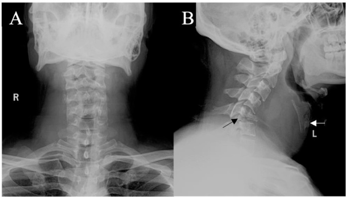
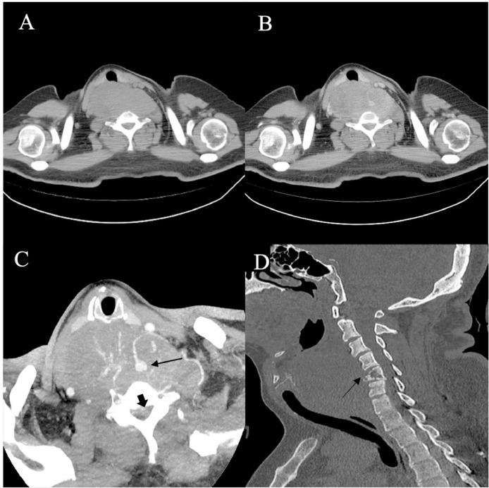
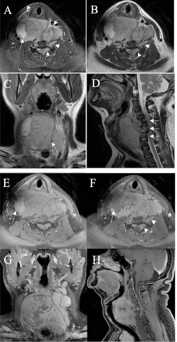
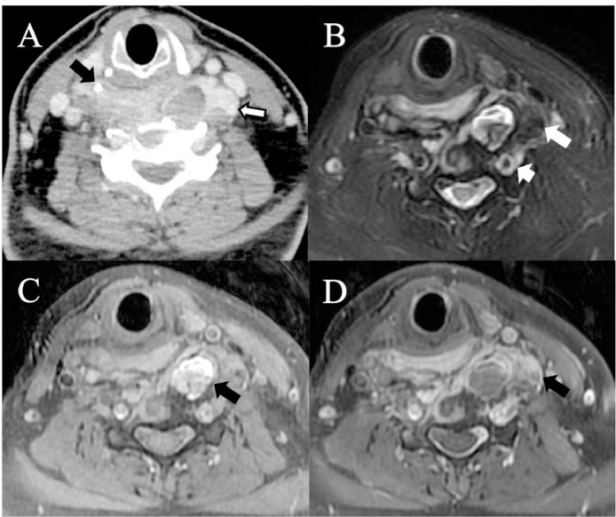
Synovial sarcoma, a rare malignant neoplasm with a poor prognosis, accounts for approximately 5%-10% of all primary soft-tissue malignancies worldwide. Typically affecting adolescents and young adults, it primarily manifests near the joints of the lower extremities. This study aimed to demonstrate that this tumor can also affect the prevertebral space. A 32-year-old male patient presented at our outpatient clinic with a 2-month history of upper limb numbness and a 1-month complaint of palpable neck mass. Imaging studies revealed a bulky, lobulated, and heterogeneous mass exhibiting heterogeneous enhancement. Furthermore, the mass caused expansion of the neuroforamen in the neck, initially suggesting a diagnosis of malignant schwannoma. However, a histopathologic examination suggested synovial sarcoma. The article provided a comprehensive review of the clinical, pathological, and radiological features of this condition. Additionally, it explored current treatment options and prognoses by referencing relevant literature.
Keywords: Imaging; Oncology; Synovial sarcoma; The retropharyngeal space.
© 2023 The Authors. Published by Elsevier Inc. on behalf of University of Washington.
Figures
Similar articles
-
Biphasic synovial sarcoma in the cervical spine: Case report.Chiropr Man Therap. 2011 May 23;19(1):12. doi: 10.1186/2045-709X-19-12. Chiropr Man Therap. 2011. PMID: 21605432 Free PMC article.
-
Primary pharyngeal synovial sarcoma in a pediatric patient: A case report.Medicine (Baltimore). 2021 Dec 30;100(52):e28411. doi: 10.1097/MD.0000000000028411. Medicine (Baltimore). 2021. PMID: 34967377 Free PMC article.
-
Synovial sarcoma in the prevertebral space of the neck: CT and MR findings.Radiat Med. 1997 Mar-Apr;15(2):121-4. Radiat Med. 1997. PMID: 9192439
-
Synovial sarcoma of the abdominal wall: Imaging findings and review of the literature.J Radiol Case Rep. 2015 Feb 28;9(2):24-30. doi: 10.3941/jrcr.v9i2.1992. eCollection 2015 Feb. J Radiol Case Rep. 2015. PMID: 25926925 Free PMC article. Review.
-
Biphasic synovial sarcoma of the epiglottis: Case report and literature review.Auris Nasus Larynx. 2018 Jun;45(3):617-621. doi: 10.1016/j.anl.2017.06.007. Epub 2017 Jul 6. Auris Nasus Larynx. 2018. PMID: 28689931 Review.
References
-
- WHO. WHO classification of tumours: soft tissue and bone tumours. 2020. ISBN: 9780511545375.
Publication types
LinkOut - more resources
Full Text Sources
