. 2023 Dec 1;325(6):G556-G569.
doi: 10.1152/ajpgi.00129.2023. Epub 2023 Sep 27.

Altered hepatic and intestinal homeostasis in a neonatal murine model of short-term total parenteral nutrition and antibiotics

Affiliations

Altered hepatic and intestinal homeostasis in a neonatal murine model of short-term total parenteral nutrition and antibiotics

Tahliyah S Mims et al. Am J Physiol Gastrointest Liver Physiol. .

Abstract

Parenteral nutrition (PN) prevents starvation and supports metabolic requirements intravenously when patients are unable to be fed enterally. Clinically, infants are frequently provided PN in intensive care settings along with exposure to antibiotics (ABX) to minimize infection during care. Unfortunately, neonates experience extremely high rates of hepatic complications. Adult rodent and piglet models of PN are well-established but neonatal models capable of leveraging the considerable transgenic potential of the mouse remain underdeveloped. Utilizing our newly established neonatal murine PN mouse model, we administered ABX or controlled drinking water to timed pregnant dams to disrupt the maternal microbiome. We randomized mouse pups to PN or sham surgery controls +/- ABX exposure. ABX or short-term PN decreased liver and brain organ weights, intestinal length, and mucosal architecture (vs. controls). PN significantly elevated evidence of hepatic proinflammatory markers, neutrophils and macrophage counts, bacterial colony-forming units, and evidence of cholestasis risk, which was blocked by ABX. However, ABX uniquely elevated metabolic regulatory genes resulting in accumulation of hepatocyte lipids, triglycerides, and elevated tauro-chenoxycholic acid (TCDCA) in serum. Within the gut, PN elevated the relative abundance of Akkermansia, Enterococcus, and Suterella with decreased Anaerostipes and Lactobacillus compared with controls, whereas ABX enriched Proteobacteria. We conclude that short-term PN elevates hepatic inflammatory stress and risk of cholestasis in early life. Although concurrent ABX exposure protects against hepatic immune activation during PN, the dual exposure modulates metabolism and may contribute toward early steatosis phenotype, sometimes observed in infants unable to wean from PN.NEW & NOTEWORTHY This study successfully established a translationally relevant, murine neonatal parenteral nutrition (PN) model. Short-term PN is sufficient to induce hepatitis-associated cholestasis in a neonatal murine model that can be used to understand disease in early life. The administration of antibiotics during PN protects animals from bacterial translocation and proinflammatory responses but induces unique metabolic shifts that may predispose the liver toward early steatosis.

Keywords: PNALD; liver; pediatrics; total parenteral nutrition.


Conflict of interest statement

J.F.P. is cofounder of Gateway Biome, Inc. There are no financial or intellectual conflicts of with this research and Gateway Biome, Inc. None of the other authors has any conflicts of interest, financial or otherwise, to disclose.

Figures

None
Graphical abstract
Figure 1.
Figure 1.
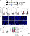
Parenteral nutrition induces hepatic inflammation and aerobic bacterial colonization, which is blocked by antibiotics. A: experimental schematic is shown where timed pregnant dams were provided antibiotic or control tap water on gestational day 14 before pup randomization to parenteral nutrition (PN) or enteral nutrition (EN) at postnatal day 9, for 72 h. B: quantitative PCR assessment of F4/80, IL-6, Tnf-α, and iNos (n = 5–8 animals/group). C: MPO- and F4/80-positive cells were quantified in tissue sections (n = 3 or 4 animals/group). D: representative immunofluorescence of myeloperoxidase (MPO) and F4/80 (macrophages), with scale bar = 100 µm. Sections were counterstained with DAPI. E: liver tissue was homogenized and plated for colony-forming units under aerobic (BHI agar) and anaerobic (Blood agar) conditions (n = 4–7 animals/group). F: 16S taxonomic identity of aerobic CFUs. G: Spearman Rank Correlation Coefficient of genus detected in PN (Orange) and PN + ABX (Green) livers under aerobic conditions. BioRender was used to generate A. ABX, antibiotics. ANOVA *P < 0.05, **P < 0.01.
Figure 2.
Figure 2.
Parenteral nutrition decreases bile salt export protein and elevates hepatic bile acid levels, whereas concurrent antibiotic exposure alters hepatic bile acid and lipid synthesis genes toward fatty acid synthesis. A: schematic representation of the classical and alternative bile acid synthesis pathways. The classical pathway begins cholesterol’s enzymatic conversion by CYP7A1, which creates intermediate products utilized by CYP8B1 to later produce CA and its taurine-conjugated form TCA. The alternative or acidic pathway begins with enzymatic conversion of cholesterol by CYP27A, which creates intermediate products used as substrate for CYP7B1 to produce CDCA and the taurine-conjugated TCDCA. cP450 or CYP2C70 will convert (U) CDCA to β-MCA in mice. In the intestines, microbe transform these primary BAs classically to DCA and alternatively to LCA, HDCA, and UDCA. B: quantitative PCR assessment of CYP genes 7a1, 8b1, 7b1, and 27a1, n = 4–7 animals/group, ANOVA, *P < 0.05, **P < 0.01, ***P < 0.001, C: quantitative PCR assessment of hepatic BA synthesis regulating genes Fxr, Shp, and Tgr5. n = 4–7 animals/group, ANOVA, **P < 0.01. D: quantitative PCR assessment of hepatic bile acid transporters, Bsep, Ntcp, Oatp, Ostα, and Ostß. n = 4–7 animals/group, ANOVA, *P < 05. E: quantitative PCR assessment of fatty acid synthesis genes Srebp1c, Fasn and Scd-1, n = 4–7 animals/group. ANOVA, *P < 0.05, **P < 0.01, ***P < 0.001. F: liver homogenate triglyceride, bile acid (BA), and cholesterol levels normalized to tissue weight. G: representative histology stained by hematoxylin and eosin staining (scale bar = 400 µm; insets = 50 µm) showing lipid droplets in ABX-exposed mice. Biorender was used to generate A. ABX, antibiotics; CA, cholic acid; CDCA, chenoxycholic acid; TCA, taurocholic acid; TCDCA, tauro-chenoxycholic acid.
Figure 3.
Figure 3.
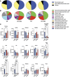
Circulating bile acid composition following parenteral nutrition and antibiotic exposure. A: pie charts show composition of serum bile acids at experimental end point, which were dominated by TCA and TβMCA. B: pie chart shows composition of remaining low abundance bile acids, excluding TCA and TβMCA. C: all individual bile acids shown by group. ANOVA *P < 0.05, **P < 0.01, ***P < 0.005, ****P < 0.001. TβMCA, tauro-β-muricholic acid; TCA, taurocholic acid.
Figure 4.
Figure 4.
Parenteral nutrition and antibiotics alter intestinal architecture and bile acid signaling. A: small intestinal and colonic length (n = 6–8 animals/group). B: mucosal architecture was assessed for villus height and crypt depths (n = 3–7 animals/group). C: quantitative PCR was used to determine expression for Fxr, Tgr5, Fgf15, Abst, iBabp, Ostα, and Ostß (n = 3–6 animals/group). ANOVA *P < 0.05, **P < 0.01. D: quantitative PCR was used to assess tight junction proteins Tjp and Ocn. E: representative histology of ZO-1 (TJP1) in the distal ileum. Counter stain was performed by DAPI. Scale bars = 100 µm.
Figure 5.
Figure 5.
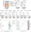
Parenteral nutrition and antibiotics alters the intestinal microbiome. A: changes in cecal microbiome community abundance are shown at the phylum level. B: α diversity of the cecal microbiome was assessed by the Shannon Diversity and Simpson’s index. C: changes at the genus level are shown for each group. D: β diversity of all experimental groups is shown on Principal Component Analysis. E: pattern search results for enriched taxa across all four experimental treatment groups. (n = 5 or 6 animals/group). ANOVA *P < 0.05, **P < 0.01, ***P < 0.005, ****P < 0.001.

References

    1. Buchman AL. Complications of long-term home total parenteral nutrition: their identification, prevention and treatment. Dig Dis Sci 46: 1–18, 2001. doi:10.1023/A:1005628121546. - DOI - PubMed
    1. Kansagra K, Stoll B, Rognerud C, Niinikoski H, Ou C-N, Harvey R, Burrin D. Total parenteral nutrition adversely affects gut barrier function in neonatal piglets. Am J Physiol Gastrointest Liver Physiol 285: G1162–G1170, 2003. doi:10.1152/ajpgi.00243.2003. - DOI - PubMed
    1. Nowak K. Parenteral nutrition-associated liver disease. Clin Liver Dis (Hoboken) 15: 59–62, 2020. doi:10.1002/CLD.888. - DOI - PMC - PubMed
    1. Omata J, Pierre JFJF, Heneghan AFAF, Tsao F, Sano Y, Jonker MAMA, Kudsk KAKA. Parenteral nutrition suppresses the bactericidal response of the small intestine. Surgery 153: 17–24, 2013. doi:10.1016/j.surg.2012.04.001. - DOI - PMC - PubMed
    1. Pierre JF, Heneghan AF, Meudt JM, Shea MP, Krueger CG, Reed JD, Kudsk KA, Shanmuganayagam D. Parenteral nutrition increases susceptibility of ileum to invasion by e coli. J Surg Res 183: 583–591, 2013. doi:10.1016/j.jss.2013.01.054. - DOI - PMC - PubMed

Publication types

Substances

LinkOut - more resources