Review
. 2023 Sep 1;13(9):867.
doi: 10.3390/bios13090867.

Electrochemical Biosensors for the Detection of Antibiotics in Milk: Recent Trends and Future Perspectives

Affiliations
Review

Electrochemical Biosensors for the Detection of Antibiotics in Milk: Recent Trends and Future Perspectives

Baljit Singh et al. Biosensors (Basel). .

Abstract

Antibiotics have emerged as ground-breaking medications for the treatment of infectious diseases, but due to the excessive use of antibiotics, some drugs have developed resistance to microorganisms. Because of their structural complexity, most antibiotics are excreted unchanged, polluting the water, soil, and natural resources. Additionally, food items are being polluted through the widespread use of antibiotics in animal feed. The normal concentrations of antibiotics in environmental samples typically vary from ng to g/L. Antibiotic residues in excess of these values can pose major risks the development of illnesses and infections/diseases. According to estimates, 300 million people will die prematurely in the next three decades (by 2050), and the WHO has proclaimed "antibiotic resistance" to be a severe economic and sociological hazard to public health. Several antibiotics have been recognised as possible environmental pollutants (EMA) and their detection in various matrices such as food, milk, and environmental samples is being investigated. Currently, chromatographic techniques coupled with different detectors (e.g., HPLC, LC-MS) are typically used for antibiotic analysis. Other screening methods include optical methods, ELISA, electrophoresis, biosensors, etc. To minimise the problems associated with antibiotics (i.e., the development of AMR) and the currently available analytical methods, electrochemical platforms have been investigated, and can provide a cost-effective, rapid and portable alternative. Despite the significant progress in this field, further developments are necessary to advance electrochemical sensors, e.g., through the use of multi-functional nanomaterials and advanced (bio)materials to ensure efficient detection, sensitivity, portability, and reliability. This review summarises the use of electrochemical biosensors for the detection of antibiotics in milk/milk products and presents a brief introduction to antibiotics and AMR followed by developments in the field of electrochemical biosensors based on (i) immunosensor, (ii) aptamer (iii) MIP, (iv) enzyme, (v) whole-cell and (vi) direct electrochemical approaches. The role of nanomaterials and sensor fabrication is discussed wherever necessary. Finally, the review discusses the challenges encountered and future perspectives. This review can serve as an insightful source of information, enhancing the awareness of the role of electrochemical biosensors in providing information for the preservation of the health of the public, of animals, and of our environment, globally.

Keywords: AMR; MIPs; antibiotics; aptamers and enzymes; biosensors; electrochemical instrumentation; immunosensors; milk; nanomaterials.


Conflict of interest statement

The authors declare no conflict of interest.

Figures

Figure 1
Figure 1
(Left) Schematic illustration of possible links between antibiotic use in agriculture and human disease. The usage of antibiotics has an impact on the incidence of resistant microorganisms. The capacity for sustained human-to-human transmission is a critical factor in the impact of infection. Arrows connecting the two populations show: (A) direct transmission of bacteria that are not adapted for transmission to humans via the food chain (e.g., campylobacter, salmonella); or (B) direct transmission of organisms that are adapted for transmission to humans; and (C) transfer of resistance genes from the agricultural setting into pathogens that are transmitted among humans. (Right) Pathway of antibiotics and antibiotic-resistant genes through agriculture and livestock.
Figure 2
Figure 2
Distribution (A) and classification (B) of the analytical methods used for antibiotics determination in food [66]. Reprinted from Ref. [66], Copyright (2010), with permission from Elsevier.
Figure 3
Figure 3
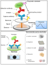
(A) Principle of electrochemical sensors (electrode) for the detection of antibiotics [68]. Reprinted from Ref. [68], Copyright (2021), with permission from Elsevier. (B(a)) Schematic of a generic biosensor device. A sample containing the target (analyte) is placed in close contact with the surface of the sensor. The bioreceptor (antibody, lectin, enzyme, aptamer, DNA, or cells), which is immobilised above the transducer (electrochemical, piezoelectric, optical, calorimetric modes, etc.), binds with the target from the sample (recognition event), after which the transducer converts the binding event into a measurable signal that is proportional to the concentration of the target (signal readout). (b) Examples of electrochemical transduction signals encompass amperometric, potentiometric, conductimetric, and impedimetric signals [69]. Reprinted from Ref. [69], Copyright (2022), with permission from Elsevier. (C) Electrochemical detection, in which the working, reference and counter electrodes are arranged in a manner allowing the sample to be in contact with all three at the same time. The range of possible surface modifications on the working electrode are shown [70]. Reproduced under the terms of the CC-BY licence from Ref. [70], Copyright 2021, The Authors, published by MDPI.
Figure 4
Figure 4
(Top) Recent advances in nanomaterial-based electrochemical detection of antibiotics (graphical representation) [72]. Reprinted from Ref. [72], Copyright (2020), with permission from Elsevier. (Middle) Schematic illustration for application of various nanomaterials and biorecognition elements in the development of antibiotic biosensors [73]. Reprinted from Ref. [73], Copyright (2020), with permission from Elsevier. (Bottom) An overview of functional carbon nanomaterials and their application in the detection of antibiotics [74]. Reproduced under the terms of the CC-BY licence from Ref. [74] [Nanomaterials], Copyright 2022, The Authors, published by MDPI.
Figure 5
Figure 5
(Top) Basic analytical principle of electrochemical immunosensors. (Bottom) Nanomaterials used as electrodes or supporting solid matrices to enhance the analytical performance of electrochemical immunosensing [84]. Reproduced under the terms of the CC-BY licence from Ref. [84], Copyright 2018, The Authors, published by MDPI.
Figure 6
Figure 6
Graphical representation/abstract of the disposable amperometric magneto-immunosensor for the direct detection of tetracycline antibiotic residues in milk [95]. Reprinted from Ref. [95], Copyright (2012), with permission from Elsevier.
Figure 7
Figure 7
Schematic display of the immunosensor developed for the detection of sulfonamide antibiotics (inset: details of the surface chemistry involved in the covalent immobilisation of the capture antibody by using EDC and Sulfo-NHS 4-ABA film formed on an SPCE) [97]. Reprinted from Ref. [97], Copyright (2012), with permission from Elsevier.
Figure 8
Figure 8
Layout of 24-site fluidic micro-array analytical device: (A) one-dimensional schematic plot of the base layer; (B) three-dimensional layered schematic plot of the whole device; (C) detection state of the Isensor; (D) preparation state of the Isensor; (E) micro-reservoir of one analysis unit in its detection state; (F) micro-reservoir of one analysis unit in its preparation state; (G) picture of the analytical device [98]. Reprinted from Ref. [98], Copyright (2018), with permission from Elsevier.
Figure 9
Figure 9
Schematic representation of the synthesis of nMoS2 NPs and the development of BSA/anti-AMP/APTES/nMoS2/ITO immunoelectrode for AMP detection [99]. Reprinted from Ref. [99], Copyright (2021), with permission from Elsevier.
Figure 10
Figure 10
(A) Schematic display of the steps involved in the affinity magnetosensor developed for β-lactam antibiotics. (B) Picture showing the SPCE and the home-made magnet holding block (1), the deposition of the modified MBs on the SPCE assembled on the magnet holding block (2) and the assembled SPCE-magnet holding block immersed in the electrochemical cell used for the amperometric measurements (3) [100]. Reproduced from Ref. [100], Copyright (2013), with permission from the Royal Society of Chemistry.
Figure 11
Figure 11
(Top) Immobilisation protocols applied in electrochemical aptasensors: (a) interaction of thiolated aptamer with AuNPs/bare Au electrode; (b) carbodiimide binding to carboxylated support; (c) electrografting with diazonium salt generated from aromatic amino group; (d) glutaraldehyde cross-binding of aminated aptamer and carrier. (Bottom) Electrochemical sensors used as transducers of aptasensors: (a) potentiometric cell: WE—working electrode, RE—reference electrode, V—voltmeter; (b) cyclic voltammetry; (c) amperometry; (d) differential pulse voltammetry (DPV) and square wave voltammetry (SWV). The red line illustrates the shape of the current peak obtained; (e) electrochemical impedance spectroscopy; the Nyquist diagram and equivalent circuit correspond to the double-electric layer. Re is electrolyte resistance, Rct is charge transfer resistance, W is Warburg impedance, and CPE is the constant phase element [111]. Reproduced under the terms of the CC-BY licence from Ref. [111] [Sensors], Copyright 2022, The Authors, published by MDPI.
Figure 12
Figure 12
Schematic representation of the sandwich-type aptasensor based on GR-3D Au and aptamer-AuNPs-HRP for the detection of oxytetracycline [117]. Reprinted from Ref. [117], Copyright (2017), with permission from Elsevier.
Figure 13
Figure 13
(A) Schematic illustration of aptasensor based on the utilisation of QDs, AuNSs, RNA-based aptamer strands, and high-affinity pairing between Bio and SA for the simultaneous detection of multiple antibiotics [119]. Reprinted from Ref. [119], Copyright (2021), with permission from Elsevier. (B) Schematic aptasensor fabrication process: (a) formation process of OMC@Ti3C2 MXene; (b) binding mode of aptamer with target [120]. Reproduced under the terms of the CC-BY licence from Ref. [120], Copyright 2022, The Authors, published by MDPI. (C) Schematic diagram of the fabrication procedure of the CeO2/CuOx@mC-based aptasensor for detecting TOB, including (i,ii) the preparation of the series of CeO2/CuOx@mC nanocomposites, (iii) the immobilisation of the aptamer strands over the CeO2/CuOx@mC composite, and (iv) TOB detection using the proposed CeO2/CuOx@mC-based aptasensor [121]. Reprinted from Ref. [121], Copyright (2019), with permission from Elsevier.
Figure 14
Figure 14
Summary of the preparation procedure, signal enhancement and sensing mechanism for MIPs in MIP-based electrochemical sensors [133]. Reprinted from Ref. [133], Copyright (2021), with permission from Elsevier.
Figure 15
Figure 15
(A) Label-free electrochemical detection of the Cloxacillin antibiotic in milk samples based on molecularly imprinted polymer and graphene oxide–gold nanocomposite [140]. Reprinted from Ref. [140], Copyright (2019), with permission from Elsevier. (B) Schematic illustration of the fabrication procedure for AuNPs/MWCNTs-CS/sol–gel-MIP/GCE [141]. Reprinted from Ref. [141], Copyright (2017), with permission from Elsevier. (C) Schematic diagram of the preparation of the aptamer-MIP nanohybride for CAP detection. Reprinted from Ref. [142], Copyright (2019), with permission from Elsevier. (D) Impedimetric ultrasensitive detection of chloramphenicol based on aptamer MIPs using a glassy carbon electrode modified by 3-ampy-RGO and silver nanoparticle [142]: (a) covering of 3-ampy-RGO on the GCE surface; (b) immobilisation of the AgNPs on the 3-ampy-RGO/GCE; (c) covalent attachment of the aptamer[CAP] complex on the AgNP/3-ampy-RGO/GCE surface; (d) electropolymerisation of resorcinol on the aptamer[CAP] complex/AgNP/3-ampy-RGO/GCE; (e) washing of the modified electrode with washing solution and removal of the CAP; (f) addition of CAP as a target and some antibiotic as interferents [142]. Reprinted from Ref. [142], Copyright (2019), with permission from Elsevier.
Figure 16
Figure 16
(A) Enzyme biosensors for biomedical applications. Schematic representation of (B) first-generation, (C) second-generation and (D) third-generation biosensors [153]. Reproduced under the terms of the CC-BY licence from Ref. [153], Copyright 2016, The Authors, published by MDPI.
Figure 17
Figure 17
Schematic depiction of the developed multi-antibiotic magnetosensor. The modified MBs (a) are commingled together (b) and incubated with the sample in the presence of fixed amounts of the three enzyme-labelled analogues, thus establishing a direct competitive assay (c); the MBs are then captured on the surface of an SPCE assembled on the magnet holding block (d) and the assembly SPCE-magnet holding block immersed in the electrochemical cell is used for the amperometric measurements (e) [161]. Reprinted from Ref. [161], Copyright (2014), with permission from Elsevier.
Figure 18
Figure 18
(Top) Schematic diagram of the experimental configuration: β-lactamases are connected to the gold electrode surface by cysteine molecules. Penicillin G molecules in solution react with the β-lactamases. (Bottom) Cyclic volammetry of a ferrocene solution, 2 mmol L−1, in KNO3, 1 mol L−1, with different electrode surfaces: bare gold; gold and cysteine SAM; gold, cysteine SAM with penicillinase; and gold, cysteine SAM with penicillinase and the analyte penicillin (inset: detail of lower currents) [155]. Reprinted from Ref. [155], Copyright (2014), with permission from Elsevier.
Figure 19
Figure 19
Schematic of a typical whole-cell-based biosensor (WCBs) [162]. Reproduced under the terms of the CC-BY licence from Ref. [162], Copyright 2017, The Authors, published by MDPI.
Figure 20
Figure 20
Electrochemical-based “antibiotsensor” for the whole-cell detection of the vancomycin-susceptible bacteria [168]. Reprinted from Ref. [168], Copyright (2021), with permission from Elsevier.

References

    1. Sharma A., Istamboulie G., Hayat A., Catanante G., Bhand S., Marty J.L. Disposable and portable aptamer functionalized impedimetric sensor for detection of kanamycin residue in milk sample. Sens. Actuators B Chem. 2017;245:507–515.
    1. Ben Y., Fu C., Hu M., Liu L., Wong M.H., Zheng C. Human health risk assessment of antibiotic resistance associated with antibiotic residues in the environment: A review. Environ. Res. 2019;169:483–493. - PubMed
    1. You Y., Silbergeld E.K. Learning from agriculture: Understanding low-dose antimicrobials as drivers of resistome expansion. Front. Microbiol. 2014;5:284. - PMC - PubMed
    1. Hong P.-Y., Al-Jassim N., Ansari M.I., Mackie R.I. Environmental and public health implications of water reuse: Antibiotics, antibiotic resistant bacteria, and antibiotic resistance genes. Antibiotics. 2013;2:367–399. doi: 10.3390/antibiotics2030367. - DOI - PMC - PubMed
    1. Hao H., Cheng G., Iqbal Z., Ai X., Hussain H.I., Huang L., Dai M., Wang Y., Liu Z., Yuan Z. Benefits and risks of antimicrobial use in food-producing animals. Front. Microbiol. 2014;5:288. - PMC - PubMed

Substances

LinkOut - more resources