

A Methylene Blue-Enhanced Nanostructured Electrochemical Immunosensor for H-FABP Myocardial Injury Biomarker
- PMID: 37754107
- PMCID: PMC10526172
- DOI: 10.3390/bios13090873
A Methylene Blue-Enhanced Nanostructured Electrochemical Immunosensor for H-FABP Myocardial Injury Biomarker
Abstract
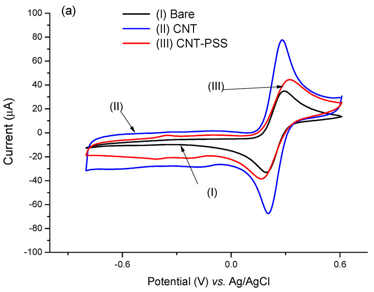
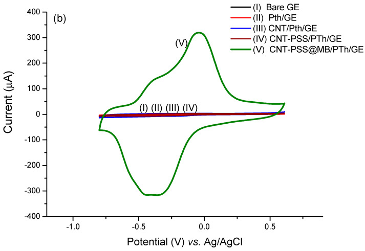
A sensitive electrochemical immunosensor for the detection of the heart-type fatty acid binding protein (HFABP), an earlier biomarker for acute myocardial infarction than Troponins, is described. The sensing platform was enhanced with methylene blue (MB) redox coupled to carbon nanotubes (CNT) assembled on a polymer film of polythionine (PTh). For this strategy, monomers of thionine rich in amine groups were electrosynthesized by cyclic voltammetry on the immunosensor's gold surface, forming an electroactive film with excellent electron transfer capacity. Stepwise sensor surface preparation was electrochemically characterized at each step and scanning electronic microscopy was carried out showing all the preparation steps. The assembled sensor platform combines MB and PTh in a synergism, allowing sensitive detection of the H-FABP in a linear response from 3.0 to 25.0 ng∙mL-1 with a limit of detection of 1.47 ng∙mL-1 HFABP that is similar to the clinical level range for diagnostics. H-FABP is a newer powerful biomarker for distinguishing between unstable angina and acute myocardial infarction.
Keywords: H-FABP; acute myocardial infarction; immunosensor; methylene blue; polythionine.
Conflict of interest statement
The authors declare no conflict of interest.
Figures


Similar articles
-
Electrochemical immunosensor development based on core-shell high-crystalline graphitic carbon nitride@carbon dots and Cd0.5Zn0.5S/d-Ti3C2Tx MXene composite for heart-type fatty acid-binding protein detection.Mikrochim Acta. 2021 May 7;188(6):182. doi: 10.1007/s00604-021-04838-6. Mikrochim Acta. 2021. PMID: 33959811
-
A sensitive sandwich-type immunosensor for the detection of galectin-3 based on N-GNRs-Fe-MOFs@AuNPs nanocomposites and a novel AuPt-methylene blue nanorod.Biosens Bioelectron. 2018 Mar 15;101:253-259. doi: 10.1016/j.bios.2017.10.026. Epub 2017 Oct 16. Biosens Bioelectron. 2018. PMID: 29096363
-
A sensitive capacitive immunosensor for direct detection of human heart fatty acid-binding protein (h-FABP).Talanta. 2015 Jan;132:37-43. doi: 10.1016/j.talanta.2014.08.067. Epub 2014 Sep 6. Talanta. 2015. PMID: 25476276
-
[Diagnostic and prognostic value of heart-type fatty acid-binding protein (H-FABP), an early biochemical marker of myocardial injury].Arch Mal Coeur Vaiss. 2005 Dec;98(12):1225-31. Arch Mal Coeur Vaiss. 2005. PMID: 16435602 Review. French.
-
Heart-type fatty acid binding protein (H-FABP) as a biomarker for acute myocardial injury and long-term post-ischemic prognosis.Acta Pharmacol Sin. 2018 Jul;39(7):1155-1163. doi: 10.1038/aps.2018.37. Epub 2018 May 17. Acta Pharmacol Sin. 2018. PMID: 29770799 Free PMC article. Review.
References
Grants and funding
LinkOut - more resources
Full Text Sources
Miscellaneous
