Review
. 2023 Sep 15;13(9):885.
doi: 10.3390/bios13090885.

Recent Uses of Paper Microfluidics in Isothermal Nucleic Acid Amplification Tests

Affiliations
Review

Recent Uses of Paper Microfluidics in Isothermal Nucleic Acid Amplification Tests

Jocelyn Reynolds et al. Biosensors (Basel). .

Abstract

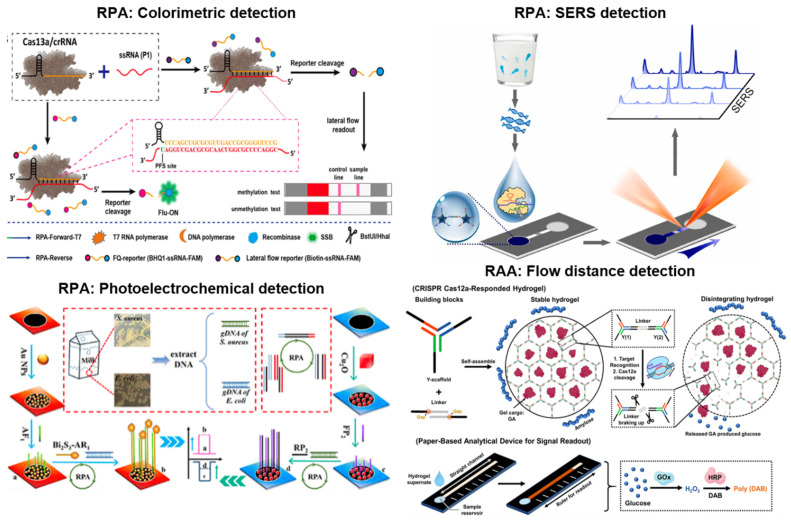
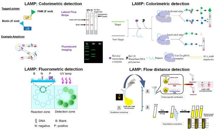
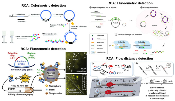
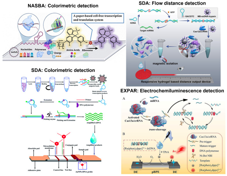
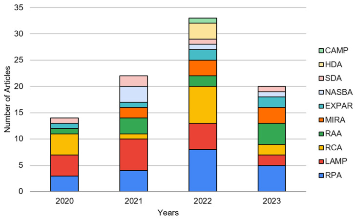
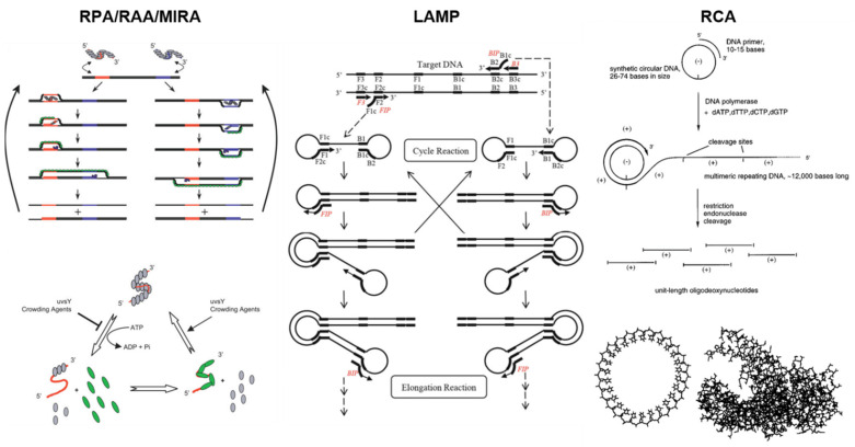
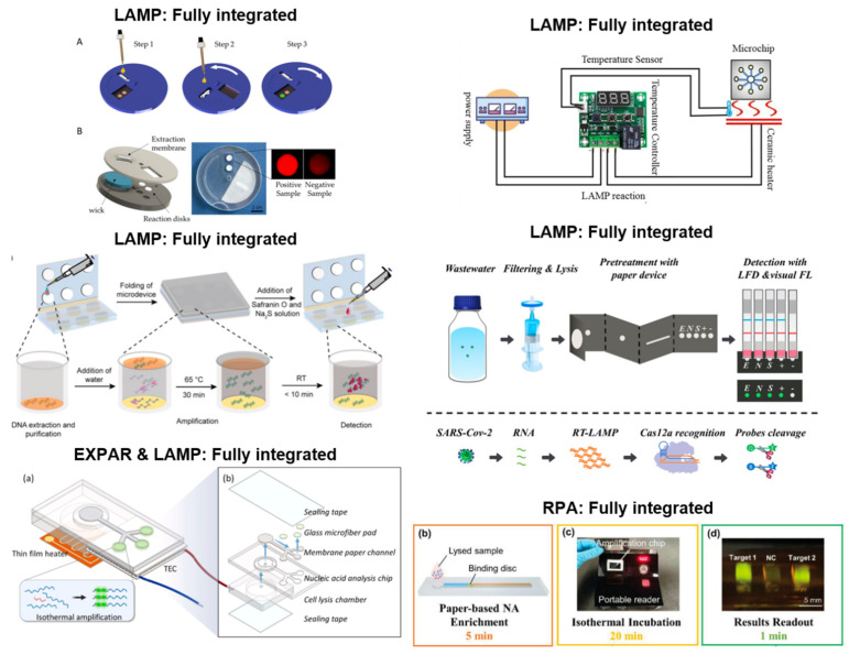
Isothermal nucleic acid amplification tests have recently gained popularity over polymerase chain reaction (PCR), as they only require a constant temperature and significantly simplify nucleic acid amplification. Recently, numerous attempts have been made to incorporate paper microfluidics into these isothermal amplification tests. Paper microfluidics (including lateral flow strips) have been used to extract nucleic acids, amplify the target gene, and detect amplified products, all toward automating the process. We investigated the literature from 2020 to the present, i.e., since the onset of the COVID-19 pandemic, during which a significant surge in isothermal amplification tests has been observed. Paper microfluidic detection has been used extensively for recombinase polymerase amplification (RPA) and its related methods, along with loop-mediated isothermal amplification (LAMP) and rolling circle amplification (RCA). Detection was conducted primarily with colorimetric and fluorometric methods, although a few publications demonstrated flow distance- and surface-enhanced Raman spectroscopic (SERS)-based detection. A good number of publications could be found that demonstrated both amplification and detection on paper microfluidic platforms. A small number of publications could be found that showed extraction or all three procedures (i.e., fully integrated systems) on paper microfluidic platforms, necessitating the need for future work.

Keywords: lateral flow immunochromatographic assay; loop-mediated isothermal amplification; microfluidic paper-based analytic device; recombinase polymerase amplification; rolling circle amplification.


Conflict of interest statement

The authors declare no conflict of interest.

Figures

Figure 3
Figure 3
Paper-based detection examples with RPA and RAA. Colorimetric detection (top left) is demonstrated using RPA and CRISPR/Cas13a to determine DNA methylation in a site-specific manner. It shows the signal amplification for methylated DNA and the elimination of the false identification of incomplete breakdown of unmethylated targets. SERS detection (top right) is demonstrated for RPA-Cas12a on a microfluidic paper-based analytical device (μPAD), increasing sensitivity in a portable, qualitative manner. Photoelectrochemical detection (bottom left) is shown on 3D-printed, paper-based electrodes to rapidly detect foodborne pathogens’ nucleic acids under 980 nm light. In the flow distance detection (bottom right), with crRNA guidance, target dsDNA becomes actuated and cleaves linkers within the DNA hydrogel. Once the linkers are broken down, signal molecules are released in the hydrogel to achieve signal transduction. As denoted in the figure, glucose is oxidized into hydrogen peroxide within the circular reservoir and converted into poly(DAB) in the straight channel via HRP. Visibly, the distance of the bar indicates the content of rRNA. Reprinted with permissions from [48], Copyright 2021 American Chemical Society; [49], Copyright 2022 Elsevier; [52], Copyright 2023 American Chemical Society; and [67], Copyright 2021 American Chemical Society. Note: [52] demonstrated amplification in addition to detection.
Figure 4
Figure 4
Paper-based detection examples with LAMP. In the colorimetric detection (top left), the primers tagging the 5′-end allow for amplification without inhibition of tags. LFIA strips with fluorescent gels characterize the LAMP products when the FAM tag attaches to the anti-FAM test line. In the other colorimetric detection (top right), primers perform reverse transcription amplification of genes. When the Cas12a-gRNA complex is activated, ssDNA is cleaved, which can be modified with a fluorescent and quenching group. Once separated, the fluorescence is observed with 480 nm light. In fluorometric detection (bottom left), fluorescence signals from the reaction and detection zones are compared, and the values are displayed on the smartphone screen. In the flow distance detection (bottom right), fluorescent SYBR-quantified LAMP products were conjugated with AuNPs on paper. Using DNA-immobilized cellulose, qualitative screening with a blue light illuminator evaluates the fluorescent migratory distance for the amounts of LAMP product. Reprinted with permissions from [81], Copyright 2020 Springer Nature; [96], Copyright 2022 American Chemical Society; and [89], Copyright 2022 American Chemical Society. Reprinted from [84] under Creative Commons Attribution License. Note: [89] demonstrated amplification in addition to detection, and [96] showcased a fully integrated system.
Figure 5
Figure 5
Paper-based detection examples with RCA. Colorimetric detection (top left) exposes the sample to a nuclease, which cleaves a selected ssDNA fragment detected in an LFIA strip. In fluorometric detection (top right), the target RNA is ligated using the padlock probe with a complementary sequence of the target RNA virus and a DNA ligase. The FnCas12a/crRNA complex cleaves targeted dsDNA regions, which emit fluorescence. The nonspecific ssDNA is also detected from a fluorescence reader. In the other fluorometric detection (bottom left), the RCA products are pre-labeled with biotin and fluorophore, efficiently capturing the fluorescence signal at high resolution and sensitivity. In distance detection (bottom right), the microfluidic paper-based analytical devices (μPADs) contain indicator and detection zones to quantify miRNAs via viscosity amplifications. The pre-amplified miRNA (a) is placed in the sample well, and the difference in the target and control flow are enhanced with surface hydrophobicity modulations (b). It is paired with smartphone auto-reading systems to improve accuracy (c). Reprinted with permissions from [106], Copyright 2021 Royal Society of Chemistry; [111], Copyright 2023 Elsevier; [114], Copyright 2020 Elsevier; and [113], Copyright 2022 Elsevier.
Figure 6
Figure 6
Paper-based detection examples with NASBA, SDA, and EXPAR. NASBA (top left): colorimetric detection is used to amplify the target RNAs to produce a visible signal that is sensitive and specific to differentiate RSV subgroups within the paper-based system. SDA (top right): flow distance detection is used, where the target miRNA is converted into dsDNA that triggers CRISPR/Cas12a cleavage activity to release trypsin. The release of trypsin hydrolyzes gelatin to increase permeability and increase the signal. SDA (bottom left): hemorrhagic fever viruses can be rapidly detected using magnetic beads and nicking enzyme-assisted isothermal strand displacement amplification. The nucleic acid sequence is amplified with a cleavage enzyme and a polymerase that extends the sequence. Once obtained, the amplified DNA is loaded on the sample pad for visual detection on the LFIA strip. EXPAR (bottom right): specific and sensitive miRNA detection can be performed using CRISPR/Cas13a, such that this system recognizes the target miRNA to initiate cleavage activity. The cleavage produces two fragments that hybridize with the template and form dsDNA that undergoes strand extension to generate more dsDNA products (A). These products can interact with the [Ru(phen)2dppz]2+ ligand to increase the luminescence collected through the photomultiplier tube (B). Reprinted with permissions from [123], Copyright 2021 Elsevier; [133], Copyright 2023 American Chemical Society; and [130], Copyright 2020 Royal Society of Chemistry. Reprinted from [139] under Creative Commons Attribution License.
Figure 1
Figure 1
The number of journal articles demonstrating paper microfluidics (including lateral flow assay) for various isothermal amplification tests, sorted by year, from 1 January 2020 to 1 July 2023.
Figure 2
Figure 2
Working principles of RPA/RAA/MIRA (left) describe the scanning of template DNA for homologous sequences via RPA recombinase and primers. Once identified, the strands are bound to gp32 (green), while the primers are lengthened via the Bsu polymerase (blue), generating one complete copy of the original template. As the process continues, this will result in increased DNA amplification. To develop a favorable environment, in the presence of ATP, the recombinase uvsX (gray) binds to oligonucleotides (red). However, during the process of ATP hydrolysis, the disassembly of the uvsX–oligonucleotide complex will initiate the binding of oligonucleotides with gp32 (green), which is a DNA-binding protein crucial to the recombinase–primer reaction. The working principle of LAMP (middle) is composed of forward inner primers (FIP) and backward inner primers (BIP) that self-elongate in a loop structure. These elongations are repeated with DNA polymerase. Strand displacement synthesis increases the amounts of DNA amplification products and complementary sequences. The working principle of RCA (right) consists of circular oligodeoxynucleotides that are templates for the DNA polymerases. They can be thousands of bases long; however, these may also be cleaved into monomer-sized oligonucleotides. For size comparison, a circular oligodeoxynucleotide 26-bases in length and a structure of DNA polymerase are depicted, specifically the Klenow fragment of E. coli. Reprinted from [32] under Creative Commons Attribution License. Reprinted with permissions from [33], Copyright 2015 Springer Nature, and [34], Copyright 1996 American Chemical Society.
Figure 7
Figure 7
Fully integrated systems with LAMP, EXPAR, and RPA. The fully integrated LAMP (top left) comprises three steps, starting with the injection of fluids to the capture membrane and subsequent absorption via wicking (A). Then, the disk rotates and undergoes an elution process before the disk counter-rotates, which induces PCR sealing, amplification, and readout (B). Another LAMP microfluidic device (top right) is also pictured, interfaced with a temperature controller and power supply. The third fully integrated LAMP device (middle left) combines DNA extraction, purification, amplification, and detection in a microdevice. The bacteria sample is loaded and captured on an FTA card before being introduced to the LAMP reagent chamber, where the DNA target is mixed with the LAMP reagents stored on the disc. After heating the microdevice, the sealant film is removed, and colorimetric detection agents are added. Another fully integrated LAMP system (middle right) detects SARS-CoV-2 genes from wastewater samples based on CRISPR/Cas12a-contained base pairs of gRNAs. Fluorescence detection is conducted with probe recognition. EXPAR and LAMP are demonstrated in a fully integrated system (bottom left), incorporating a sensor cartridge with a thin-film heater and a thermoelectric cooler for amplification (a). The cartridge layers are expanded to show the individual layers wit channels, chambers, microfiber pads, and analysis chips (b). The heater heats the lysis chamber, which evaporates and absorbs into the paper strip moving to the reaction chamber. The sample is collected on cellulose, where the microfiber filter pad reduces the fluorescence signal. RPA with nucleic acid enrichment on the LFIA strip (bottom right) is shown to extract the nucleic acids onto an FTA card (b). The amplification chip is loaded with the FTA card, primers, probes, and RPA reagents and incubated at 40 °C for 20 min (c). The results were observed under 488 nm blue light excitation once the chip was placed into the detection chamber (d). The portable reader visualizes the fluorescence results compatible with smartphone applications. Reprinted from [91,94] under Creative Commons Attribution License. Reprinted with permissions from [95], Copyright 2022 Elsevier; [96], Copyright 2022 American Chemical Society; and [92], Copyright 2023 Elsevier. Reprinted from [51] under Creative Commons Attribution License.

Similar articles

Cited by

References

    1. Geng L., Deng D.D., Wubben M.J., Jenkins J.N., McCarty J.C., Jr., Abdurakhmonov I. A High-Throughput Standard PCR-Based Genotyping Method for Determining Transgene Zygosity in Segregating Plant Populations. Front. Plant Sci. 2017;8:1252. doi: 10.3389/fpls.2017.01252. - DOI - PMC - PubMed
    1. Ahn H., Batule B.S., Seok Y., Kim M.-G. Single-Step Recombinase Polymerase Amplification Assay Based on a Paper Chip for Simultaneous Detection of Multiple Foodborne Pathogens. Anal. Chem. 2018;90:10211–10216. doi: 10.1021/acs.analchem.8b01309. - DOI - PubMed
    1. D’Agata R., Breveglieri G., Zanoli L.M., Borgatti M., Spoto G., Gambari R. Direct Detection of Point Mutations in Nonamplified Human Genomic DNA. Anal. Chem. 2011;83:8711–8717. doi: 10.1021/ac2021932. - DOI - PubMed
    1. Mullis K.B., Faloona F.A. [21] Specific Synthesis of DNA in Vitro via a Polymerase-Catalyzed Chain Reaction. Meth. Enzymol. 1987;155:335–350. doi: 10.1016/0076-6879(87)55023-6. - DOI - PubMed
    1. Louie M., Louie L., Simor A.E. The Role of DNA Amplification Technology in the Diagnosis of Infectious Diseases. Can. Med. Assoc. J. 2000;163:301–309. doi: 10.1016/s1381-1169(00)00220-x. - DOI - PMC - PubMed

LinkOut - more resources