Review
. 2023 Sep 19;13(9):892.
doi: 10.3390/bios13090892.

An Updated Review on Electrochemical Nanobiosensors for Neurotransmitter Detection

Affiliations
Review

An Updated Review on Electrochemical Nanobiosensors for Neurotransmitter Detection

Hye Kyu Choi et al. Biosensors (Basel). .

Abstract

Neurotransmitters are chemical compounds released by nerve cells, including neurons, astrocytes, and oligodendrocytes, that play an essential role in the transmission of signals in living organisms, particularly in the central nervous system, and they also perform roles in realizing the function and maintaining the state of each organ in the body. The dysregulation of neurotransmitters can cause neurological disorders. This highlights the significance of precise neurotransmitter monitoring to allow early diagnosis and treatment. This review provides a complete multidisciplinary examination of electrochemical biosensors integrating nanomaterials and nanotechnologies in order to achieve the accurate detection and monitoring of neurotransmitters. We introduce extensively researched neurotransmitters and their respective functions in biological beings. Subsequently, electrochemical biosensors are classified based on methodologies employed for direct detection, encompassing the recently documented cell-based electrochemical monitoring systems. These methods involve the detection of neurotransmitters in neuronal cells in vitro, the identification of neurotransmitters emitted by stem cells, and the in vivo monitoring of neurotransmitters. The incorporation of nanomaterials and nanotechnologies into electrochemical biosensors has the potential to assist in the timely detection and management of neurological disorders. This study provides significant insights for researchers and clinicians regarding precise neurotransmitter monitoring and its implications regarding numerous biological applications.

Keywords: electrochemistry; nanobiosensors; nanomaterials; neurological diseases; neurotransmitter.


Conflict of interest statement

The authors declare no conflict of interest.

Figures
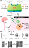
Figure 2
Figure 2
Representative neurotransmitters and their detection for early diagnosis and pathophysiological monitoring. (a) Electrochemical dopamine detection achieved via the conversion between dopamine and dopamine-o-quinone (reprinted with permission from [26]; Copyright © 2021 by the authors. Licensee: MDPI). (b) Role of acetylcholine in the immunomodulation by T cells (reprinted with permission from [30]; Copyright © 2019, The Association for the Publication of the Journal of Internal Medicine). (c) Electrochemical glutamate biosensor composed of glutamate dehydrogenase (GLDH), poly(amidoamine) dendrimer-encapsulated platinum nanoparticles (Pt-PAMAM) and carbon nanotubes (CNTs) (reprinted with permission from [37]; Copyright © 2007, Elsevier B.V.).
Figure 3
Figure 3
Recent types of electrochemical nanobiosensors used for neurotransmitter detection. (a) Structure of the amperometric biosensor for the detection of various catecholamines (reprinted with permission from [54]; Copyright © 2022 by the authors. Licensee: MDPI). (b) Plot of the peak current of the cyclic voltammogram of carbon quantum dot/copper oxide nanocomplex-modified glassy carbon electrode in 0.4 mM epinephrine solution as a function of the square root of scan rate (reprinted with permission from [64]; Copyright © 2022, Elsevier B.V.). (c) Schematic of the assembly process of the dual-response MIP-sensing membrane and electrochemical detection of dopamine and adenine (reprinted with permission from [73]; Copyright © 2022, Elsevier B.V.). (d) Schematic of the assembly process of the dual-response MIP-sensing membrane and electrochemical detection of dopamine and adenine (reprinted with permission from [78]; Copyright © 2023, Elsevier B.V.).
Figure 4
Figure 4
In vitro neurotransmitter detection system from neuronal cells and stem cells using electrochemical sensors: (a) Schematic of the sensor probe developed using Au-nanorattles (AuNRTs) and reduced graphene oxide (rGO) (AuNRTs-rGO) nanocomposite based on the serotonin detection mechanism (reprinted with permission from [90]; Copyright © 2022, Elsevier B.V.). (b) Scanning electron microscopy image and Raman spectra of three-dimensional fuzzy graphene microelectrode (top) and color plot (bottom left) and background-subtracted cyclic voltammogram showing reduction and oxidation peaks of injected dopamine and serotonin (reprinted with permission from [93]; Copyright © 2022 Elsevier B.V.). (c) Schematic representation of the conversion of hNSCs into dopaminergic (DAergic) and non-DAergic neurons (top) and cyclic voltammogram of cells undergoing differentiation into DAergic neurons (DA neurons) (bottom) (reprinted with permission from [17]; Copyright © 2015 WILEY-VCH Verlag GmbH & Co. KGaA, Weinheim). (d) Schematic of p3D-carbon where the cells differentiate at the bottom or between pillars (top), calculated average charge related to the amount of detected dopamine released by human neural stem cells (hNSCs) (bottom left), and characteristic current–time trace recorded during amperometric detection of dopamine (bottom right) (reprinted with permission from [102]; Copyright © 2014 WILEY-VCH Verlag GmbH & Co. KGaA, Weinheim).
Figure 5
Figure 5
In vivo neurotransmitter monitoring system using electrochemical sensors: (a) photograph of the crucian fish brain, the field-effect transistor biosensor setup on the fish brain (left), and the Ids output signal of the crucian fish brain monitored by the biosensor (right) (reprinted with permission from [105]; Copyright © 2021, Elsevier B.V.). (b) Schematic of aptCA-functionalized carbon fiber electrodes (CFEs) for in vivo dopamine sensing (left), and the current responses of aptCA-functionalized CFEs in the rat nucleus accumbens upon electrical stimulation of the rat’s medial forebrain bundle (right) (reprinted with permission from [109]; Copyright © 2020, Wiley-VCH GmbH). (c) Amperogram of stimulated glutamate release in the subthalamic nucleus of a rat brain slice (left) and stimulated rat brain in vivo (right) (reprinted with permission from [110]; Copyright © 2019, Elsevier B.V.). (d) Schematic of the solid-phase microextraction-based approach used for quantitative measurements of multiple neurotransmitters (reprinted with permission from [114]; Copyright © 2019, American Chemical Society).
Figure 1
Figure 1
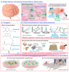
Schematic of electrochemical nanobiosensors used for neurotransmitter detection. (i) Detection of neurotransmitters is important, since a problem in the production and transmission of neurotransmitters could potentially have fatal consequences in the signal transmission process in nerves and may cause cranial nerve-related diseases. To achieve monitoring of (ii) the neurotransmitters sensitively and selectively, (iii) various nanomaterials and nanotechnologies have been applied in the development of electrochemical biosensors. Additionally, utilizing fabricated biosensors, the neurotransmitters in (iv) neuronal cells, stem cells, and animal models can be monitored.

References

    1. Edwards R.H. The neurotransmitter cycle and quantal size. Neuron. 2007;55:835–858. doi: 10.1016/j.neuron.2007.09.001. - DOI - PubMed
    1. Hnasko T.S., Edwards R.H. Neurotransmitter corelease: Mechanism and physiological role. Annu. Rev. Physiol. 2012;74:225–243. doi: 10.1146/annurev-physiol-020911-153315. - DOI - PMC - PubMed
    1. Spitzer N.C. Neurotransmitter Switching in the Developing and Adult Brain. Annu. Rev. Neurosci. 2017;40:1–19. doi: 10.1146/annurev-neuro-072116-031204. - DOI - PubMed
    1. Hornykiewicz O. Brain neurotransmitter changes in Parkinson’s disease. In: Marsden C.D., Fahn S., editors. Movement Disorders. Butterworth-Heinemann; Oxford, UK: 1981. pp. 41–58.
    1. Reinikainen K.J., Soininen H., Riekkinen P.J. Neurotransmitter changes in Alzheimer’s disease: Implications to diagnostics and therapy. J. Neurosci. Res. 1990;27:576–586. doi: 10.1002/jnr.490270419. - DOI - PubMed

LinkOut - more resources