Case Reports
. 2023 Sep 28;389(13):1203-1210.
doi: 10.1056/NEJMoa2307798.

Death after High-Dose rAAV9 Gene Therapy in a Patient with Duchenne's Muscular Dystrophy

Affiliations
Case Reports

Death after High-Dose rAAV9 Gene Therapy in a Patient with Duchenne's Muscular Dystrophy

Angela Lek et al. N Engl J Med. .

Abstract

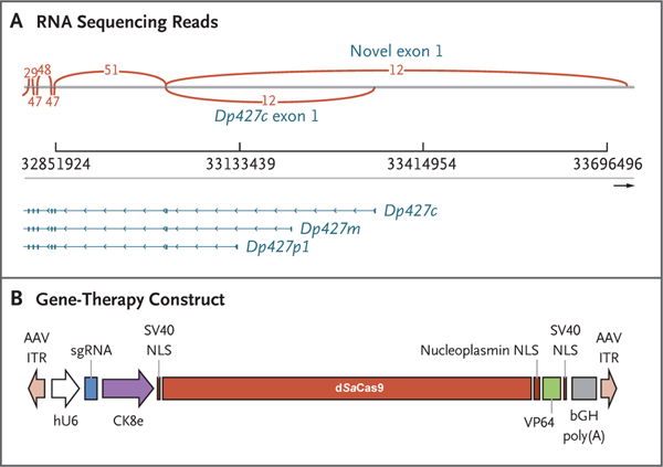
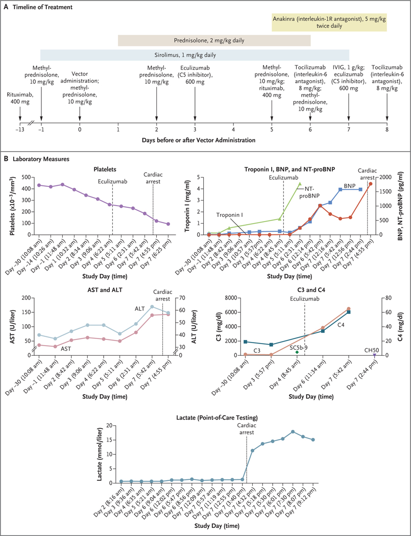
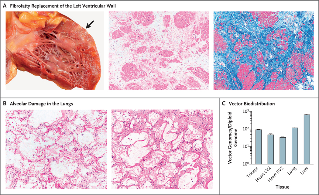
We treated a 27-year-old patient with Duchenne's muscular dystrophy (DMD) with recombinant adeno-associated virus (rAAV) serotype 9 containing dSaCas9 (i.e., "dead" Staphylococcus aureus Cas9, in which the Cas9 nuclease activity has been inactivated) fused to VP64; this transgene was designed to up-regulate cortical dystrophin as a custom CRISPR-transactivator therapy. The dose of rAAV used was 1×10¹⁴ vector genomes per kilogram of body weight. Mild cardiac dysfunction and pericardial effusion developed, followed by acute respiratory distress syndrome (ARDS) and cardiac arrest 6 days after transgene treatment; the patient died 2 days later. A postmortem examination showed severe diffuse alveolar damage. Expression of transgene in the liver was minimal, and there was no evidence of AAV serotype 9 antibodies or effector T-cell reactivity in the organs. These findings indicate that an innate immune reaction caused ARDS in a patient with advanced DMD treated with high-dose rAAV gene therapy. (Funded by Cure Rare Disease.).


Figures

Figure 1.
Figure 1.. Design of the CRISPR–Transactivator Therapeutic.
Panel A shows RNA sequencing reads, including exons 1 through 7 (right to left) of the full-length DMD transcripts Dp427c (cortical), Dp427m (muscle), and Dp427p1 (Purkinje). A novel exon 1 (GRCh38 chrX: 33,726,502–33,726,715) that has expression similar to or higher than that of Dp427c was also detected by RNA sequencing. Arcs indicate the numbers of reads that span exons. Panel B shows the therapeutic construct that was cloned into a plasmid backbone with adeno-associated virus (AAV) serotype 2 inverted terminal repeats (ITRs) (Addgene plasmid 99680). The single guide RNA (sgRNA) expression is regulated by a human U6 (hU6) promoter, and the expression of the “dead” Staphylococcus aureus Cas9 (dSaCas9)–VP64 fusion protein is regulated by a CK8e promoter (Hauschka Laboratory, University of Washington), which was engineered from the regulatory elements from mouse muscle-type creatine kinase. The abbreviation bGH poly(A) denotes bovine growth hormone polyadenylation signal, and NLS nuclear localization sequence.
Figure 2.
Figure 2.. Data from the Clinical Study.
Panel A shows the timeline of treatment administered. IVIG denotes intravenous immune globulin. Days on the timeline indicate days before or after vector administration (which occurred on day 0). Panel B shows cardiac, complement, and liver measures as assessed during the clinical study (beginning 30 days before vector administration). Days on the x axis in each graph in Panel B are based on the individual protocol convention, in which the day of treatment is day 1 rather than day 0. In the graph of C3 and C4, the SC5b-9 level shown is 453 ng per milliliter (normal value, ≤244) and the CH50 level shown is less than 10 U per milliliter (normal range, 31 to 60). Additional laboratory results from the periods before and after gene therapy are shown in Table S3. ALT denotes alanine aminotransferase, AST aspartate aminotransferase, BNP B-type natriuretic peptide, and NT-proBNP N-terminal pro-BNP.
Figure 3.
Figure 3.. Postmortem Analysis of Tissue.
Panel A shows fibrofatty replacement of the left ventricular wall (arrow) in the patient’s heart. Histologic evaluation of this area shows marked interstitial fibrosis and fatty replacement with residual cardiac myocytes; there is no histologic evidence of myocarditis or thrombotic microangiopathy (hematoxylin–eosin and Masson’s trichrome staining). The findings are consistent with severe cardiomyopathy. Panel B shows a microscopic image of diffuse alveolar damage in the patient’s lungs, characterized by hyaline membrane deposition with interstitial and intraalveolar edema (hematoxylin–eosin and periodic acid–Schiff staining). The images in Panel B are shown at 2.5 times the magnification of the middle and right images in Panel A. Panel C shows vector biodistribution in patient tissues. Vector genomes were quantified by quantitative polymerase chain reaction and calculated to indicate the number of vector genomes per diploid genome for each tissue. Analyses were performed in technical triplicates; data are reported as means, and T bars indicate the standard deviation. LV2 and RV2 are specific sites within the left and right ventricles, respectively.

Comment in

References

    1. Gao QQ, McNally EM. The dystrophin complex: structure, function, and implications for therapy. In: Comprehensive physiology. 2015;1223–39. - PMC - PubMed
    1. Duan D, Goemans N, Takeda S, Mercuri E, Aartsma-Rus A. Duchenne muscular dystrophy. Nat Rev Dis Primers 2021;7: 13. - PMC - PubMed
    1. Lapidos KA, Kakkar R, McNally EM. The dystrophin glycoprotein complex: signaling strength and integrity for the sarcolemma. Circ Res 2004;94:1023–31. - PubMed
    1. Wilson DGS, Tinker A, Iskratsch T. The role of the dystrophin glycoprotein complex in muscle cell mechanotransduction. Commun Biol 2022;5:1022. - PMC - PubMed
    1. Li C, Samulski RJ. Engineering adeno-associated virus vectors for gene therapy. Nat Rev Genet 2020;21:255–72. - PubMed

Publication types

LinkOut - more resources