. 2023 Sep 27;11(5):e0127123.
doi: 10.1128/spectrum.01271-23. Online ahead of print.

Streptomyces albidoflavus Q antifungal metabolites inhibit the ergosterol biosynthesis pathway and yeast growth in fluconazole-resistant Candida glabrata: phylogenomic and metabolomic analyses

Affiliations

Streptomyces albidoflavus Q antifungal metabolites inhibit the ergosterol biosynthesis pathway and yeast growth in fluconazole-resistant Candida glabrata: phylogenomic and metabolomic analyses

Celia Bautista-Crescencio et al. Microbiol Spectr. .

Abstract

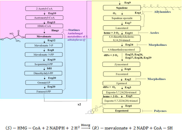
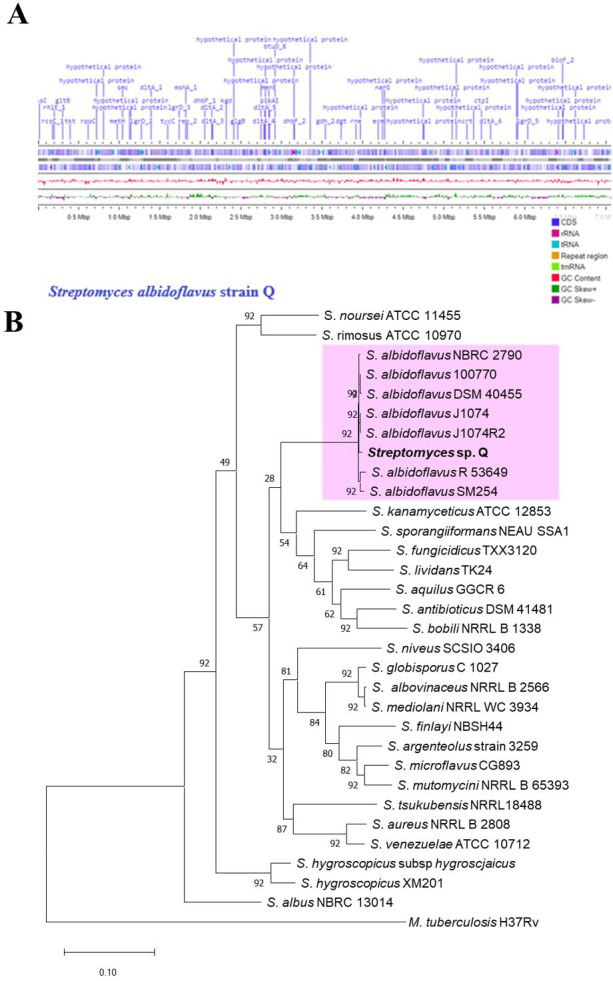
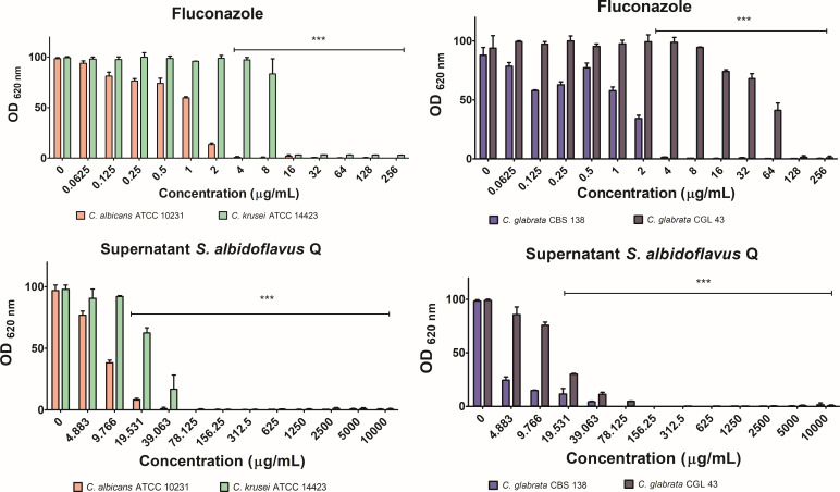
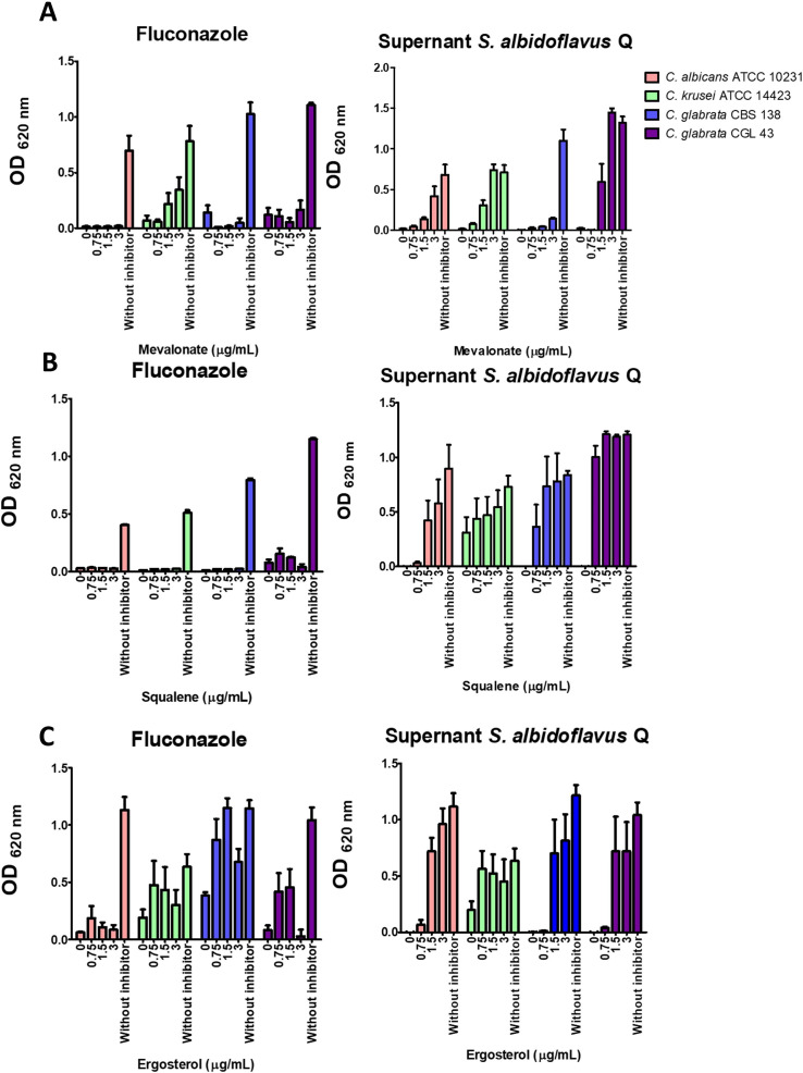
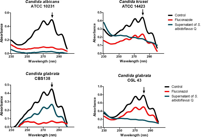
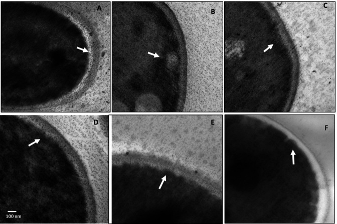
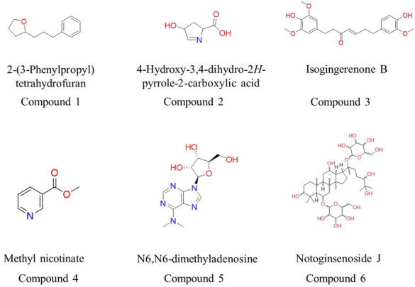
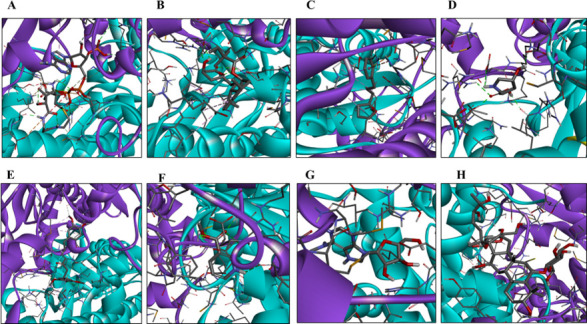
There is an urgent need to develop new antifungals due to the increasing prevalence of multidrug-resistant fungal infections and the recent emergence of COVID-19-associated candidiasis. A good study model for evaluating new antifungal compounds is Candida glabrata, an opportunistic fungal pathogen with intrinsic resistance to azoles (the most common clinical drugs for treating fungal infections). The aim of the current contribution was to conduct in vitro tests of antifungal metabolites produced by the bacteria Streptomyces albidoflavus Q, identify their molecular structures, and utilize several techniques to provide evidence of their therapeutic target. S. albidoflavus was isolated from maize rhizospheric soil in Mexico and identified by phylogenomic analysis using a 92-gene core. Of the 66 metabolites identified in S. albidoflavus Q by a liquid chromatography-high resolution mass spectrometry (LC-HRMS) metabolomic analysis of the lyophilized supernatant, six were selected by the Way2drug server based on their in silico binding to the likely target, 3-hydroxy-3-methyl-glutaryl-CoA reductase (HMGR, the key enzyme in the ergosterol biosynthesis pathway). Molecular modeling studies show a relatively high binding affinity for the CgHMGR enzyme by two secondary metabolites: isogingerenone B (diaryl heptanoid) and notoginsenoside J (polycyclic triterpene). These secondary metabolites were able to inhibit ergosterol synthesis and affect yeast viability in vitro. They also caused alterations in the ultrastructure of the yeast cytoplasmic membrane, as evidenced by transmission electron microscopy. The putative target of isogingerenone B and notoginsenoside J is distinct from that of azole drugs (the most common clinical antifungals). The target for the latter is the lanosterol 14 alpha-demethylase enzyme (Erg11). IMPORTANCE Multidrug resistance has emerged among yeasts of the genus Candida, posing a severe threat to global health. The problem has been exacerbated by the pandemic associated with COVID-19, during which resistant strains of Candida auris and Candida glabrata have been isolated from patients infected with the SARS-CoV-2 virus. To confront this challenge, the World Health Organization has invoked scientists to search for new antifungals with alternative molecular targets. This study identified 66 metabolites produced by the bacteria Streptomyces albidoflavus Q, 6 of which had promising properties for potential antifungal activity. The metabolites were tested in vitro as inhibitors of ergosterol synthesis and C. glabrata growth, with positive results. They were also found to damage the cytoplasmic membrane of the fungus. The corresponding molecular structures and their probable therapeutic target were established. The target is apparently distinct from that of azole drugs.

Keywords: Candida glabrata; HMGR (EC 1.1.1.34); Streptomyces albidoflavus; WGS; actinobacteria; actinomycete; antifungal; cytoplasmic membrane; ergosterol; metabolomics; multi-drug resistance; plant-associated metabolites.


Conflict of interest statement

The authors declare no conflict of interest.

Figures

Fig 1
Fig 1
The biosynthetic pathway of ergosterol in yeasts, divided into three modules: the mevalonate pathway (in pink), the farnesyl pyrophosphate pathway (in blue), and the last step leading to ergosterol (in yellow). Enzymes, intermediates, inhibitors, and the requirements for oxygen, heme, and iron are indicated. Inhibitors are listed with their respective target: statins bind with HMGR, allylamines with Erg1, azoles with Erg11, morpholines with Erg2 and Erg24, and polyenes with ergosterol. 3-Hydroxy-3-methyl-glutaryl-CoA reductase (HMGR) is the proposed target for secondary metabolites produced by Streptomyces albidoflavus. Below the boxes, the reaction described is catalyzed by the HMGR enzyme with NADPH as a cofactor.
Fig 2
Fig 2
Phenotypic characteristics of Streptomyces albidoflavus Q. The strain was inoculated onto solid GAE medium and incubated at 28°C for 15 days. (A) Its colonial morphology is characteristic of actinobacteria. (B) The microscopic morphology corresponds to a Gram-positive bacteria with filamentous structures (1,000×). (C) S. albidoflavus metabolites inhibited C. glabrata growth. The supernatant (SN) of S. albidoflavus Q was concentrated by lyophilization and resuspended. Fluconazole (Flu) and water served as the positive and negative controls, respectively, for the inhibition of yeast growth (the modified M44 CLSI method).
Fig 3
Fig 3
General features of the Streptomyces albidoflavus Q genome and the phylogenetic tree. (A) A linear representation of S. albidoflavus Q genome contigs was obtained with Proksee (https://proksee.ca). The scale is expressed in megabases (Mbp), and two dark blue lines denote forward and reverse strand CDSs, respectively. Some genes are portrayed in violet with the default setting of Proksee. The tRNA (blue arrows), rRNA (magenta arrows), and tmRNA (green arrows) are shown with violet lines. The content (in pink) and skew (in dark green and violet) of gene clusters are illustrated. (B) The phylogenomic tree of S. albidoflavus Q was inferred by concatenated alignment of 92 core genes (UBCGs). Gene support indices (GSIs) and percentage bootstrap values are given at branching points. Bars represent 0.10 substitution per position.
Fig 4
Fig 4
Inhibition of the growth of Candida spp. While growth without any inhibitor served as the negative control, fluconazole was used as the positive control of inhibitory activity. The optical density was determined in a Thermo Scientific Multiskan FC microplate photometer at 620 nm (OD620) after incubation at 37°C for 24 h. The quantification of growth was expressed as the average ± standard deviation (SD) of optimal density values from three independent assays. Significant differences were analyzed by two-way ANOVA. ***P < 0.001.
Fig 5
Fig 5
A Candida spp. growth rescue assay was carried out. The yeasts were first treated with fluconazole or the metabolites of S. albidoflavus at sublethal concentrations, followed by the addition of exogenous mevalonate, squalene, or ergosterol to the culture medium. Following both initial treatments, ergosterol caused Candida spp. to undergo growth recovery. Squalene and mevalonate promoted growth recovery of Candida spp. after treatment with S. albidoflavus metabolites but not after treatment with fluconazole. The yeasts without antifungal treatment served as the control. Yeast growth was quantified by optical density in a Thermo Scientific Multiskan FC microplate photometer at 620 nm (OD620) upon completion of incubation at 37°C for 24 h. The values are expressed as the average of three independent assays ± SD. The basal growth value was established with the control (the yeast without any inhibitor). Significant differences were analyzed by two-way ANOVA. ***P < 0.001.
Fig 6
Fig 6
Effect of the lyophilized supernatant of Streptomyces albidoflavus Q on the concentration of ergosterol in Candida spp. species. The different species were grown in YPD liquid yeast medium and treated with sublethal concentrations of the inhibitors. The control was the growth of the yeasts in the absence of any inhibitor. The yeasts were incubated at 37°C for 18 h under constant shaking at 200  rpm. The concentration of the extracted sterols (in the n-heptane layer) was determined by spectrophotometrically scanning them (from 230 to 300  nm). The extraction of total sterols was performed on a 100 mg sample of yeast.
Fig 7
Fig 7
Image of Candida glabrata treated with the S. albidoflavus Q supernatant, taken with a transmission electron microscope. Micrographs: (A–C) of C. glabrata CBS 138 and (D–F) of C. glabrata CGL 43. (A and D) Without treatment (negative control); (B and E) treated with fluconazole (positive control); (C and F) treated with the lyophilized supernatant of S. albidoflavus Q. The arrows indicate the cytoplasmic membrane with (C and F) and without (A and D) treatment. The yeasts were each treated with sublethal concentrations of the antifungals at 37°C for 18 h in YPD liquid yeast medium.
Fig 8
Fig 8
Structures of metabolites with potential inhibitory activity on CgHMGR. First, 66 secondary metabolites were found in the lyophilized supernatant of S. albidoflavus Q through metabolomic analysis. Subsequently, they were examined by using the web resource PASS Online, deposited in server Way2Drug (http://way2drug.com/passonline/), which selected the most probable inhibitors.
Fig 9
Fig 9
For each of the metabolites identified as potential inhibitors of HMGR, a schematic illustration portrays the binding mode of the ligand with the CgHMGR enzyme. The predicted binding mode of HMG-CoA (A), simvastatin (B), compound 1 (C), compound 2 (D), compound 3 (E), compound 4 (F), compound 5 (G), and compound 6 (H). The α-helix and β-strand structures are depicted as ribbons, colored in cyan (subunit a) and purple (subunit b). The amino acids that interact with the ligand and the ligand itself are represented as sticks. The figure was created by Celia-Esthela Bautista-Crescencio with Discovery Studio 2021 client software.

Similar articles

Cited by

References

    1. Lai C-C, Chen S-Y, Ko W-C, Hsueh P-R. 2021. Increased antimicrobial resistance during COVID-19 pandemic. Int J Antimicrob Agents 57:106324. doi:10.1016/j.ijantimicag.2021.106324 - DOI - PMC - PubMed
    1. Pristov KE, Ghannoum MA. 2019. Resistance of Candida to azoles and echinocandins worldwide. Clin Microbiol Infect 25:792–798. doi:10.1016/j.cmi.2019.03.028 - DOI - PubMed
    1. Tortorano AM, Prigitano A, Morroni G, Brescini L, Barchiesi F. 2021. Candidemia: evolution of drug resistance and novel therapeutic approaches. Infect Drug Resist 14:5543–5553. doi:10.2147/IDR.S274872 - DOI - PMC - PubMed
    1. Barka EA, Vatsa P, Sanchez L, Gaveau-Vaillant N, Jacquard C, Meier-Kolthoff JP, Klenk H-P, Clément C, Ouhdouch Y, van Wezel GP. 2016. Taxonomy physiology, and natural products of actinobacteria. Microbiol Mol Biol Rev 80:1–43. doi:10.1128/MMBR.00019-15 - DOI - PMC - PubMed
    1. Xu L, Ye K-X, Dai W-H, Sun C, Xu L-H, Han B-N. 2019. Comparative genomic insights into secondary metabolism biosynthetic gene cluster distributions of marine Streptomyces. Mar Drugs 17:498. doi:10.3390/md17090498 - DOI - PMC - PubMed

LinkOut - more resources