. 2023 Sep 21;9(3):52.
doi: 10.3390/ijns9030052.

Whole-Genome Sequencing Can Identify Clinically Relevant Variants from a Single Sub-Punch of a Dried Blood Spot Specimen

Affiliations

Whole-Genome Sequencing Can Identify Clinically Relevant Variants from a Single Sub-Punch of a Dried Blood Spot Specimen

David J McBride et al. Int J Neonatal Screen. .

Abstract

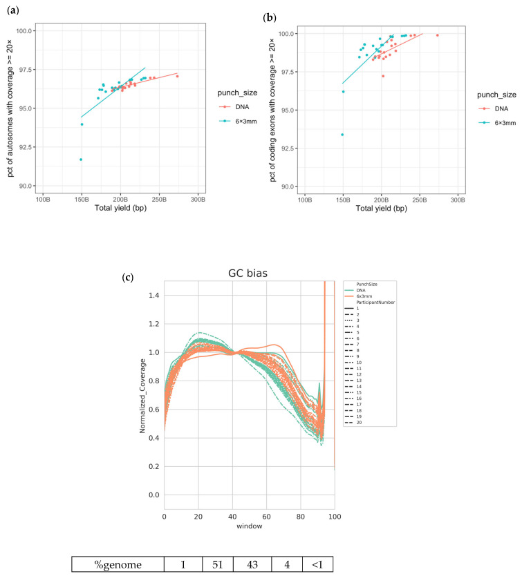
The collection of dried blood spots (DBS) facilitates newborn screening for a variety of rare, but very serious conditions in healthcare systems around the world. Sub-punches of varying sizes (1.5-6 mm) can be taken from DBS specimens to use as inputs for a range of biochemical assays. Advances in DNA sequencing workflows allow whole-genome sequencing (WGS) libraries to be generated directly from inputs such as peripheral blood, saliva, and DBS. We compared WGS metrics obtained from libraries generated directly from DBS to those generated from DNA extracted from peripheral blood, the standard input for this type of assay. We explored the flexibility of DBS as an input for WGS by altering the punch number and size as inputs to the assay. We showed that WGS libraries can be successfully generated from a variety of DBS inputs, including a single 3 mm or 6 mm diameter punch, with equivalent data quality observed across a number of key metrics of importance in the detection of gene variants. We observed no difference in the performance of DBS and peripheral-blood-extracted DNA in the detection of likely pathogenic gene variants in samples taken from individuals with cystic fibrosis or phenylketonuria. WGS can be performed directly from DBS and is a powerful method for the rapid discovery of clinically relevant, disease-causing gene variants.

Keywords: DNA sequencing; cystic fibrosis; dried blood spots; newborn screening; phenylketonuria; whole genome sequencing.


Conflict of interest statement

David J. McBride, Claire Fielding, Taksina Newington, Alexandra Vatsiou, Harry Fischl, Maya Bajracharya, Vicki S. Chambers, Louise Fraser, Pauline A. Fujita, Jennifer Becq, Zoya Kingsbury, and Mark T. Ross are either current or previous full-time employees of Illumina Inc. Stuart J. Moat and Sian Morgan declare no conflicts of interest.

Figures

Figure 1
Figure 1
Good-quality WGS data achieved from DBS and extracted DNA. (a) Percentage of autosomes with coverage ≥20× vs. total yield sequenced; (b) percentage of coding exons with coverage ≥20× vs. total yield sequenced; (c) normalized coverage across various windows of GC content in the human genome, grouped by participant. The numbers below the plot indicate the percentage of the human reference genome sequence included in each window of GC content.
Figure 2
Figure 2
WGS data generated from a variety of DBS punch inputs. (a) Qubit values measured post-DBS lysis protocol for participants 9 to 20 are shown above the sequencing library quantifications determined by quantitative PCR (qPCR) for each input type. (b) Percentage of autosomes with coverage ≥20× vs. total yield sequenced; (c) percentage of coding exons with coverage ≥20× vs. total yield sequenced; (d) normalized coverage across various windows of GC content in the human genome, grouped by sample input type and averaged for the 20 participants. Numbers below the plot indicate the percentages of genomic sequences included in each window of GC content; (e) fragment length median for each input type for participants 9 to 20. Ranges of values are summarized by box and whisker plots, with dotted lines link data points from the same participants.
Figure 3
Figure 3
Identification of CFTR variants in samples from individuals with cystic fibrosis. IGV screenshots of likely pathogenic variants discovered in three participants with cystic fibrosis. Tracks from DNA and DBS (6 × 3 mm) inputs are shown. Data from participant 21 (extracted DNA) are shown for comparison. (a) Homozygous Phe508del variant seen in participant 6. (b) Asn1303Lys and Phe508del variants seen in participant 7. (c) Gly542Ter and Phe508del variants seen in participant 8.
Figure 3
Figure 3
Identification of CFTR variants in samples from individuals with cystic fibrosis. IGV screenshots of likely pathogenic variants discovered in three participants with cystic fibrosis. Tracks from DNA and DBS (6 × 3 mm) inputs are shown. Data from participant 21 (extracted DNA) are shown for comparison. (a) Homozygous Phe508del variant seen in participant 6. (b) Asn1303Lys and Phe508del variants seen in participant 7. (c) Gly542Ter and Phe508del variants seen in participant 8.
Figure 4
Figure 4
Equivalent performance of a DBS punch (6 mm) and DNA extracted via EDTA blood tubes. (a) Percentage of autosomal regions with ≥20× coverage for each sample; (b) percentage of exome regions with ≥20× coverage for each sample; (c) normalized coverage across various windows of GC content in the human genome, grouped by sample input type; (d) IGV screenshot of likely pathogenic gene variants discovered in participant with PKU. Tracks from the DNA and DBS inputs are shown. Data from participant 6 (extracted DNA) are shown for comparison (e) Likely pathogenic variants discovered in samples taken from a second participant with PKU. Cartoon representation of the variant location on chromosome 12 and zoomed-in IGV screenshots from the DBS taken directly from the patient. Asterix highlights the presence of the SNV shown in lower panel.
Figure 4
Figure 4
Equivalent performance of a DBS punch (6 mm) and DNA extracted via EDTA blood tubes. (a) Percentage of autosomal regions with ≥20× coverage for each sample; (b) percentage of exome regions with ≥20× coverage for each sample; (c) normalized coverage across various windows of GC content in the human genome, grouped by sample input type; (d) IGV screenshot of likely pathogenic gene variants discovered in participant with PKU. Tracks from the DNA and DBS inputs are shown. Data from participant 6 (extracted DNA) are shown for comparison (e) Likely pathogenic variants discovered in samples taken from a second participant with PKU. Cartoon representation of the variant location on chromosome 12 and zoomed-in IGV screenshots from the DBS taken directly from the patient. Asterix highlights the presence of the SNV shown in lower panel.

References

    1. Therrell B.L., Padilla C.D., Loeber J.G., Kneisser I., Saadallah A., Borrajo G.J., Adams J. Current status of newborn screening worldwide: 2015. Semin. Perinatol. 2015;39:171–187. doi: 10.1053/j.semperi.2015.03.002. - DOI - PubMed
    1. Rehm H.L., Bale S.J., Bayrak-Toydemir P., Berg J.S., Brown K.K., Deignan J.L., Friez M.J., Funke B.H., Hegde M.R., Lyon E. ACMG clinical laboratory standards for next-generation sequencing. Anesth. Analg. 2013;15:733–747. doi: 10.1038/gim.2013.92. - DOI - PMC - PubMed
    1. Deignan J.L., Astbury C., Cutting G.R., del Gaudio D., Gregg A.R., Grody W.W., Monaghan K.G., Richards S. CFTR variant testing: A technical standard of the American College of Medical Genetics and Genomics (ACMG) Anesth. Analg. 2020;22:1288–1295. doi: 10.1038/s41436-020-0822-5. - DOI - PMC - PubMed
    1. The 100,000 Genomes Project Pilot Investigators 100,000 Genomes Pilot on Rare-Disease Diagnosis in Health Care—Preliminary Report. N. Engl. J. Med. 2021;385:1868–1880. doi: 10.1056/NEJMoa2035790. - DOI - PMC - PubMed
    1. Gross A.M., Ajay S.S., Rajan V., Brown C., Bluske K., Burns N.J., Chawla A., Coffey A.J., Malhotra A., Scocchia A., et al. Copy-number variants in clinical genome sequencing: Deployment and interpretation for rare and undiagnosed disease. Anesth. Analg. 2018;21:1121–1130. doi: 10.1016/j.cancergen.2018.04.058. - DOI - PMC - PubMed

LinkOut - more resources