. 2023 Sep 1;9(9):903.
doi: 10.3390/jof9090903.

Axenic Long-Term Cultivation of Pneumocystis jirovecii

Affiliations

Axenic Long-Term Cultivation of Pneumocystis jirovecii

Diana Riebold et al. J Fungi (Basel). .

Abstract

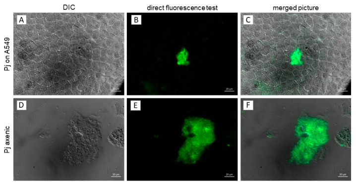
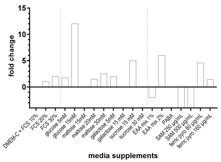
Pneumocystis jirovecii, a fungus causing severe Pneumocystis pneumonia (PCP) in humans, has long been described as non-culturable. Only isolated short-term experiments with P. jirovecii and a small number of experiments involving animal-derived Pneumocystis species have been published to date. However, P. jirovecii culture conditions may differ significantly from those of animal-derived Pneumocystis, as there are major genotypic and phenotypic differences between them. Establishing a well-performing P. jirovecii cultivation is crucial to understanding PCP and its pathophysiological processes. The aim of this study, therefore, was to develop an axenic culture for Pneumocystis jirovecii. To identify promising approaches for cultivation, a literature survey encompassing animal-derived Pneumocystis cultures was carried out. The variables identified, such as incubation time, pH value, vitamins, amino acids, and other components, were trialed and adjusted to find the optimum conditions for P. jirovecii culture. This allowed us to develop a medium that produced a 42.6-fold increase in P. jirovecii qPCR copy numbers after a 48-day culture. Growth was confirmed microscopically by the increasing number and size of actively growing Pneumocystis clusters in the final medium, DMEM-O3. P. jirovecii doubling time was 8.9 days (range 6.9 to 13.6 days). In conclusion, we successfully cultivated P. jirovecii under optimized cell-free conditions in a 70-day long-term culture for the first time. However, further optimization of the culture conditions for this slow grower is indispensable.

Keywords: A549; DMEM; Pneumocystis; Pneumocystis jirovecii; axenic; culture; human lung carcinoma cells.


Conflict of interest statement

The authors declare no conflict of interest. The funders had no role in the design of the study; in the collection, analyses, or interpretation of data; in the writing of the manuscript; or in the decision to publish the results.

Figures

Figure 1
Figure 1
Processing of broncho-alveolar lavage fluid (BALF) samples from patients with Pneumocystis pneumonia (PCP). The results of the optimization experiments with co-culture on A549 cells are presented in the Supplementary Materials. Pj—P. jirovecii.
Figure 2
Figure 2
P. jirovecii clusters in cultures using A549 feeder cells and in axenic P. jirovecii cultures (patient 5, 14 days after start of culture). (A,D)—DIC, (B,E)—FITC channel after staining with commercial direct fluorescence test; (C,F)—merged photos. Clusters of P. jirovecii can be seen clearly in the axenic culture (DF), while small P. jirovecii clusters tend to disappear behind the layer of A549 cells (A) and are only visible when stained with DFT (B,C). The larger clusters that can be seen in later stages of P. jirovecii culture are also clearly visible in the co-cultures with A549 cells. bar = 50 µm.
Figure 3
Figure 3
P. jirovecii trophic forms and asci (cysts) in initial BALF samples (A): DiffQuick stain showing trophic forms, some with visible nuclei (BALF from patient 1); (B): Grocott methenamine silver stain (GMS) with asci (BALF from patient 1). GMS stains the walls of intermediate and mature asci in gray, making it easy to detect asci even at low magnifications. However, no trophic forms or spores can be detected using this method. DiffQuick or Giemsa stain permit the detection of trophic forms and unripe asci. Therefore, the methods should be used in combination. C-E: large cluster of trophic forms and developing asci examined with DIC microscopy (C) and stained with DFT (D). Photos (C,D) are merged in (E) (BALF from patient 5).
Figure 4
Figure 4
(A,B): REM pictures of P. jirovecii (A) asci with microdots (arrows) and trophic form (double arrow) and (B) empty and ruptured asci (arrow), partially with spores (double arrow). In both REM pictures (A,B), filopodia (rough structures visible on the surface of most asci) are clearly visible. (C) Filopodia are also visible in TEM pictures (black arrow). (C,D) Direct interaction between trophic forms with membrane blurring (large black arrow, TEM, (D)) = detail of (C); (E) interaction between trophic forms and host cells (A549) (arrow). (F) and mitochondria (M) are visible in the trophic forms, but the number of mitochondria per cell cannot be counted with TEM. Structures in P. jirovecii asci (G) and trophic forms (H): nuclei (N) and some other organelle residues are visible, but some structures were destroyed by the fixation process, as indicated by white structures associated with shrinking of the cytoplasm (CS).
Figure 5
Figure 5
P. jirovecii culture with A549 cells and axenic culture without feeder cells using P. jirovecii from a single patient’s BALF samples. The inoculum number (mtLSU qPCR copies/mL BALF) was equal for cultures with and without A549 cells but differed between the patient samples (A). The maximal increase in P. jirovecii mtLSU copy numbers during culture was 4.5 × 10⁷ copies/mL culture, or 2770% of the P. jirovecii inoculum in sample 1, cultured on A549 cells (B,C). All cultures with a P. jirovecii inoculum below 1 × 10⁶ copies/mL or above 4 × 10⁶ copies/mL showed slow growth (isolates of patients 2 and 3), steady state (isolate of patient 5), or a decline (isolate of patient 6) in P. jirovecii copy numbers. This was observed independently in cultures using A549 cells and in axenic cultures.
Figure 6
Figure 6
Culture supplements and concentrations tested for improving the growth of P. jirovecii with DMEM-C medium in axenic culture (for culture optimization on A549 cells, see Supplementary Materials). P. jirovecii was grown in 24-well plates for 10–14 days and quantified by qPCR. Growth was examined microscopically every 2nd day. EAA—essential amino acid mix 50×, NEAA—non-essential amino acid mix; SAM—S-adenosyl-L-methionine, PABA—para aminobenzoic acid; ferric pyro—ferric pyrophosphate.
Figure 7
Figure 7
Culture supplements and concentrations were tested for their effect on the growth of P. jirovecii in DMEM-O2 medium (with 30% FCS, 25 mM HEPES, and 4 mM GlutaMax instead of glutamine) in axenic culture only. P. jirovecii was grown in 24-well plates for 10–14 days. P. jirovecii growth was quantified by qPCR. Growth was examined microscopically every 2nd day. ferric pyro—ferric pyrophosphate.
Figure 8
Figure 8
P. jirovecii cluster from in vitro culture in DMEM-O2 medium, axenic culture day 10, stained with LiveDead staining. (A): P. jirovecii clusters without L-alanine showed vital (bright green) and potentially inactive/dead P. jirovecii (orange). The trophic forms were located mainly in the inner parts of the huge clusters (black arrow), while some developing asci were located in the outer parts (white arrows). (B): P. jirovecii clusters with 20 mg/L L-alanine showed many vital (bright green) and only a few potentially inactive/dead P. jirovecii (orange). Compared to A, the cluster was the same size, but the P. jirovecii organisms inside the cluster seemed to be better nourished than those without additional L-alanine and only exhibited a few ascii (arrow).
Figure 9
Figure 9
Growth of P. jirovecii in five of 24 axenic long-term flask cultures in DMEM-O2 medium (for complete graphs of all flask cultures, see Figure S8). The starting volume was 10 mL/flask, and all cultures were incubated at 37 °C and 5% CO2 for 22 to 57 days. Culture conditions, except the three factors (1) patient samples for inoculum, (2) duration of culture, and (3) final culture volume, were not altered. Samples for qPCR analysis were taken, and volumes of fresh medium were added on top of the old medium to dilute the growing P. jirovecii to appropriate densities in the flask (fed-batch culture). As seen after the first five cultures, P. jirovecii growth ceased when the starting inoculum was too dense (A). A start inoculum of 1 to 2 × 10⁶ copies/mL culture medium volume was optimal, while all cultures with a higher or lower inoculum showed decreasing P. jirovecii copy numbers/mL medium. The culture duration was increased from 24 days to up to 57 days with an optimum of 48 days (B). These alterations increased the P. jirovecii harvest from 0.6 × 10⁶ P. jirovecii copies/mL medium in V1 to 3.7 × 10⁷ P. jirovecii copies/mL medium in V22 (C), which was a 42.6-fold increase in this culture (D). The doubling time ranged from 6.94 days in V11 to 13.6 days in V20, with a mean doubling time of 10.1 days in the optimal cultures (E). A medium increase of up to 60% during culture and a duration of 48 days resulted in the largest increase of P. jirovecii, with an optimum doubling time of 6.9 to 8.8 days and a 42.6-fold P. jirovecii increase.
Figure 10
Figure 10
Schematic overview of the P. jirovecii culture optimization setup from our initial DMEM-C to DMEM-O1 and DMEM-O2 mediums and ongoing optimization with DMEM-O3 medium. FCS—fetal calf serum; SAM—S-adenosyl-L-meth3ionine sulphate; PABA—para aminobenzoic acid; pen/strep—penicillin + streptomycin; EAA—essential amino acids; NEAA—non-essential amino acids. Light grey—initial supplements and concentrations derived from the literature for (axenic) culture of rat-derived P. carinii; changing colors—substitution of supplements; gradients from light to dark—concentration increase in the supplement.
Figure 11
Figure 11
Growth of P. jirovecii clusters isolated on days 35 (single replicate: O3, upper line) and 55 (biological triplicates: O3-A, -B, and -C, lower line), which were cultured in chamber slides with DMEM-O3 medium for 15 days. P. jirovecii clusters were counted on days 0, 5, 10, and 15 (A), and the cluster surface was measured (B) from the same well on each experiment day (well of day 15) to confirm P. jirovecii growth microscopically (see Supplementary Materials Section S12, Figures S9 and S10). To analyze P. jirovecii copy numbers/well (C), mtLSU qPCR was performed in technical replicates on consecutive wells. Cluster numbers, cluster surfaces, and qPCR copy numbers increased in all four culture attempts over time. Nevertheless, qPCR showed a slower increase in copy numbers in all three replicates of experiment 2, starting at day 55 of the initial P. jirovecii culture.
Figure 12
Figure 12
P. jirovecii clusters from the experiment with DMEM-O3 medium, stained with a P. jirovecii-specific antibody (Merifluor IVD kit, FITC-Pj mAb), HOECHST 33,342 to visualize trophozoite nuclei (A), and additionally with an anti-β-1,3-glucan antibody to stain the ascii (C). In this experiment, all visible stained clusters consisted of trophic forms but did not contain ascii, as indicated by size and morphological features, and grew relatively flat with a maximum height of 200 μm (B,D).

References

    1. Burghi G., Biard L., Roux A., Valade S., Robert-Gangneux F., Hamane S., Maubon D., Debourgogne A., Le Gal S., Dalle F., et al. Characteristics and outcome according to underlying disease in non-AIDS patients with acute respiratory failure due to Pneumocystis pneumonia. Eur. J. Clin. Microbiol. Infect. Dis. 2021;40:1191–1198. doi: 10.1007/s10096-020-04118-w. - DOI - PubMed
    1. Nejmi H., Ziati J., Tijani A., Charaf K., Eladib A.R., Younous S., Samkaoui M.A., Zougari L. Pneumocystis jiroveci pneumonia in an immunocompetent female patient. Med. Mal. Infect. 2010;40:241–242. doi: 10.1016/j.medmal.2009.08.006. - DOI - PubMed
    1. Cushion M.T., Tisdale-Macioce N., Sayson S.G., Porollo A. The persistent challenge of Pneumocystis growth outside the mammalian lung: Past and future approaches. Front. Microbiol. 2021;12:681474. doi: 10.3389/fmicb.2021.681474. - DOI - PMC - PubMed
    1. Skalski J.H., Kottom T.J., Limper A.H. Pathobiology of Pneumocystis pneumonia: Life cycle, cell wall, and cell signal transduction. FEMS Yeast Res. 2015;15:fov046. doi: 10.1093/femsyr/fov046. - DOI - PubMed
    1. Hauser P.M., Burdet F.X., Cisse O.H., Keller L., Taffe P., Sanglard D., Pagni M. Comparative genomics suggests that the fungal pathogen Pneumocystis is an obligate parasite scavenging amino acids from its host’s lungs. PLoS ONE. 2010;5:e15152. doi: 10.1371/journal.pone.0015152. - DOI - PMC - PubMed

LinkOut - more resources