

Study of the Antimicrobial Potential of the Arthrospira platensis, Planktothrix agardhii, Leptolyngbya cf. ectocarpi, Roholtiella mixta nov., Tetraselmis viridis, and Nanofrustulum shiloi against Gram-Positive, Gram-Negative Bacteria, and Mycobacteria
- PMID: 37755105
- PMCID: PMC10532822
- DOI: 10.3390/md21090492
Study of the Antimicrobial Potential of the Arthrospira platensis, Planktothrix agardhii, Leptolyngbya cf. ectocarpi, Roholtiella mixta nov., Tetraselmis viridis, and Nanofrustulum shiloi against Gram-Positive, Gram-Negative Bacteria, and Mycobacteria
Abstract
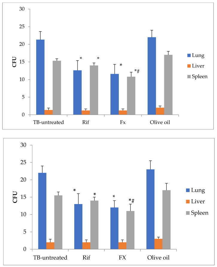
The incidence of diseases brought on by resistant strains of micro-organisms, including tuberculosis, is rising globally as a result of the rapid rise in pathogenic micro-organism resistance to antimicrobial treatments. Secondary metabolites with potential for antibacterial activity are produced by cyanobacteria and microalgae. In this study, gram-positive (S. aureus, E. faecalis) and gram-negative (K. pneumoniae, A. baumannii, P. aeruginosa) bacteria were isolated from pulmonary tuberculosis patients receiving long-term antituberculosis therapy. The antimicrobial potential of extracts from the cyanobacteria Leptolyngbya cf. ectocarpi, Planktothrix agardhii, Arthrospira platensis, Rohotiella mixta sp. nov., Nanofrustulum shiloi, and Tetraselmis (Platymonas) viridis Rouchijajnen was evaluated. On mouse splenocytes and peritoneal macrophages, extracts of cyanobacteria and microalgae had inhibitory effects. In vitro studies have shown that cyanobacteria and microalgae extracts suppress the growth of bacteria and mycobacteria. At the same time, it has been demonstrated that cyanobacterial and microalgal extracts can encourage bacterial growth in a test tube. Additionally, the enhanced fucoxanthin fraction significantly reduced the development of bacteria in vitro. In a mouse experiment to simulate tuberculosis, the mycobacterial load in internal organs was considerably decreased by fucoxanthin. According to the information gathered, cyanobacteria and microalgae are potential sources of antibacterial compounds that can be used in the manufacturing of pharmaceutical raw materials.
Keywords: antimicrobial potential; cyanobacter; diatoms; gram-positive and gram-negative bacteria; green microalgae; mycobacteria.
Conflict of interest statement
The authors declare no conflict of interest.
Figures
Similar articles
-
Bioprospecting of Microalgae Isolated from the Adriatic Sea: Characterization of Biomass, Pigment, Lipid and Fatty Acid Composition, and Antioxidant and Antimicrobial Activity.Molecules. 2022 Feb 12;27(4):1248. doi: 10.3390/molecules27041248. Molecules. 2022. PMID: 35209036 Free PMC article.
-
Antimicrobial Activity of Arthrospira (Former Spirulina) and Dunaliella Related to Recognized Antimicrobial Bioactive Compounds.Int J Mol Sci. 2024 May 19;25(10):5548. doi: 10.3390/ijms25105548. Int J Mol Sci. 2024. PMID: 38791586 Free PMC article. Review.
-
Cyanobacteria and Eukaryotic Microalgae as Emerging Sources of Antibacterial Peptides.Molecules. 2020 Dec 9;25(24):5804. doi: 10.3390/molecules25245804. Molecules. 2020. PMID: 33316949 Free PMC article. Review.
-
Antimicrobial Potential of the Green Microalgae Isolated from the Persian Gulf.Iran J Public Health. 2022 May;51(5):1134-1142. doi: 10.18502/ijph.v51i5.9428. Iran J Public Health. 2022. PMID: 36407722 Free PMC article.
-
Evaluation of microalgae and cyanobacteria as potential sources of antimicrobial compounds.Saudi Pharm J. 2020 Dec;28(12):1834-1841. doi: 10.1016/j.jsps.2020.11.010. Epub 2020 Nov 26. Saudi Pharm J. 2020. PMID: 33424272 Free PMC article.
Cited by
-
Application of microbial pigments in the pharmaceutical industry: current status and opportunities.Arch Microbiol. 2025 Mar 31;207(5):104. doi: 10.1007/s00203-025-04261-y. Arch Microbiol. 2025. PMID: 40164794 Review.
References
-
- Mitishev A., Kurdyukov E., Rodina O., Semenova E., Moiseeva I., Fadeeva T. Microalgae as a new source of biologically active compounds with antibacterial activity. Probl. Biol. Med. Pharm. Chem. 2021;24:24–29. doi: 10.29296/25877313-2021-07-04. - DOI
-
- Grubišić M., Šantek B., Zorić Z., Čošić Z., Vrana I., Gašparović B., Čož-Rakovac R., Ivančić Šantek M. Bioprospecting of Microalgae Isolated from the Adriatic Sea: Characterization of Biomass, Pigment, Lipid and Fatty Acid Composition, and Antioxidant and Antimicrobial Activity. Molecules. 2022;27:1248. doi: 10.3390/molecules27041248. - DOI - PMC - PubMed
LinkOut - more resources
Full Text Sources
