. 2023 Oct 3;120(40):e2215421120.
doi: 10.1073/pnas.2215421120. Epub 2023 Sep 27.

Externalized histones fuel pulmonary fibrosis via a platelet-macrophage circuit of TGFβ1 and IL-27

Affiliations

Externalized histones fuel pulmonary fibrosis via a platelet-macrophage circuit of TGFβ1 and IL-27

Dennis R Riehl et al. Proc Natl Acad Sci U S A. .

Abstract

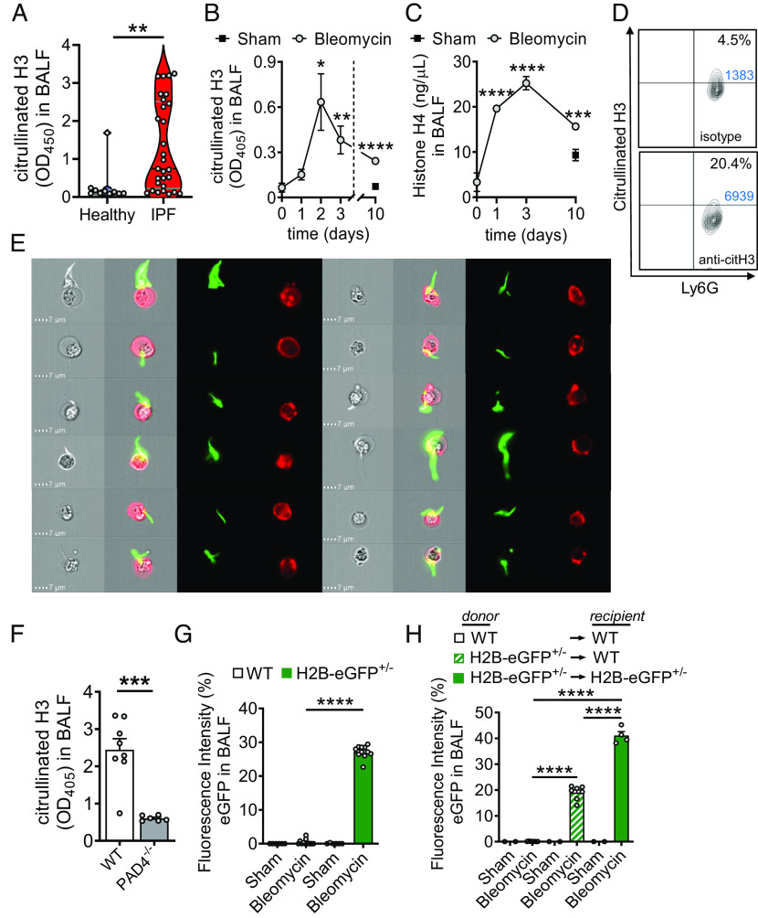
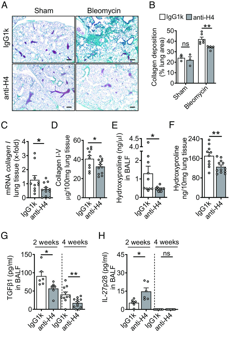
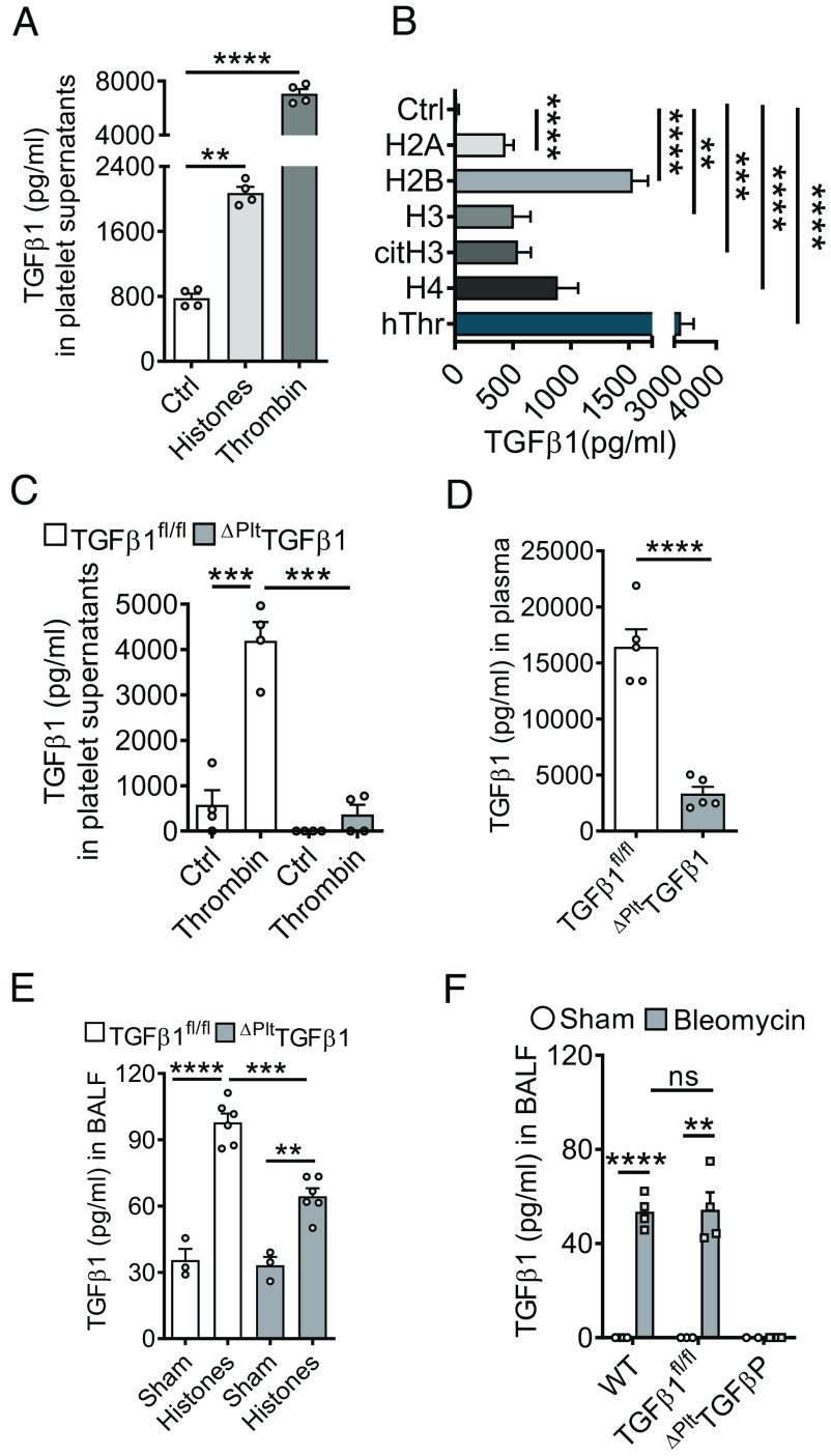
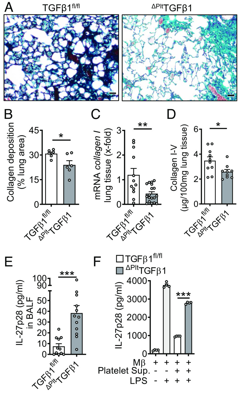
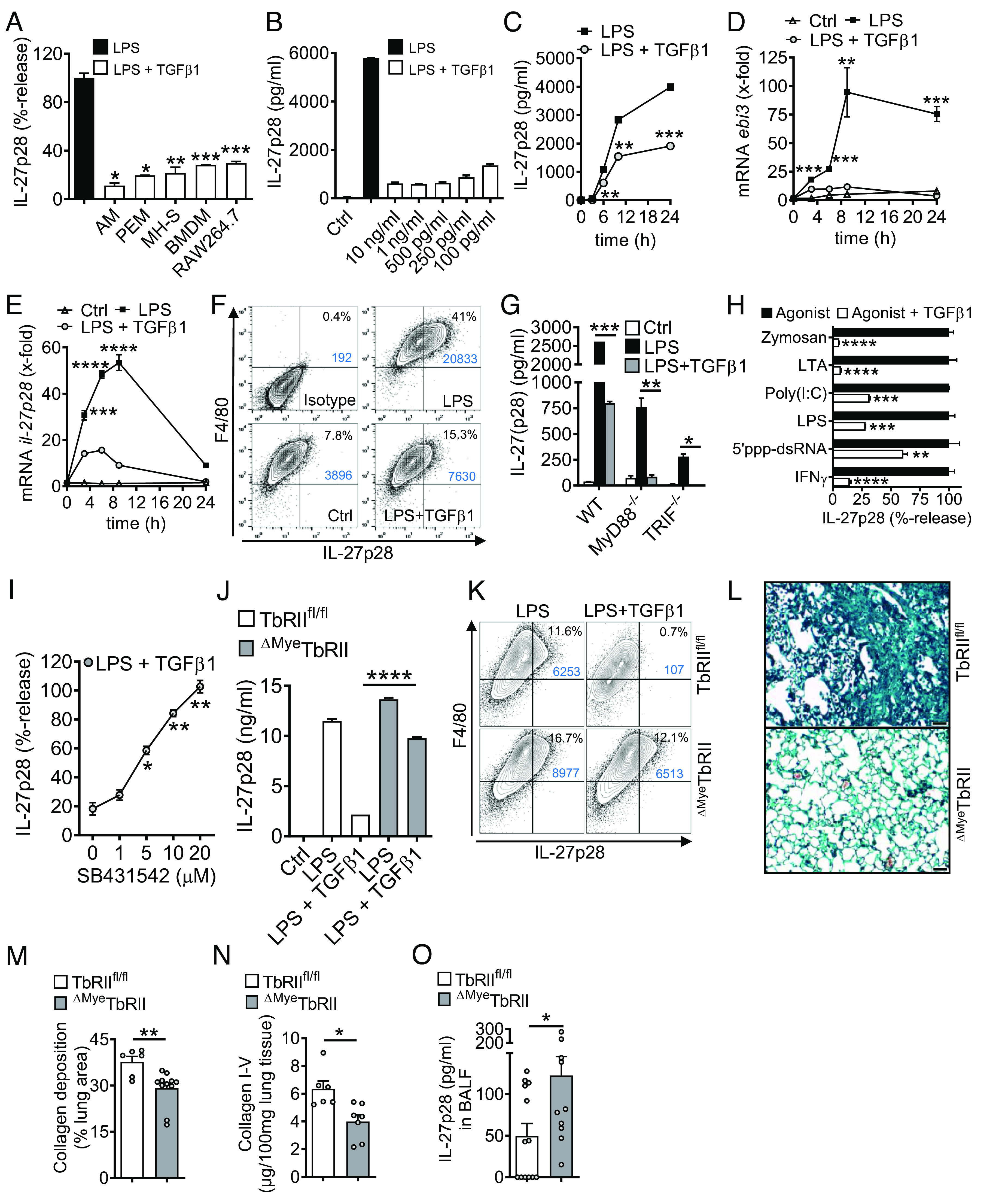
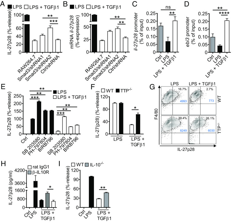
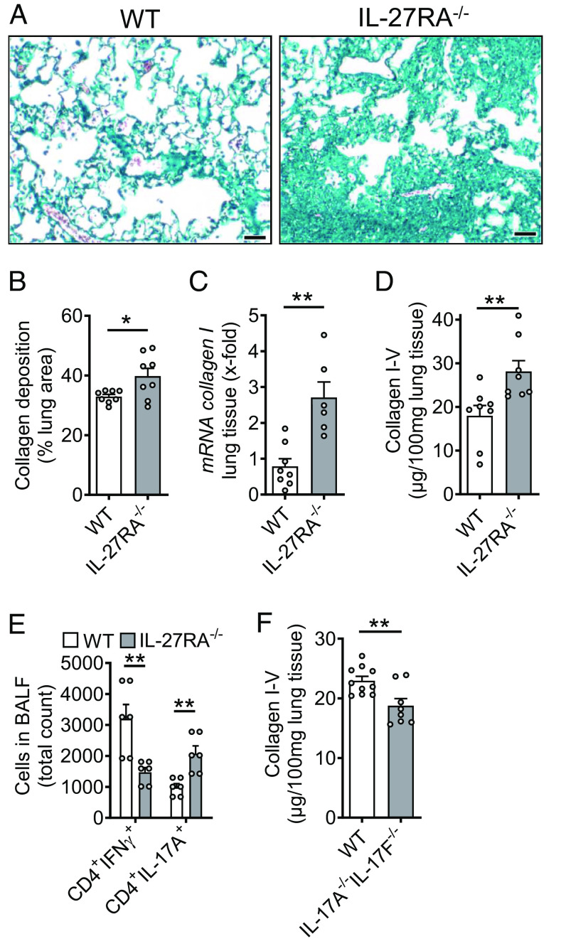
Externalized histones erupt from the nucleus as extracellular traps, are associated with several acute and chronic lung disorders, but their implications in the molecular pathogenesis of interstitial lung disease are incompletely defined. To investigate the role and molecular mechanisms of externalized histones within the immunologic networks of pulmonary fibrosis, we studied externalized histones in human and animal bronchoalveolar lavage (BAL) samples of lung fibrosis. Neutralizing anti-histone antibodies were administered in bleomycin-induced fibrosis of C57BL/6 J mice, and subsequent studies used conditional/constitutive knockout mouse strains for TGFβ and IL-27 signaling along with isolated platelets and cultured macrophages. We found that externalized histones (citH3) were significantly (P < 0.01) increased in cell-free BAL fluids of patients with idiopathic pulmonary fibrosis (IPF; n = 29) as compared to healthy controls (n = 10). The pulmonary sources of externalized histones were Ly6G+CD11b+ neutrophils and nonhematopoietic cells after bleomycin in mice. Neutralizing monoclonal anti-histone H2A/H4 antibodies reduced the pulmonary collagen accumulation and hydroxyproline concentration. Histones activated platelets to release TGFβ1, which signaled through the TGFbRI/TGFbRII receptor complex on LysM+ cells to antagonize macrophage-derived IL-27 production. TGFβ1 evoked multiple downstream mechanisms in macrophages, including p38 MAPK, tristetraprolin, IL-10, and binding of SMAD3 to the IL-27 promotor regions. IL-27RA-deficient mice displayed more severe collagen depositions suggesting that intact IL-27 signaling limits fibrosis. In conclusion, externalized histones inactivate a safety switch of antifibrotic, macrophage-derived IL-27 by boosting platelet-derived TGFβ1. Externalized histones are accessible to neutralizing antibodies for improving the severity of experimental pulmonary fibrosis.

Keywords: cytokines; innate immunity; macrophages; platelets; pulmonology.


Conflict of interest statement

The authors declare no competing interest.

Figures

Fig. 1.
Fig. 1.
Cellular sources of externalized histones during pulmonary fibrosis. (A) Presence of externalized citrullinated histone H3 in BALF samples of healthy human controls (n = 10) and patients with IPF (n = 29), ELISA with optical density (OD) measurements at 405 nm. (B and C) Time course of citrullinated histone H3 (B) and Histone H4 (C) in cell-free BALF of C57BL/6 J mice (n ≥ 4/group for each time point) after treatment with bleomycin i.t. or NaCl 0.9% i.t. (sham; negative control), ELISA. (D) NET formation detected as externalized citrullinated histone H3 on nonpermeabilized Ly6G+CD11b+ neutrophils in BALF of C57BL/6 J wild-type (WT) mice 2 d after bleomycin i.t. (n = 6 mice/group), flow cytometry with pregating on CD11b. Absolute cell numbers in the quadrants of interest are shown in blue font. (E) Visualization of NETs from BALF cells, which were obtained 2 d after bleomycin i.t. and stained for citrullinated externalized histone H3 (green) together with Ly6G (red) for neutrophils, ImageStreamX Mark II imaging flow cytometry. (F) PAD4−/− mice and WT mice were subjected to bleomycin-induced lung injury and citrullinated H3 was detected in cell-free BALF after 2 d. (G) Detection of green fluorescence in cell-free BALF of histone H2B-eGFP fusion protein reporter mice (H2B-eGFP+/−) 2 d after bleomycin administration as compared to WT control mice. Fluorescence of a FITC-labeled control antibody was used for normalization (=100% value). (H) Chimeric mice were generated by transplantation of H2B-eGFP+/− donor bone marrow into irradiated WT recipients. In the control groups, syngeneic bone marrow was transplanted. After 5 wk, all mice received bleomycin i.t. and H2B-eGFP fluorescence was detected in cell-free BALF another 2 d later. Numbers of mice in frames F–H are indicated by circles; A: Mann–Whitney test, B and C: one-way ANOVA, F–H: Student’s t test, *P < 0.05, **P < 0.01, ***P < 0.001, ****P < 0.0001.
Fig. 2.
Fig. 2.
Externalized histones promote pulmonary fibrosis. (A) Histology of lungs from WT mice which received either monoclonal anti-H2A/H4 antibodies (clone: BWA3, 250 µg/mouse i.v.) or mouse IgG1κ isotype control antibodies simultaneously with i.t. bleomycin (n = 3–6/group). Masson–Goldner’s trichrome staining of lungs was performed after 4 wk (Scale bar, 50 μm). (B) Quantification of lung collagen deposition from scanned whole slides from mice in frame A. (C) ECM expression as evaluated by RT-PCR for collagen I (col1a2) mRNA in lung homogenates during bleomycin-induced fibrosis. (D) SIRCOL assay for collagen I–V in lung tissue of anti-histone antibody-treated mice or control IgG1k-treated mice after bleomycin i.t. (E and F) Hydroxyproline concentrations in BALF and lung tissue of anti-histone antibody-treated mice as compared to IgG1k controls during fibrosis, 4 wk. (G and H) TGFβ1 and IL-27p28 concentrations in BALF of anti-histone antibody-treated mice as compared to IgG1k controls at 2 wk or 4 wk after bleomycin, ELISA. Horizontal dashed lines indicate groups of samples which were analyzed on separate days and ELISA plates. Experiments in all frames were done with C57BL/6 J (WT) mice and analyzed 4 wk (frame A–F) or 2 wk (stated in frame G and H) after bleomycin i.t. (1 U/kg body weight). Numbers of mice in frames B–H are indicated by circles; Student’s t test or one-way ANOVA; *P < 0.05, **P < 0.01, ns: not significant.
Fig. 3.
Fig. 3.
TGFβ1 is released by platelets in response to externalized histones. (A) Platelets were isolated from C57BL/6 J mice and activated with purified histones (50 µg/mL) or thrombin (100 ng/mL) for 10 min before quantification of TGFβ1 in supernatants. (B) TGFβ1 release from platelets after incubation with recombinant human histones (H2A, H2B, H2, H4), citrullinated histone H3, human thrombin, and untreated (Ctrl), 10 min. (C) Isolated platelets from mice with platelet-specific gene deletion of TGFβ1 (ΔPltTGFβ1) mice and littermate controls (TGFβ1fl/fl) were activated with thrombin (100 ng/mL) for 10 min before quantification of TGFβ1 in supernatants. (D) Plasma concentrations of TGFβ1 in mice with platelet-specific deletion compared to littermate controls, 4 wk after bleomycin. (E) ΔPltTGFβ1 mice and TGFβ1fl/fl control mice received purified histones i.t. (100 µg/mouse) or PBS i.t. (sham) and TGFβ1 was detected in BALF after 8 h. (F) TGFβ1 in BALF of mice with platelet-specific deletion compared to TGFβ1fl/fl littermate controls and C57BL/6 J (WT) mice, 24 h after bleomycin. TGFβ1 was measured by ELISA in all frames. Frames A–C are representative of three independent experiments and frames D–F were done with numbers of mice as indicated by circles; Student’s t test or one-way ANOVA; **P < 0.01, ***P < 0.001, ****P < 0.0001, ns: not significant.
Fig. 4.
Fig. 4.
Platelet-derived TGFβ1 drives pulmonary fibrosis. (A) Histology of lung sections 4 wk after bleomycin-induced fibrosis. ΔPltTGFβ1 lungs were compared to TGFβ1fl/fl control lungs, Masson–Goldner’s trichrome staining (Scale bar, 50 µm), n = 6/group. (B) Quantification of lung collagen deposition from scanned whole slides from mice in frame A. (C) RT-PCR for collagen I gene expression in lung homogenates of ΔPltTGFβ1 mice and TGFβ1fl/fl control mice during bleomycin-induced fibrosis. (D) Collagen I–V protein content in lungs of ΔPltTGFβ1 mice and TGFβ1fl/fl mice, SIRCOL assay. (E) IL-27p28 in BALF of ΔPltTGFβ1 and TGFβ1fl/fl mice during bleomycin-induced lung fibrosis, ELISA. (F) Supernatants of activated washed platelets from ΔPltTGFβ1 mice or TGFβ1fl/fl control mice were transferred to cell cultures of C57BL/6 J bone marrow-derived macrophages (BMDM) before stimulation with LPS for 24 h and quantification of IL-27p28 release by ELISA. Frames B–E (4 wk of bleomycin i.t.) were done with numbers of mice as indicated by circles and frame F is representative of three independent experiments; Student’s t test or one-way ANOVA; *P < 0.05, **P < 0.01, ***P < 0.001.
Fig. 5.
Fig. 5.
TGFβ receptor type I/II signaling mediates suppression of IL-27p28 in macrophages. (A) Mononuclear phagocytic cells were incubated with LPS (100 ng/mL) ± TGFβ1 (10 ng/mL) followed by detection of IL-27p28 in supernatants; C57BL/6 J-derived AM and PEM, mouse AM cell line (MH-S), C57BL/6 J BMDM and RAW264.7 macrophage cell line, 24 h. LPS was set as 100% for each cell type. (B) TLR4/LPS-activated BMDM were incubated with different concentrations of TGFβ1 before quantification of IL-27p28, 24 h. (C) Time course of IL-27p28 release by LPS-activated BMDM ± TGFβ1. (D) RT-PCR for ebi3 mRNA after LPS ± TGFβ1 in BMDM. (E) RT-PCR for il-27p28 mRNA after LPS ± TGFβ1 in BMDM. (F) Flow cytometry for intracellular IL-27p28 in BMDM, 12 h, pregated on CD11b+ cells. (G) Inhibition of IL-27p28 release by TGFβ1 in BMDM from C57BL/6 J (WT), MyD88−/− or TRIF−/− mice, 24 h. (H) Suppression of IL-27p28 release by TGFβ1 in BMDM activated with several agonists; Zymosan (TLR2), LTA (TLR2), Poly(I:C) (TLR3), LPS (TLR4), 5′ppp-dsRNA (RIG-I) and IFNγ, 24 h. (I) Reversal of TGFβ1-mediated IL-27p28 suppression by increasing concentrations of the TGFβ receptor I signaling inhibitor, SB431542, in TLR4/LPS-activated BMDM, 24 h. LPS alone was used for normalization (=100% value). (J) IL-27p28 from BMDM of TGFβ receptor II floxed (TbRIIfl/fl) mice and ΔMyeTbRII mice after LPS ± TGFβ1, 24 h. (K) Flow cytometry of intracellular IL-27p28 in BMDM from TbRIIfl/fl or ΔMyeTbRII mice, 12 h. (L) Lung histology of bleomycin-induced fibrosis in TbRIIfl/fl or ΔMyeTbRII mice, 4 wk, Masson–Goldner’s trichrome staining (Scale bar, 50 µm). (M) Quantification of lung collagen deposition from scanned whole slides from mice in frame L. (N) Collagen I–V in lungs of TbRIIfl/fl mice and ΔMyeTbRII mice after bleomycin i.t, 4 wk, SIRCOL assay. (O) IL-27p28 in BALF of bleomycin-treated TbRIIfl/fl mice and ΔMyeTbRII mice, 4 wk. Frames A-CG-JO: ELISA. Representatives of three independent experiments (frames A–K) or n ≥ 6 mice/group (frames L–O); Student’s t test or one-way ANOVA; *P < 0.05, **P < 0.01, ***P < 0.001, ****P < 0.0001.
Fig. 6.
Fig. 6.
TGFβ1-mediated antagonism of IL-27p28 release via SMAD3, p38, TTP, and IL-10. (A) IL-27p28 release with reduced activity of TGFβ1 in lentiviral transduced RAW264.7 macrophages with shRNA knockdown of SMAD3 (two independent shRNAs) as compared to nontarget shRNA (Ctrl), 24 h, ELISA. (B) TGFβ1 partially fails to suppress il-27p28 mRNA expression in transduced RAW264.7 macrophages after treatments with LPS ± TGFβ1, 6 h, RT-PCR. (C) TGFβ1 restores SMAD3 binding to the il-27p28 promoter region, BMDM, 3 h, ChIP assay. (D) TGFβ1 restores SMAD3 binding to the ebi3 promoter region, BMDM, 3 h, ChIP assay. (E) Effects of several p38 MAPK inhibitors (SB 203580 [10 µM], PH-797804 [10 nM], BIRB796 [50 nM]) on IL-27p28 release in BMDM after LPS ± TGFβ1, 24 h, ELISA. (F) Reduced capacity of TGFβ1 to suppress IL-27p28 release in BMDM from tristetraprolin-deficient (TTP−/−) mice as compared to WT mice, ELISA, 24 h. (G) Flow cytometry of F4/80+CD11b+IL-27p28+ BMDM from TTP−/− mice and WT mice after LPS ± TGFβ1, 12 h, pregated on CD11b. (H) IL-27p28 release from BMDM incubated with LPS ± TGFβ1 in the copresence of blocking IL-10 receptor antibodies (α-IL10R; 10 µg/mL) or rat IgG1 isotype control antibodies (10 µg/mL), ELISA. (I) Defective suppression of IL-27p28 by TGFβ1 in BMDM from IL-10−/− mice as compared to WT mice. BMDM were from C57BL/6 J (WT) mice in all frames unless specified otherwise; LPS (100 ng/mL), TGFβ1 (10 ng/mL). Data in each frame are representative of three independent experiments; Student’s t test or one-way ANOVA; *P < 0.05, **P < 0.01, ***P < 0.001, ****P < 0.0001; ns: not significant.
Fig. 7.
Fig. 7.
Protective role of IL-27RA during pulmonary fibrosis. (A) Lung sections of IL-27RA−/− mice and WT mice during bleomycin-induced pulmonary fibrosis, Masson–Goldner’s trichrome staining, n ≥ 6/group (Scale bar, 50 µm). (B) Quantification of collagen deposition using scanned whole slides from mice in frame A. (C) Expression of collagen I mRNA in lung tissue of IL-27RA−/− and WT mice after bleomycin, RT-PCR. (D) SIRCOL protein assay for collagen I–V in lung tissue of the experiment described under C. (E) Flow cytometry of CD4+ T cells in BALF of bleomycin i.t.-treated IL-27RA−/− mice as compared to WT mice. (F) Collagen accumulation after bleomycin-induced pulmonary fibrosis in IL-17A−/−IL-17F−/− double-knockout mice as compared to WT mice, SIRCOL assay. All frames were analyzed at 4 wk after bleomycin and frames B–F were done with numbers of mice as indicated by circles; Student’s t test or one-way ANOVA; *P < 0.05, **P < 0.01.

References

    1. Sgalla G., Kulkarni T., Antin-Ozerkis D., Thannickal V. J., Richeldi L., Update in pulmonary fibrosis 2018. Am. J. Respir. Crit. Care Med. 200, 292–300 (2019). - PubMed
    1. Wynn T. A., Integrating mechanisms of pulmonary fibrosis. J. Exp. Med. 208, 1339–1350 (2011). - PMC - PubMed
    1. Smigiel K. S., Parks W. C., Macrophages, wound healing, and fibrosis: Recent insights. Curr. Rheumatol. Rep. 20, 17 (2018). - PubMed
    1. Leask A., Abraham D. J., TGF-beta signaling and the fibrotic response. FASEB J. 18, 816–827 (2004). - PubMed
    1. Massague J., TGFbeta signalling in context. Nat. Rev. Mol. Cell Biol. 13, 616–630 (2012). - PMC - PubMed

Publication types

LinkOut - more resources