. 2023 Sep 27;15(715):eadg5567.
doi: 10.1126/scitranslmed.adg5567. Epub 2023 Sep 27.

A MERS-CoV antibody neutralizes a pre-emerging group 2c bat coronavirus

Affiliations

A MERS-CoV antibody neutralizes a pre-emerging group 2c bat coronavirus

Longping V Tse et al. Sci Transl Med. .

Abstract

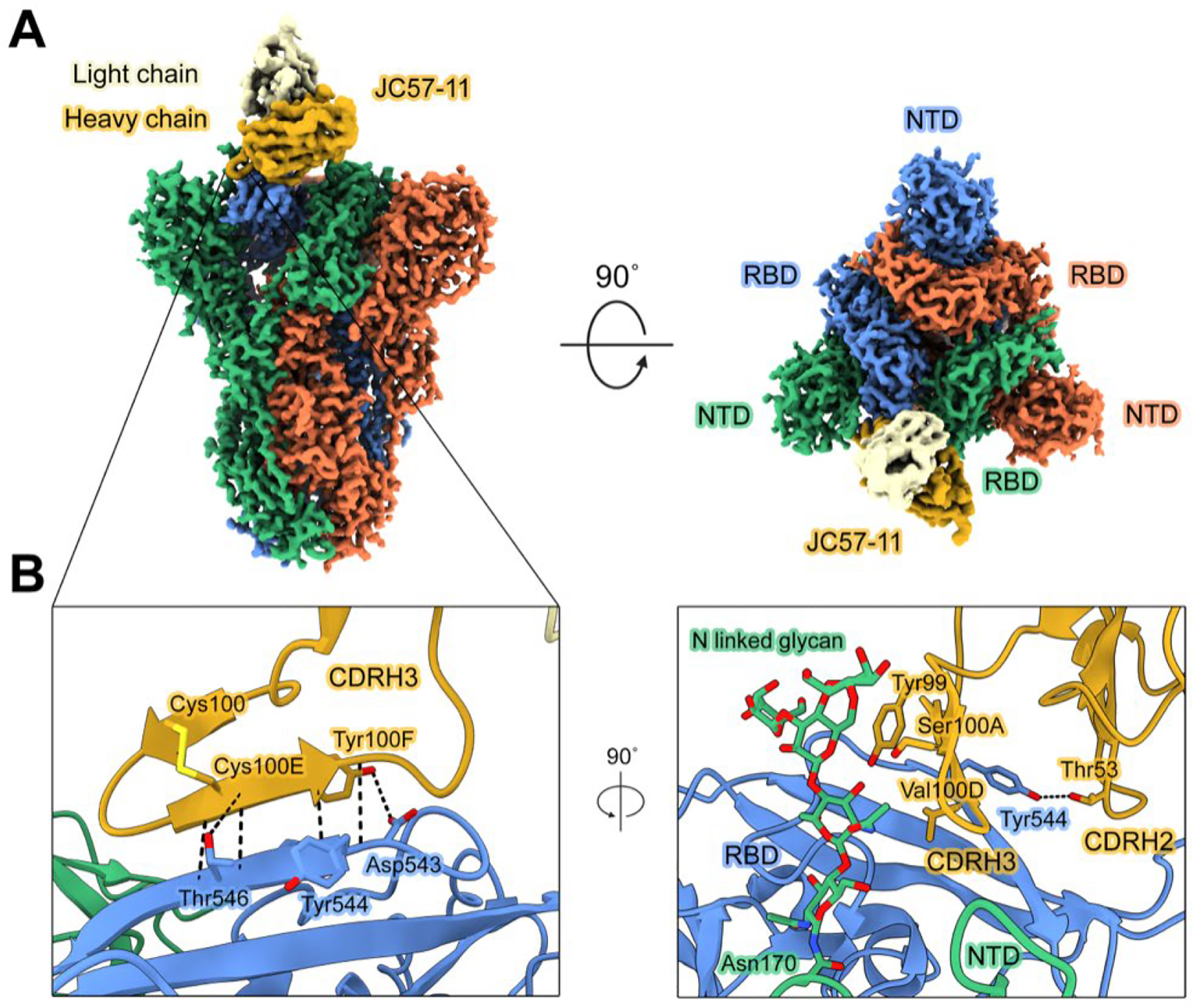
The repeated emergence of zoonotic human betacoronaviruses (β-CoVs) dictates the need for broad therapeutics and conserved epitope targets for countermeasure design. Middle East respiratory syndrome (MERS)-related coronaviruses (CoVs) remain a pressing concern for global health preparedness. Using metagenomic sequence data and CoV reverse genetics, we recovered a full-length wild-type MERS-like BtCoV/li/GD/2014-422 (BtCoV-422) recombinant virus, as well as two reporter viruses, and evaluated their human emergence potential and susceptibility to currently available countermeasures. Similar to MERS-CoV, BtCoV-422 efficiently used human and other mammalian dipeptidyl peptidase protein 4 (DPP4) proteins as entry receptors and an alternative DPP4-independent infection route in the presence of exogenous proteases. BtCoV-422 also replicated efficiently in primary human airway, lung endothelial, and fibroblast cells, although less efficiently than MERS-CoV. However, BtCoV-422 shows minor signs of infection in 288/330 human DPP4 transgenic mice. Several broad CoV antivirals, including nucleoside analogs and 3C-like/Mpro protease inhibitors, demonstrated potent inhibition against BtCoV-422 in vitro. Serum from mice that received a MERS-CoV mRNA vaccine showed reduced neutralizing activity against BtCoV-422. Although most MERS-CoV-neutralizing monoclonal antibodies (mAbs) had limited activity, one anti-MERS receptor binding domain mAb, JC57-11, neutralized BtCoV-422 potently. A cryo-electron microscopy structure of JC57-11 in complex with BtCoV-422 spike protein revealed the mechanism of cross-neutralization involving occlusion of the DPP4 binding site, highlighting its potential as a broadly neutralizing mAb for group 2c CoVs that use DPP4 as a receptor. These studies provide critical insights into MERS-like CoVs and provide candidates for countermeasure development.


Conflict of interest statement

Competing Interests: R.S.B. is a member of the Scientific Advisory Board of Invivyd and VaxArt and has consulted on virus-related countermeasures with Gilead, Moderna, Takeda, BioNet and Janssen Bio that are unrelated to this project.

Figures

Figure 1.
Figure 1.. BtCoV-422 recombinant and reporter viruses were generated using reverse genetics.
(A) Shown is a phylogenetic tree of coronavirus spike proteins. (B) Structural alignment of spike proteins is shown for the indicated viruses. Magenta denotes amino acid variations and cyan denotes the RDB of spike protein. (C) Shown are the putative proteolytic cleavage sites in S1/S2 site and S2’ site in multiple group 2C CoVs, including BtCoV-422 and MERS-CoV. The red text indicates furin cleavage sites. The underlined text indicates fusion peptides. (D) Shown is the design of BtCoV-422 infectious clones, including the wildtype clone, BtCoV-422-WT, the GFP reporter virus, BtCoV-422-GFP, and the nLuc reporter virus, BtCoV-422-nLuc. The size of each fragment, the restriction sites for cloning and the T7 promoter are shown. (E) Viral yields are shown for the BtCoV-422-WT virus and reporter viruses. The data are presented as mean ± SD. (F) Plaque morphologies are shown for BtCoV-422-WT and reporter viruses on Vero cells stained with neutral red.
Figure 2.
Figure 2.. BtCoV-422 infects distal but not proximal primary human airway epithelial cultures.
(A) Shown are viral growth kinetics and representative fluorescent images of primary human nasal epithelial cells (NEs) from 4 different donors infected with BtCoV-422 or BtCoV-422-GFP. (B) Shown are viral growth kinetics and representative fluorescent images of primary human NEs from 4 different donors infected with MERS-CoV or MERS-CoV-RFP. Scale bars indicate 200 μm. (C) Shown are viral growth kinetics of BtCoV-422, BtCoV-422-GFP, and MERS-CoV-RFP in LAEs from 4 different donors. (D and E) Shown are growth kinetics and representative fluorescent images of BtCoV-422-GFP in primary human microvascular endothelial cells (MVEs) (D) and primary human lung fibroblasts (FBs) (E). Scale bars indicate 200 μm. The data are presented as mean ± SD.
Figure 3.
Figure 3.. BtCoV-422 and MERS-CoV use similar species profile of DPP4 as functional receptors.
(A and B) Shown are representative images of BtCoV-422 (A) and MERS-CoV (B) infection of Vero-DPP4 KO cells, Vero-DPP4 WT cells, and Vero-DPP4 KO cells stably expressing a panel of seven DPP4 orthologs from other species, including bat (P. Pipistrellus), camel, ferret, guinea pig, human, hamster, and mouse. Vector control is also shown. Infected cells were stained with anti-N antibodies. The scale bars indicate 200 μm. (C and D) Shown is quantitative analysis of BtCoV-422-nLuc (C) and MERS-CoV-nLuc (D) infecting the same panel of DPP4 cell lines. Relative light units (RLUs) were compared using one-way ANOVA to DPP4-KO cells. N.S., not significant; ***P < 0.001; ****P < 0.0001. The data are presented as mean ± SD.
Figure 4.
Figure 4.. External proteases enhance BtCoV-422 replication and syncytia formation.
(A to C) Infection of BtCoV-422-nLuc (MOI: 0.02) was measured in Caco-2 (A), Calu3 (B), and Vero (C) cells in the presence of trypsin, elastase, or thermolysin. Nano luciferase activity were measured at 24hpi and reported as RLUs. Bkgd indicates background luminescence and is represented by the dotted horizontal line. RLUs were compared using one-way ANOVA to no protease control. N.S., not significant; **P< 0.01; ****P < 0.0001. (D and E) Growth kinetics were measured for BtCoV-422-WT (D) and BtCoV-422-GFP (E) in Vero cells with or without addition of trypsin. (F) Representative images are shown for BtCoV-422-GFP-infected Vero cells at 24 hours with or without addition of trypsin (Top row: no bright field; Bottom row: with bright field). Syncytia formation is observed in the trypsin-containing cultures. Scale bars indicate 200 μm. The data are presented as mean ± SD.
Figure 5.
Figure 5.. BtCoV-422 uses alternative, DPP4-independent pathways to enter cells in the presence of trypsin.
(A and B) Representative brightfield and BtCoV-422-GFP fluorescence images (A) and growth curves (B) are shown for BtCoV-422-GFP infection of in Vero DPP4 KO or WT cells, with or without trypsin. (C) Infection of Vero DPP4 KO or WT cells by BtCoV-422-nLuc, with or without trypsin, was measured by luciferase activity at 24 hours post infection. (D and E) Representative brightfield and MERS-CoV-RFP fluorescent images (D) and growth curves (E) are shown for MERS-CoV-RFP infection of Vero DPP4 KO or WT cells, with or without trypsin. (F) Infection of Vero DPP4 KO or WT cells by MERS-CoV-nLuc with or without trypsin, was measured by luciferase activity at 24 h post infection. Fluorescent images were taken at 48hpi with an MOI of 0.05. Scale bars indicate 200 μm RLUs were compared using non-parametric t-test compared with the corresponding background values background signal in the corresponding condition. N.S., not significant; **P< 0.01; ***P < 0.001; ****P < 0.0001. The data are presented as mean ± SD. (G) Shown are biolayer interferometry measurements of MERS-CoV, SARS-CoV-2 Omicron BA.1, and BtCoV-422 spike proteins on a streptavidin biosensor dipped into human ACE2 for 180 seconds.
Figure 6.
Figure 6.. In vitro potencies of MERS-CoV-vaccinated mouse serum, neutralizing mAbs, and antivirals against BtCoV-422 in Huh7.5 cells.
(A) Inhibition concentration 50 (IC50) values are shown for BtCoV-422 and MERS-CoV replication in the presence of remdesivir, molnupiravir, and nirmatrelvir in Huh7.5 cells. Quantitation of BtCoV-422 replication was measured by nLuc activity in technical triplicates. IC50 values are shown above. (B and C) Nano-luciferase based neutralization assays of MERS-CoV-nLuc and BtCoV-422-nLuc was performed using serum from mice immunized with a MERS-CoV-S2P mRNA vaccine (B) and a panel of neutralizing mAbs against MERS-CoV spike protein (C). Data are presented in (B and C) as IC50 values. The numbers in (B) indicate individual mice vaccinated with different doses. Serum from mock PBS-vaccinated mice were used as a control in (B). LoD indicates limit of detection at 40 μg/ml, unless specified in the graph. IC50 between BtCoV-422 and MERS-CoV were compared using Student’s t-test. **P< 0.01; ****P < 0.0001. Horizontal bars indicate mean values.
Figure 7.
Figure 7.. Cryo-EM structure of BtCoV-422 RBD and JC57–11 complex.
(A) Shown is the composite cryo-EM map of JC57–11 (EMD-40272, PDB 8SAK) bound to the RBD of the BtCoV-422 spike protein viewed from the side (left) and down toward the membrane (right). Spike protein protomers are colored orange, blue, and green. The heavy chain of JC75–11 is colored yellow, and the light chain is colored tan. (B) Shown is a model of the binding interfaces between JC57–11 and BtCoV-422 RBD. Interacting side chains are depicted as sticks. Hydrogen bonds are depicted as black dotted lines. Oxygen atoms are colored red, nitrogen atoms are colored blue, and sulfur atoms are colored yellow.

References

    1. Zhou P, Yang X-L, Wang X-G, Hu B, Zhang L, Zhang W, Si H-R, Zhu Y, Li B, Huang C-L, Chen H-D, Chen J, Luo Y, Guo H, Jiang R-D, Liu M-Q, Chen Y, Shen X-R, Wang X, Zheng X-S, Zhao K, Chen Q-J, Deng F, Liu L-L, Yan B, Zhan F-X, Wang Y-Y, Xiao G-F, Shi Z-L, A pneumonia outbreak associated with a new coronavirus of probable bat origin. Nature (2020), doi:10.1038/s41586-020-2012-7. - DOI - PMC - PubMed
    1. Updates on COVID-19 Variants of Concern (VOC).
    1. Zaki AM, van Boheemen S, Bestebroer TM, Osterhaus ADME, Fouchier RAM, Isolation of a Novel Coronavirus from a Man with Pneumonia in Saudi Arabia. New England Journal of Medicine. 367, 1814–1820 (2012). - PubMed
    1. Baric RS, Sullivan E, Hensley L, Yount B, Chen W, Persistent Infection Promotes Cross-Species Transmissibility of Mouse Hepatitis Virus. J Virol. 73, 638 (1999). - PMC - PubMed
    1. Arabi YM, Arifi AA, Balkhy HH, Najm H, Aldawood AS, Ghabashi A, Hawa H, Alothman A, Khaldi A, al Raiy B, Clinical course and outcomes of critically ill patients with Middle East respiratory syndrome coronavirus infection. Ann Intern Med. 160, 389–397 (2014). - PubMed

Substances