. 2023 Dec;12(32):e2301472.
doi: 10.1002/adhm.202301472. Epub 2023 Oct 13.

Microphysiological Modeling of Gingival Tissues and Host-Material Interactions Using Gingiva-on-Chip

Affiliations

Microphysiological Modeling of Gingival Tissues and Host-Material Interactions Using Gingiva-on-Chip

Giridharan Muniraj et al. Adv Healthc Mater. 2023 Dec.

Abstract

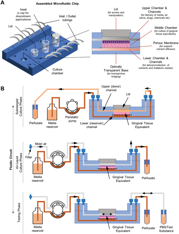
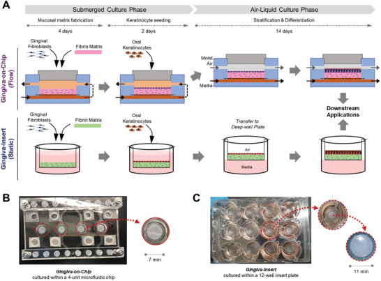
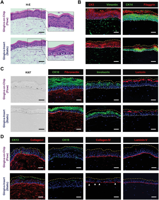
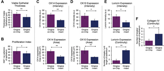
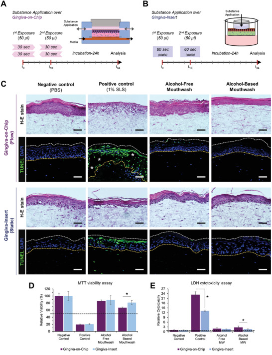
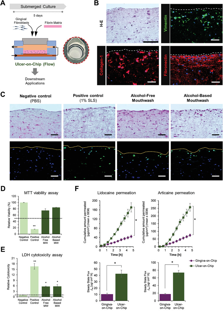
Gingiva plays a crucial barrier role at the interface of teeth, tooth-supporting structures, microbiome, and external agents. To mimic this complex microenvironment, an in vitro microphysiological platform and biofabricated full-thickness gingival equivalents (gingiva-on-chip) within a vertically stacked microfluidic device is developed. This design allowed long-term and air-liquid interface culture, and host-material interactions under flow conditions. Compared to static cultures, dynamic cultures on-chip enabled the biofabrication of gingival equivalents with stable mucosal matrix, improved epithelial morphogenesis, and barrier features. Additionally, a diseased state with disrupted barrier function representative of gingival/oral mucosal ulcers is modeled. The apical flow feature is utilized to emulate the mechanical action of mouth rinse and integrate the assessment of host-material interactions and transmucosal permeation of oral-care formulations in both healthy and diseased states. Although the gingiva-on-chip cultures have thicker and more mature epithelium, the flow of oral-care formulations induced increased tissue disruption and cytotoxic features compared to static conditions. The realistic emulation of mouth rinsing action facilitated a more physiological assessment of mucosal irritation potential. Overall, this microphysiological system enables biofabrication of human gingiva equivalents in intact and ulcerated states, providing a miniaturized and integrated platform for downstream host-material and host-microbiome applications in gingival and oral mucosa research.

Keywords: biocompatibility; drug permeation; gingiva; microfluidics; organ-on-a-chip; periodontal disease.


Conflict of interest statement

The authors would also like to disclose that two of the authors, M.A. and G.S., are co‐founders of Revivo Biosystems Pte Ltd., and the microfluidic device used in this study is based on a patent application (WO2018030958A1) licensed to the company.

Figures

Figure 1
Figure 1
Schematic representation of microfluidic gingiva‐on‐chip device. A) Schematic view of the assembled device that shows the different features such as microchannels, chambers, inlets, and outlets micromilled into polymethyl methacrylate sheets that are vertically stacked and thermally bonded. B) Schematic representation of fluidic circuit used for perfusion of media, air, and test substances at different phases of the organotypic culture.
Figure 2
Figure 2
Biofabrication of organotypic full‐thickness gingival equivalents underflow and static conditions. A) Schematic representation of the organotypic culture under flow within the microfluidic device (gingiva‐on‐chip) and under static conditions using porous culture inserts (gingiva‐insert). The culture under both conditions includes mucosal matrix fabrication, keratinocyte seeding, and air‐liquid interface culture followed by their downstream applications. Macroscopic views of the gingival tissues fabricated within the B) microfluidic device and C) in static insert culture systems. The gingiva‐on‐chip equivalents within the microfluidic device B) are visible and easily accessible through the lid opening in the upper chamber (inset).
Figure 3
Figure 3
Characterization of the full‐thickness gingiva equivalents reconstructed underflow and static conditions. A) Hematoxylin‐eosin H–E) stained sections demonstrate the multi‐layered, orthokeratinized gingival epithelium over fibroblast‐populated lamina propria‐like matrix. B–D) Immunostained sections show the expression of cytokeratins (CK5, CK14, CK10, CK13, and CK19), stratified epithelial differentiation markers (involucrin, filaggrin, loricrin), proliferation marker (Ki67), vimentin, matrix proteins (collagen‐I and fibronectin) and basement membrane markers (collagen‐IV, laminin‐V) (Scale bar: 50 µm).
Figure 4
Figure 4
Morphometric analysis of the full‐thickness gingiva equivalents reconstructed underflow and static conditions. The bar graphs show the quantification and comparison of A) thickness of viable epithelium, B) Ki67 proliferation index, C–E) intensity and area of CK14, CK10, and loricrin expression, and E,F) number of breaks in the expression of loricrin and collagen IV among the gingival equivalents cultured under flow and static conditions. Area of CK14 expression corresponds to the area of viable epithelium (without the cornified layers), while the area of CK10 expression corresponds to suprabasal layers including the cornified layers. The number of breaks in loricrin and collagen IV are normalized to unit length (400 µm) of the epithelium. (n≥3; *p<0.05, **p<0.01, Student t‐test).
Figure 5
Figure 5
Application and comparison of gingival equivalents for in vitro assessment of oral‐care products underflow and static conditions. Schematic representation of exposure of the surface of gingival equivalents to oral‐care formulations under A) flow and B) static conditions on the gingival‐on‐chip and gingiva‐insert respectively. C) Hematoxylin‐eosin H–E) and TUNEL‐stained sections of the gingival equivalents after exposure to oral‐care formulations and controls (Scale bar: 50 µm; * represent epithelial disruption). D) Relative cell viability and E) relative LDH release following exposure to oral‐care formulations. Dotted lines represent the respective thresholds. (n≥3; *p<0.05).
Figure 6
Figure 6
Application of gingiva‐on‐chip for disease modeling and drug permeation studies. A) Schematic representation of the fabrication of ulcer‐on‐chip equivalents under flow conditions, and macroscopic view of its shiny, wet surface. B) Hematoxylin‐eosin and immunostained sections show the expression of vimentin and matrix proteins (collagen‐I and fibronectin). C) Hematoxylin‐eosin and TUNEL‐stained sections, D) relative cell viability, and E) relative LDH release from the ulcer‐on‐chip equivalents after exposure to oral‐care formulations and controls (Scale bar: 50 µm). Dotted lines represent the respective thresholds. F,G) Line and bar graphs show the permeation profiles of lidocaine HCl and articaine HCl permeation through gingiva‐on‐chip and ulcer‐on‐chip equivalents. (n≥3; *p<0.05).

References

    1. Klausner M., Ayehunie S., Breyfogle B. A., Wertz P. W., Bacca L., Kubilus J., Toxicol. In Vitro 2007, 21, 938. - PubMed
    1. Bierbaumer L., Schwarze U. Y., Gruber R., Neuhaus W., Tissue Barriers 2018, 6, 1479568. - PMC - PubMed
    1. Viñuela‐Prieto J. M., Sánchez‐Quevedo M. C., Alfonso‐Rodríguez C. A., Oliveira A. C., Scionti G., Martín‐Piedra M. A., Moreu G., Campos A., Alaminos M., Garzón I., J. Periodontal Res. 2015, 50, 658. - PubMed
    1. Presland R. B., Dale B. A., Crit. Rev. Oral Biol. Med. 2000, 11, 383. - PubMed
    1. Ibrahim M. S., El‐Wassefy N. A., Farahat D. S., Biomaterials for Oral and Dental Tissue Engineering, Woodhead Publishing, Cambridge: 2017.

Publication types

LinkOut - more resources