. 2023 Sep 27;14(1):6040.
doi: 10.1038/s41467-023-41721-9.

AlphaFold-Multimer predicts cross-kingdom interactions at the plant-pathogen interface

Affiliations

AlphaFold-Multimer predicts cross-kingdom interactions at the plant-pathogen interface

Felix Homma et al. Nat Commun. .

Abstract

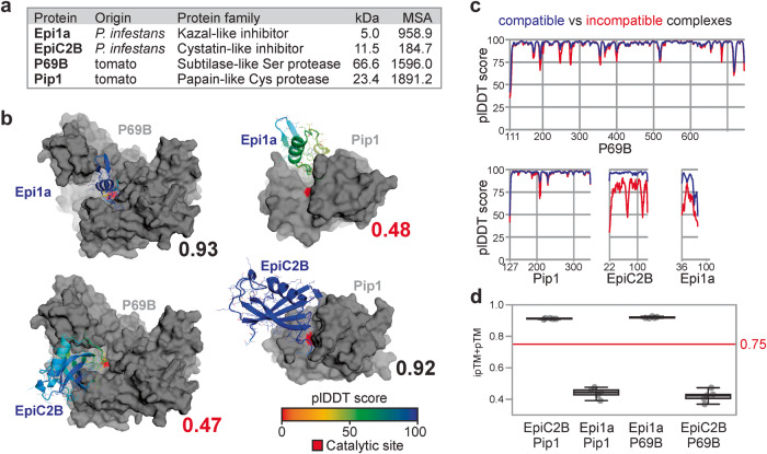
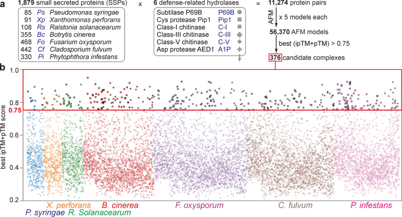
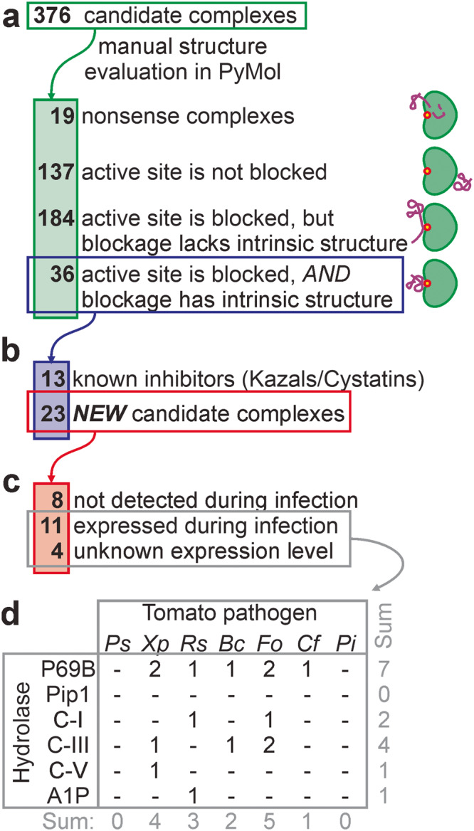
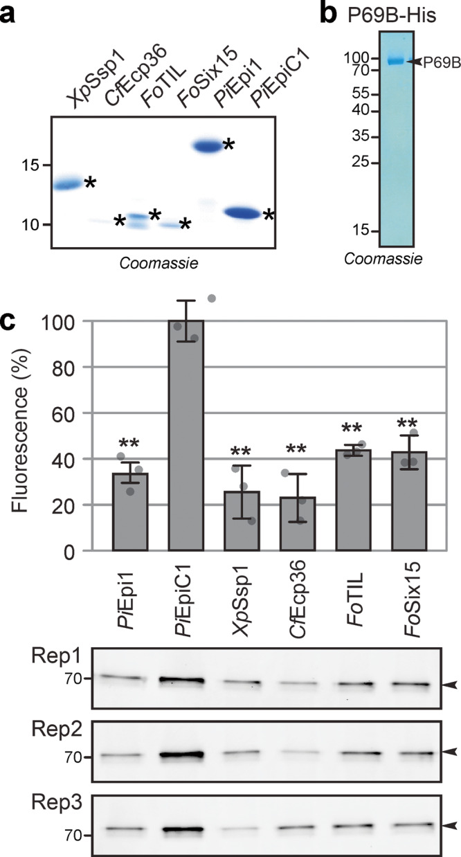
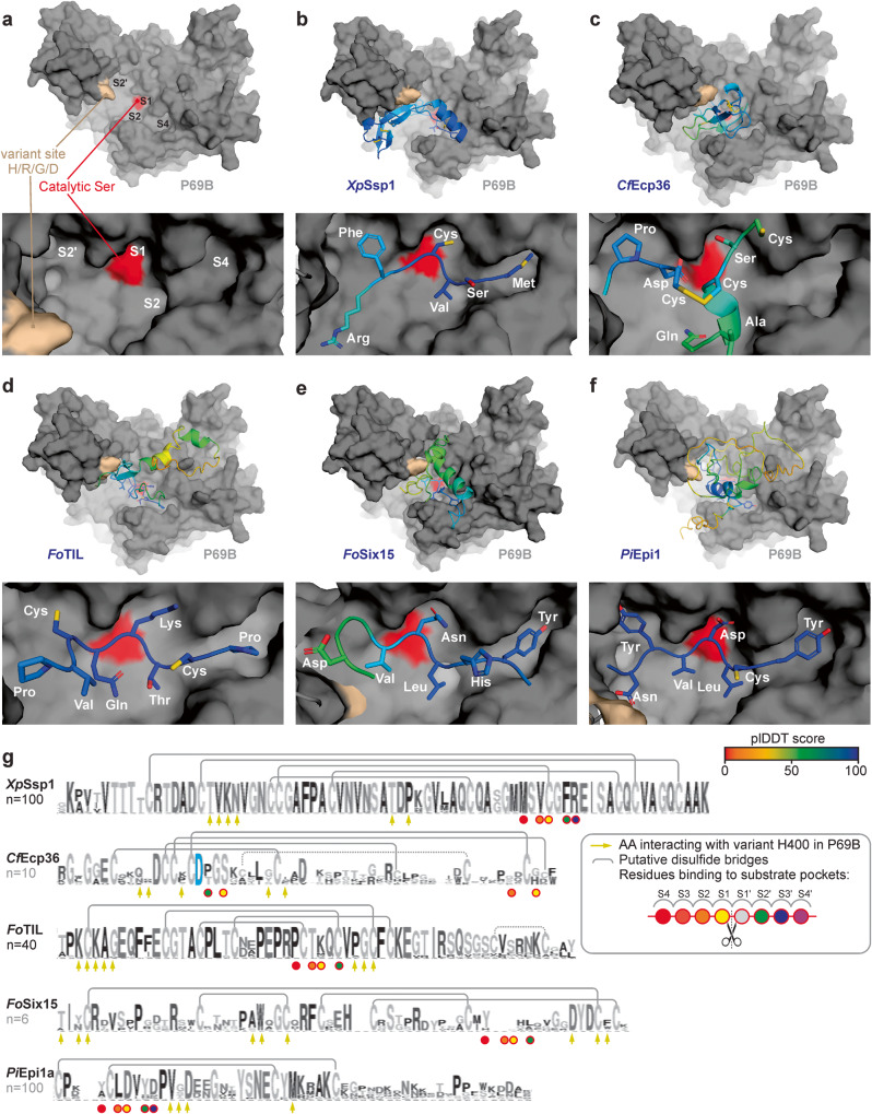
Adapted plant pathogens from various microbial kingdoms produce hundreds of unrelated small secreted proteins (SSPs) with elusive roles. Here, we used AlphaFold-Multimer (AFM) to screen 1879 SSPs of seven tomato pathogens for interacting with six defence-related hydrolases of tomato. This screen of 11,274 protein pairs identified 15 non-annotated SSPs that are predicted to obstruct the active site of chitinases and proteases with an intrinsic fold. Four SSPs were experimentally verified to be inhibitors of pathogenesis-related subtilase P69B, including extracellular protein-36 (Ecp36) and secreted-into-xylem-15 (Six15) of the fungal pathogens Cladosporium fulvum and Fusarium oxysporum, respectively. Together with a P69B inhibitor from the bacterial pathogen Xanthomonas perforans and Kazal-like inhibitors of the oomycete pathogen Phytophthora infestans, P69B emerges as an effector hub targeted by different microbial kingdoms, consistent with a diversification of P69B orthologs and paralogs. This study demonstrates the power of artificial intelligence to predict cross-kingdom interactions at the plant-pathogen interface.


Conflict of interest statement

The authors declare no competing interests.

Figures

Fig. 1
Fig. 1. AFM correctly distinguishes existing from non-existing hydrolase-inhibitor complexes.
a Used inhibitors and their target proteases with their origin, mature molecular weight (MW, in kDa) and depth of mean non-gap multiple sequence alignment (MSA) detected for proteins in compatible complexes. b Best structures predicted by AFM for existing and non-existing inhibitor-hydrolase complexes, with their ipTM + pTM scores ranging from 0 (worst) to 1 (best). Pip1 and P69B are shown in gray, with their catalytically active residue in red. EpiC2B and Epi1a are colored using a rainbow scheme based on their plDDT scores, which range from 0 (worst) to 100 (best). PDB files of these modeals are provided in Supplementary Data 3. c plDDT scores within the four proteins in predicted compatible (blue) and incompatible (red) complexes. d ipTM + pTM quality scores for each of the n = 5 five models for each of the protein pairs, showing the median, 25th and 75th percentiles, and whiskers representing 1.5 times the interquartile range. The raw data are provided in Supplementary Data 6.
Fig. 2
Fig. 2. AFM screen between 1879 SSPs and 6 hydrolases identifies 376 candidate complexes.
a 1879 proteins from seven tomato pathogens that are likely secreted and small (<35 kDa) were screened for complexes with six secreted defense-related hydrolases of tomato using AlphaFold-Multimer (AFM). The best of the five generated AFM models for each of the 11,274 protein pairs were selected if the ipTM + pTM score was 0.75 or higher, resulting in 376 putative complexes. b best ipTM + pTM scores for all the 11,274 complexes involving 1879 small secreted proteins (SSPs) of the seven tomato pathogens listed on the bottom. Symbols for complexes with the six different hydrolases (explained in (a)) highlight the 376 candidate complexes with ipTM + pTM ≥ 0.75. The best and all ipTM + pTM values for each protein pair used for this figure are provided in Supplementary Data 7 and Supplementary Data 8, respectively.
Fig. 3
Fig. 3. Selection of candidate complexes.
a The 376 candidate complexes were manually screened for complexes where the SSP blocks the active site of the hydrolase with an intrinsic fold. b The remaining 36 candidate complexes included 13 complexes between Kazal-like inhibitors and P69B and cystatin-like inhibitors with Pip1, and 23 candidate complexes with other candidate inhibitors. c Of the 23 remaining candidate complexes, transcriptome analysis of infected plants showed that genes encoding eight SSPs are not expressed at the tested conditions, whereas genes encoding 11 SSPs are expressed during infection. No transcriptomic datasets were available for Xp (4 SSPs). d Distribution of the final 15 candidate hydrolase-inhibitor complexes over the tested tomato pathogens and target hydrolases.
Fig. 4
Fig. 4. Activity labeling of P69B is suppressed by four inhibitors.
a Purified candidate inhibitors. Candidate inhibitors and Epi1a (positive control) and EpiC1 (negative control) were expressed in E. coli as fusion proteins with N-terminal His-MBP-TEV. The fusion proteins were purified over Ni-NTA and amylose resin, subsequently, and then cleaved by TEV protease. See Supplementary Fig. 2 for the full gel. His-TEV protease and purification tags were subsequently removed using Ni-NTA and MW cut off filter and SSPs were used for inhibition assays in (c). Purification of candidate inhibitors was repeated at least once for each candidate independently. b P69B-His was transiently expressed in Nicotiana benthamiana by agroinfiltration and purified over Ni-NTA from apoplastic fluids isolated at 5 days-post-agroinfiltration. The eluate was analysed on protein gel stained with Coomassie (shown here) and used for inhibition assays (in (c)). Purification of P69B-His was repeated twice, not including experiments for a previous publication. c All four candidate inhibitors and the Epi1 but not EpiC1 suppress activity-based labeling of P69B with FP-TAMRA. Purified P69B-His was pre-incubated with purified (candidate) inhibitors at a 1:100 molar ratio and then labeled with FP-TAMRA in n = 3 replicates using the same purified proteins. Proteins were separated on protein gels and scanned for fluorescence. Fluorescence was quantified and the signal intensity of the negative control (EpiC1) was set at 100% labeling to calculate the relative labeling upon preincubation with the positive control (Epi1) and the four candidate inhibitors. Error bars represent STDEV of n = 3 replicates. **p < 0.01 (p-values from two-sided, pairwise t-tests were adjusted for multiple testing using the Benjamini–Hochberg procedure). These p-values are 0.00065; 0.00091; 0.00063; 0.00046; and 0.0010 for comparing EpiC1 with PiEpi1; XpSSP1; CfEcp36; FoTIL and FoSix15, respectively. MW makers are listed in kDa. A similar suppression of labeling was observed at 2-fold higher candidate inhibitor concentrations and in a repeat experiment using independently purified proteins. Original images for the gels are provided in Supplementary Data 9 and the raw quantification data in Supplementary Data 6.
Fig. 5
Fig. 5. P69B is an effector hub targeted by five pathogen-derived inhibitors.
AFM-predicted models of P69B without inhibitor (a), or with XpSsp1; (b) CfEcp36; (c) FoTIL; (d) FoSix15 (e) and PiEpi1 (f). P69B is shown in a gray surface representation with the hyper-variant residue (crème) and the active site (red), in the substrate binding groove that has substrate binding pockets (S4-S2-S1-S2’) that bind to substrate/inhibitor residues P4, P2, P1 and P2’, respectively. The inhibitors are shown as cartoons and sticks and colored using a rainbow scheme based on their plDDT scores, which range from 0 (worst) to 100 (best). The zoomed image (bottom) shows the predicted occupation of the substrate binding pockets in P69B by different residues of the inhibitor. g Sequence conservation between homologs of the identified P69B inhibitors. Shown is the sequence logo for n = x close homologs from other plant pathogen species, identified by BLAST searches in the NCBI database and presented in Weblogo. Highlighted are the residues that probably interact with the substrate binding pockets in P69B (circles); the conserved Asp residue in CfEcp36 that interacts with the catalytic site (blue); the residues that might interact with the variant residue in P69B (arrows); and putative disulfide brides observed in the AFM model (gray lines). PDB files for the shown models are available in Supplementary Data 3.

References

    1. Doehlemann G, Hemetsberger C. Apoplastic immunity and its suppression by filamentous plant pathogens. New Phytol. 2013;198:1001–1016. doi: 10.1111/nph.12277. - DOI - PubMed
    1. Darino M, Kanyuka K, Hammond-Kosack KE. Apoplastic and vascular defences. Essays Biochem. 2022;66:595–605. doi: 10.1042/EBC20220159. - DOI - PubMed
    1. van Loon LC, Rep M, Pieterse CM. Significance of inducible defense-related proteins in infected plants. Annu. Rev. Phytopathol. 2006;44:135–162. doi: 10.1146/annurev.phyto.44.070505.143425. - DOI - PubMed
    1. Jordá L, Coego A, Conejero V, Vera P. A genomic cluster containing four differentially regulated subtilisin-like processing protease genes is in tomato plants. J. Biol. Chem. 1999;274:2360–2365. doi: 10.1074/jbc.274.4.2360. - DOI - PubMed
    1. Jordá L, Conejero V, Vera P. Characterization of P69E and P69F, two differentially regulated genes encoding new members of the subtilisin-like proteinase family from tomato plants. Plant Physiol. 2000;122:67–74. doi: 10.1104/pp.122.1.67. - DOI - PMC - PubMed

Publication types