. 2023 Sep 27;8(1):141.
doi: 10.1038/s41541-023-00742-7.

Duration of SARS-CoV-2 mRNA vaccine persistence and factors associated with cardiac involvement in recently vaccinated patients

Affiliations

Duration of SARS-CoV-2 mRNA vaccine persistence and factors associated with cardiac involvement in recently vaccinated patients

Aram J Krauson et al. NPJ Vaccines. .

Abstract

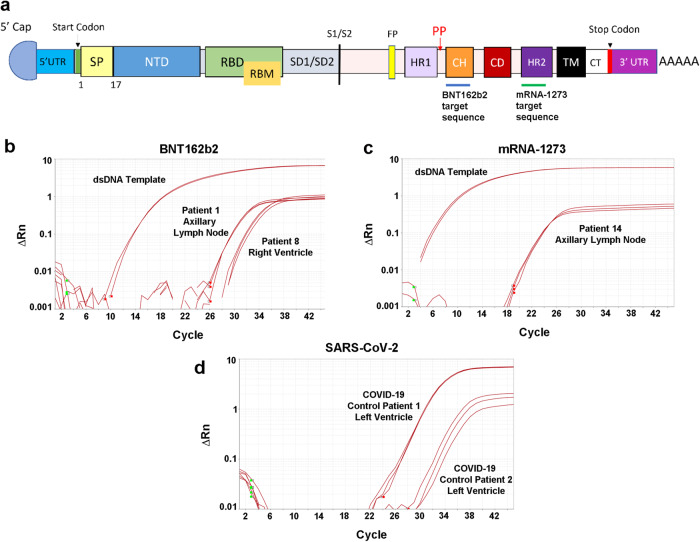
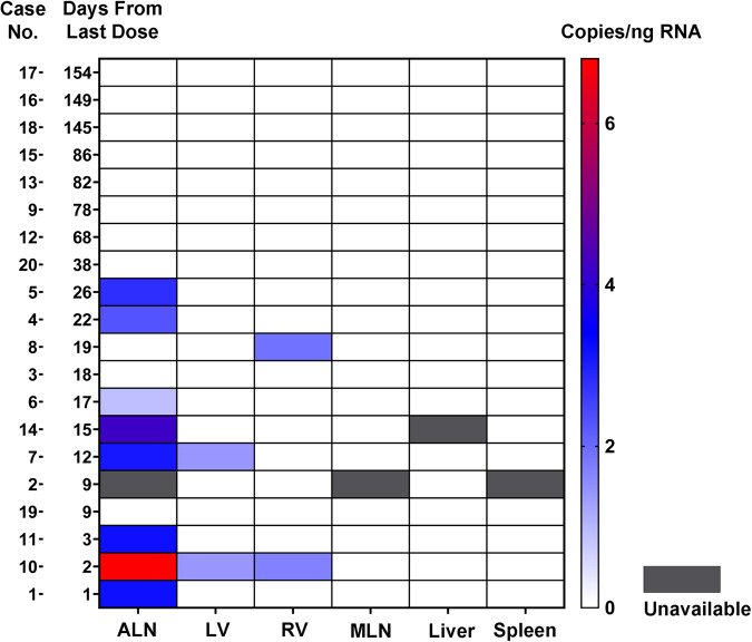
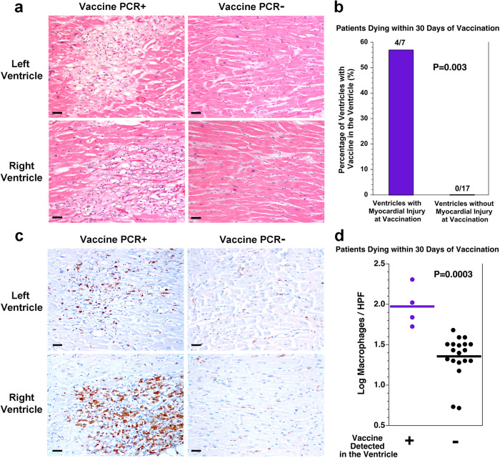
At the start of the COVID-19 pandemic, the BNT162b2 (BioNTech-Pfizer) and mRNA-1273 (Moderna) mRNA vaccines were expediently designed and mass produced. Both vaccines produce the full-length SARS-CoV-2 spike protein for gain of immunity and have greatly reduced mortality and morbidity from SARS-CoV-2 infection. The distribution and duration of SARS-CoV-2 mRNA vaccine persistence in human tissues is unclear. Here, we developed specific RT-qPCR-based assays to detect each mRNA vaccine and screened lymph nodes, liver, spleen, and myocardium from recently vaccinated deceased patients. Vaccine was detected in the axillary lymph nodes in the majority of patients dying within 30 days of vaccination, but not in patients dying more than 30 days from vaccination. Vaccine was not detected in the mediastinal lymph nodes, spleen, or liver. Vaccine was detected in the myocardium in a subset of patients vaccinated within 30 days of death. Cardiac ventricles in which vaccine was detected had healing myocardial injury at the time of vaccination and had more myocardial macrophages than the cardiac ventricles in which vaccine was not detected. These results suggest that SARS-CoV-2 mRNA vaccines routinely persist up to 30 days from vaccination and can be detected in the heart.


Conflict of interest statement

The authors declare no competing interests.

Figures

Fig. 1
Fig. 1. Screening autopsies for mRNA vaccines with designed RT-qPCR assays.
Detection of the SARS-CoV-2 mRNA vaccines in autopsies requires devising sequence-specific regions for RT-qPCR assays within the spike gene structure. a The functional domains of the mRNA vaccine SARS-CoV-2 spike protein includes the assay targets for the BNT162b2 mRNA vaccine in the central helix (CH) domain (blue line below). For detection of the mRNA-1273 vaccine (green line), the assay detects a sequence within the heptad repeat 2 (HR2) segment (see Table 2). UTR untranslated region, SP signal peptide, NTD N-terminal domain, RBD receptor-binding domain, RBM receptor-binding motif, SD subdomain, FP fusion peptide, HR1 heptad repeat 1, CD connector domain, TM transmembrane, CT cytoplasmic tail. Also marked is the start codon (green line), stop codon (red line), S1/S2 cleavage site (black line), the vaccines’ two Proline substitutions (red P’s), and the polyA tail at the end. Sample amplification plots of RT-qPCR assays to detect b BNT162b2 and c mRNA-1273 SARS-CoV-2 vaccines in axillary lymph node autopsy samples. Double-stranded DNA templates (dsDNA) were used as positive controls. d All vaccine-positive samples were further screened for SARS-CoV-2 E gene. The viral gene was detected only in the two SARS-CoV-2 infected cardiac left ventricle samples (positive controls).
Fig. 2
Fig. 2. Biodistribution and persistence of SARS-CoV-2 mRNA vaccines.
Depicted is a heat map for vaccine detection in the tissues with the patients arranged by the interval in days from last vaccination to death. The scale at the right indicates the copies of vaccine mRNA per ng of RNA. Vaccine mRNA was detected in bilateral axillary lymph nodes (ALN), cardiac left ventricle (LV), and cardiac right ventricle (RV), within 30 days of vaccination in a subset of patients, but not in mediastinal lymph nodes (MLN), liver or spleen.
Fig. 3
Fig. 3. SARS-CoV-2 vaccine in the heart is associated with healing myocardial injury at the time of vaccination and macrophage infiltration in the myocardium.
a Depicted are histologic images of the myocardium from both the left and right ventricles of the heart with either vaccine detected in the sample or not detected in the sample. For the two samples in which vaccine was detected, there was healing myocardial injury (left), compared with no healing myocardial injury in the two samples in which vaccine was not detected (right). Scale bars indicate 40 microns. b For the 12 patients dying within 30 days of vaccination, myocardial injury was present at the time of vaccination in 4 of the 7 ventricular samples with vaccine in the heart but none of the 17 ventricular samples without vaccine in the heart (P = 0.003, Fisher exact test). c Depicted are immunohistochemical stains for the macrophage marker CD68 on the myocardium from both the left and right ventricles of the heart with either vaccine detected in the sample or not detected in the sample. For the two samples in which vaccine was detected, there are more macrophages (left, brown stain), compared with the two samples in which vaccine was not detected (right). Scale bars indicate 40 microns. d Quantitation of the macrophages revealed more macrophages per 400× high power field (HPF) in the ventricular samples in which vaccine was detected than in the ventricular samples in which vaccine was not detected (P = 0.0003, t test, n = 4 vs n = 19, t = 4.3782, df = 21, difference between means = 0.62, 95% CI: 0.21–0.91). Tissue from one ventricle without vaccine detection and without myocyte injury was not available for CD68 immunohistochemistry.

Similar articles

Cited by

References

    1. McColl ER, et al. COVID-19 vaccines and the virus: impact on drug metabolism and pharmacokinetics. Drug Metab. Dispos. 2023;51:130–141. doi: 10.1124/dmd.122.000934. - DOI - PMC - PubMed
    1. Watson OJ, et al. Global impact of the first year of COVID-19 vaccination: a mathematical modelling study. Lancet Infect. Dis. 2022;22:1293–1302. doi: 10.1016/S1473-3099(22)00320-6. - DOI - PMC - PubMed
    1. Kwon S, Kwon M, Im S, Lee K, Lee H. mRNA vaccines: the most recent clinical applications of synthetic mRNA. Arch. Pharm. Res. 2022;45:245–262. doi: 10.1007/s12272-022-01381-7. - DOI - PMC - PubMed
    1. Szebeni J, et al. Applying lessons learned from nanomedicines to understand rare hypersensitivity reactions to mRNA-based SARS-CoV-2 vaccines. Nat. Nanotechnol. 2022;17:337–346. doi: 10.1038/s41565-022-01071-x. - DOI - PubMed
    1. Andries O, et al. N(1)-methylpseudouridine-incorporated mRNA outperforms pseudouridine-incorporated mRNA by providing enhanced protein expression and reduced immunogenicity in mammalian cell lines and mice. J. Control. Release. 2015;217:337–344. doi: 10.1016/j.jconrel.2015.08.051. - DOI - PubMed