

Sensitivity of an AI method for [18F]FDG PET/CT outcome prediction of diffuse large B-cell lymphoma patients to image reconstruction protocols
- PMID: 37758869
- PMCID: PMC10533444
- DOI: 10.1186/s13550-023-01036-8
Sensitivity of an AI method for [18F]FDG PET/CT outcome prediction of diffuse large B-cell lymphoma patients to image reconstruction protocols
Abstract
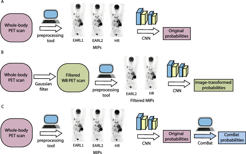
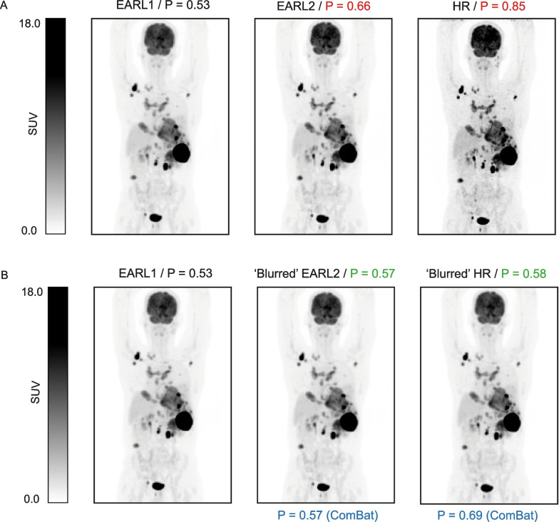
Background: Convolutional neural networks (CNNs), applied to baseline [18F]-FDG PET/CT maximum intensity projections (MIPs), show potential for treatment outcome prediction in diffuse large B-cell lymphoma (DLBCL). The aim of this study is to investigate the robustness of CNN predictions to different image reconstruction protocols. Baseline [18F]FDG PET/CT scans were collected from 20 DLBCL patients. EARL1, EARL2 and high-resolution (HR) protocols were applied per scan, generating three images with different image qualities. Image-based transformation was applied by blurring EARL2 and HR images to generate EARL1 compliant images using a Gaussian filter of 5 and 7 mm, respectively. MIPs were generated for each of the reconstructions, before and after image transformation. An in-house developed CNN predicted the probability of tumor progression within 2 years for each MIP. The difference in probabilities per patient was then calculated between both EARL2 and HR with respect to EARL1 (delta probabilities or ΔP). We compared these to the probabilities obtained after aligning the data with ComBat using the difference in median and interquartile range (IQR).
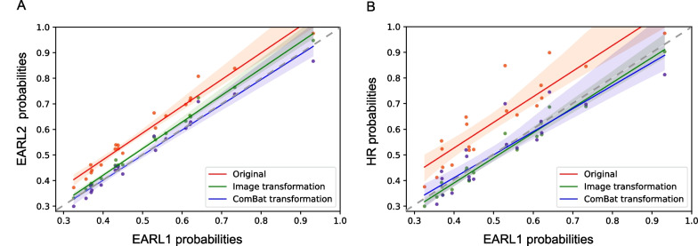
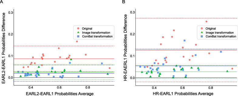
Results: CNN probabilities were found to be sensitive to different reconstruction protocols (EARL2 ΔP: median = 0.09, interquartile range (IQR) = [0.06, 0.10] and HR ΔP: median = 0.1, IQR = [0.08, 0.16]). Moreover, higher resolution images (EARL2 and HR) led to higher probability values. After image-based and ComBat transformation, an improved agreement of CNN probabilities among reconstructions was found for all patients. This agreement was slightly better after image-based transformation (transformed EARL2 ΔP: median = 0.022, IQR = [0.01, 0.02] and transformed HR ΔP: median = 0.029, IQR = [0.01, 0.03]).
Conclusion: Our CNN-based outcome predictions are affected by the applied reconstruction protocols, yet in a predictable manner. Image-based harmonization is a suitable approach to harmonize CNN predictions across image reconstruction protocols.
Keywords: Convolutional neural networks; Diffuse large B-cell lymphoma; PET; Reconstruction.
© 2023. Springer-Verlag GmbH Germany, part of Springer Nature.
Conflict of interest statement
This work was financially supported by the Hanarth Fonds Fund and the Dutch Cancer Society (#VU-2018–11648). M.C.F., S.S.V.G, J.J.E., B.M.d.V., S.E.W., G.J.C.Z., S.P. and R.B. declare no competing financial interests. J.M.Z. received research funding from Roche and received honoraria for advisory boards from Takeda, Gilead, BMS and Roche. No other potential conflicts of interest relevant to this article exist.
Figures
Similar articles
-
Quantitative and clinical implications of the EARL2 versus EARL1 [18F]FDG PET-CT performance standards in head and neck squamous cell carcinoma.EJNMMI Res. 2023 Oct 25;13(1):91. doi: 10.1186/s13550-023-01042-w. EJNMMI Res. 2023. PMID: 37878160 Free PMC article.
-
Convolutional neural networks for automatic image quality control and EARL compliance of PET images.EJNMMI Phys. 2022 Aug 9;9(1):53. doi: 10.1186/s40658-022-00468-w. EJNMMI Phys. 2022. PMID: 35943622 Free PMC article.
-
Combatting the effect of image reconstruction settings on lymphoma [18F]FDG PET metabolic tumor volume assessment using various segmentation methods.EJNMMI Res. 2022 Jul 29;12(1):44. doi: 10.1186/s13550-022-00916-9. EJNMMI Res. 2022. PMID: 35904645 Free PMC article.
-
Sensitivity of 18F-fluorodihydrotestosterone PET-CT to count statistics and reconstruction protocol in metastatic castration-resistant prostate cancer.EJNMMI Res. 2019 Jul 30;9(1):70. doi: 10.1186/s13550-019-0531-8. EJNMMI Res. 2019. PMID: 31363939 Free PMC article.
-
Quantitative implications of the updated EARL 2019 PET-CT performance standards.EJNMMI Phys. 2019 Dec 26;6(1):28. doi: 10.1186/s40658-019-0257-8. EJNMMI Phys. 2019. PMID: 31879795 Free PMC article.
Cited by
-
Prognostic impact of metabolic tumor volume using the SUV4.0 segmentation threshold in 1,960 lymphoma patients from prospective LYSA trials.Eur J Nucl Med Mol Imaging. 2025 Jul;52(9):3180-3189. doi: 10.1007/s00259-025-07176-4. Epub 2025 Mar 20. Eur J Nucl Med Mol Imaging. 2025. PMID: 40108044
-
Enhanced HoVerNet Optimization for Precise Nuclei Segmentation in Diffuse Large B-Cell Lymphoma.Diagnostics (Basel). 2025 Aug 4;15(15):1958. doi: 10.3390/diagnostics15151958. Diagnostics (Basel). 2025. PMID: 40804921 Free PMC article.
References
-
- Eertink JJ, van de Brug T, Wiegers SE, Zwezerijnen GJC, Pfaehler EAG, Lugtenburg PJ, et al. (18)F-FDG PET baseline radiomics features improve the prediction of treatment outcome in diffuse large B-cell lymphoma. Eur J Nucl Med Mol Imaging. 2022;49(3):932–942. doi: 10.1007/s00259-021-05480-3. - DOI - PMC - PubMed
Grants and funding
LinkOut - more resources
Full Text Sources
