. 2023 Sep 27;13(1):16214.
doi: 10.1038/s41598-023-43503-1.

Computer-aided diagnosis for screening of lower extremity lymphedema in pelvic computed tomography images using deep learning

Affiliations

Computer-aided diagnosis for screening of lower extremity lymphedema in pelvic computed tomography images using deep learning

Yukihiro Nomura et al. Sci Rep. .

Abstract

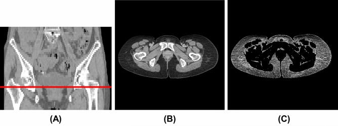
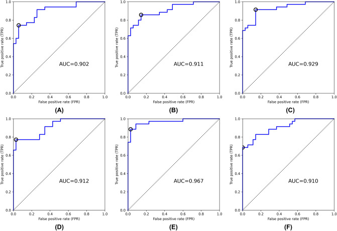
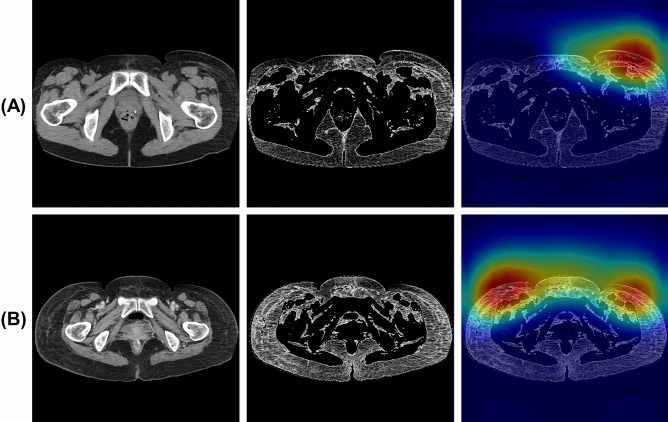
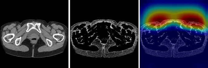
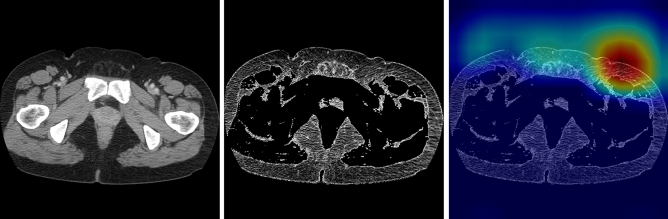
Lower extremity lymphedema (LEL) is a common complication after gynecological cancer treatment, which significantly reduces the quality of life. While early diagnosis and intervention can prevent severe complications, there is currently no consensus on the optimal screening strategy for postoperative LEL. In this study, we developed a computer-aided diagnosis (CAD) software for LEL screening in pelvic computed tomography (CT) images using deep learning. A total of 431 pelvic CT scans from 154 gynecological cancer patients were used for this study. We employed ResNet-18, ResNet-34, and ResNet-50 models as the convolutional neural network (CNN) architecture. The input image for the CNN model used a single CT image at the greater trochanter level. Fat-enhanced images were created and used as input to improve classification performance. Receiver operating characteristic analysis was used to evaluate our method. The ResNet-34 model with fat-enhanced images achieved the highest area under the curve of 0.967 and an accuracy of 92.9%. Our CAD software enables LEL diagnosis from a single CT image, demonstrating the feasibility of LEL screening only on CT images after gynecologic cancer treatment. To increase the usefulness of our CAD software, we plan to validate it using external datasets.


Conflict of interest statement

The authors declare no competing interests.

Figures

Figure 1
Figure 1
Example of CT image at the greater trochanter level. (A) Coronal section (red line: greater trochanter level), (B) original image (window level, 50 HU; window width, 400 HU), (C) fat-enhanced image.
Figure 2
Figure 2
ROC curves for test set for each input image and model [(A) original image, ResNet-18, (B) original image, ResNet-34, (C) original image, ResNet-50, (D) fat-enhanced image, RedNet-18, (E) fat enhanced image, ResNet-34, (F) fat-enhanced image, ResNet-50]. The circle indicates the cutoff point chosen using Youden’s index. AUC: area under the curve.
Figure 3
Figure 3
Examples of correctly classified as positive [(A) affected side: left, (B) affected side: both]. From left to right, original image, fat-enhanced image, and Grad-CAM heatmap overlaid on the fat-enhanced image.
Figure 4
Figure 4
Example of incorrectly classified as positive (normal case). From left to right, original image, fat-enhanced image, and Grad-CAM heatmap overlaid on the fat-enhanced image.
Figure 5
Figure 5
Example of incorrectly classified as negative (affected side: right). From left to right, original image, fat-enhanced image, and Grad-CAM heatmap overlaid on the fat-enhanced image.

Similar articles

Cited by

References

    1. Grada AA, Phillips TJ. Lymphedema: Diagnostic workup and management. J. Am. Acad. Dermatol. 2017;77:995–1006. doi: 10.1016/j.jaad.2017.03.021. - DOI - PubMed
    1. Grada AA, Phillips TJ. Lymphedema: Pathophysiology and clinical manifestations. J. Am. Acad. Dermatol. 2017;77:1009–1020. doi: 10.1016/j.jaad.2017.03.022. - DOI - PubMed
    1. Manrique OJ, et al. Overview of lymphedema for physicians and other clinicians: A review of fundamental concepts. Mayo Clin. Proc. 2022;97:1920–1935. doi: 10.1016/j.mayocp.2020.01.006. - DOI - PubMed
    1. Kim WJ, Kim J, Kang M, Park DH, Jeon JY. Usefulness of computed tomography venography in gynecologic cancer patients with lower extremity edema. Medicine. 2020;99:e21818. doi: 10.1097/MD.0000000000021818. - DOI - PMC - PubMed
    1. Wong M, et al. The prevalence of undiagnosed postoperative lower limb lymphedema among gynecological oncology patients. Eur. J. Surg. Oncol. 2022;48:1167–1172. doi: 10.1016/j.ejso.2021.12.464. - DOI - PubMed