. 2023 Oct;622(7983):545-551.
doi: 10.1038/s41586-023-06567-7. Epub 2023 Sep 27.

Uniquely preserved gut contents illuminate trilobite palaeophysiology

Affiliations

Uniquely preserved gut contents illuminate trilobite palaeophysiology

Petr Kraft et al. Nature. 2023 Oct.

Abstract

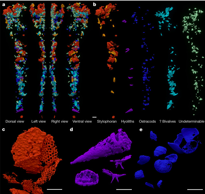
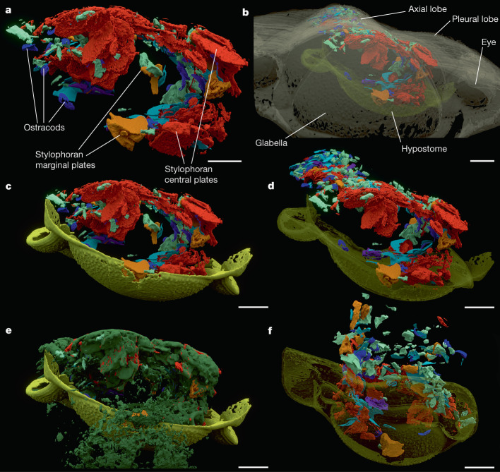
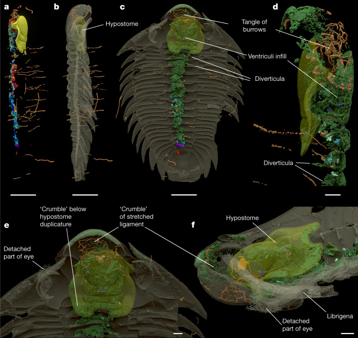
Trilobites are among the most iconic of fossils and formed a prominent component of marine ecosystems during most of their 270-million-year-long history from the early Cambrian period to the end Permian period1. More than 20,000 species have been described to date, with presumed lifestyles ranging from infaunal burrowing to a planktonic life in the water column2. Inferred trophic roles range from detritivores to predators, but all are based on indirect evidence such as body and gut morphology, modes of preservation and attributed feeding traces; no trilobite specimen with internal gut contents has been described3,4. Here we present the complete and fully itemized gut contents of an Ordovician trilobite, Bohemolichas incola, preserved three-dimensionally in a siliceous nodule and visualized by synchrotron microtomography. The tightly packed, almost continuous gut fill comprises partly fragmented calcareous shells indicating high feeding intensity. The lack of dissolution of the shells implies a neutral or alkaline environment along the entire length of the intestine supporting digestive enzymes comparable to those in modern crustaceans or chelicerates. Scavengers burrowing into the trilobite carcase targeted soft tissues below the glabella but avoided the gut, suggesting noxious conditions and possibly ongoing enzymatic activity.


Conflict of interest statement

The authors declare no competing interests.

Figures

Fig. 1
Fig. 1. Bohemolichas incola (Barrande, 1872).
a, Internal mould of specimen (inventory no. 8) in the nodule (coated with ammonium chloride). bd, Scan model of the same specimen in dorsal (b), ventral (c) and left lateral (d) view. Exoskeleton in cream, hypostome in gold, digestive tract contents in shades of red and blue. The red dotted line indicates an anomalous position of segments five and six. Voxel size, 11.35 µm (applies for all figures and extended data). Scale bar, 10 mm.
Fig. 2
Fig. 2. Digestive tract content composition.
a, Scan model of the full digestive tract infill. b, Digestive tract infill separated by content in series of dorsal views (stylophoran marginal plates in yellow, stylophoran central plates in red). c, Detail of stylophoran echinoderm central plates. d, Detail of hyolithid conch in lateral view (top), cross-section (bottom left) and a fragment of hyolithid operculum with cardinal processes in different views (bottom right). e, Detail of ostracod shells. Scale bars, 1 mm (a,b), 500 µm (ce).
Fig. 3
Fig. 3. Details of contents in the anterior digestive tract.
ae, Anterior oblique view of the head region: ventriculi (a); with transparent exoskeleton and hypostome (b); with hypostome (c); with hypostome transparent (d); with hypostome and ‘crumble’ (e). f, Posterior oblique view of the head region, hypostome transparent. Colour coding as in Fig. 1, ‘crumble’ in dark green; labelling in a and b for orientation. Scale bars, 1 mm.
Fig. 4
Fig. 4. Associated ichnofossils.
a,b, Distribution of trace fossils in right lateral view with hypostome (a) and transparent exoskeleton (b). c, Ventral view with ‘crumble’. df, Details of head region in left lateral (d); ventral (e) and oblique ventrolateral (f) views. Colour coding as in Fig. 1, trace fossils in brown, exoskeleton and hypostome transparent. Scale bars, 5 mm (ac), 1 mm (df).
Fig. 5
Fig. 5. Reconstruction of the digestive tract of Bohemolichas incola.
a, In ventral view. b,c, Left lateral view of scan model of exoskeleton with hypostome reconstructed in life position (b) and reconstruction of the digestive tract (c). Locomotory (including spines) and respiratory appendages suppressed for clarity. Hypostome in a and c is transparent; exoskeleton in c is transparent.
Extended Data Fig. 1
Extended Data Fig. 1. Bohemolichas incola (Barrande, 1872).
a, Scan model of exoskeleton in ventral view with digestive tract contents; b, and hypostome. c, Right lateral view. d, Sagittal section; eg, transverse sections from the scan. Position of the sections indicated on the scan models by red lines. Colour coding as in Fig. 1. (applies for all Extended Data figures). Scale bars 10 mm.
Extended Data Fig. 2
Extended Data Fig. 2. Scan model of the anterior part of the digestive tract infill with exoskeleton.
a1, oblique right lateroventral view; a2, with ‘crumble’; a3, and associated trace fossils. b1, in oblique ventral view; b2, with ‘crumble’. c, In right lateroventral view with ‘crumble’. Scale bars 1 mm.
Extended Data Fig. 3
Extended Data Fig. 3. Scan model of the anterior part of the digestive tract infill on transparent exoskeleton.
a, ‘Crumble’ and trace fossils in oblique left ventrolateral view; b, in oblique right ventrolateral view. c1, In oblique left ventrolateral view with transparent hypostome; c2, with tract infill contents; c3, and ‘crumble’. d1, In oblique right ventrolateral view with transparent hypostome; d2, with tract infill contents; d3, and ‘crumble’. Scale bars 1 mm.
Extended Data Fig. 4
Extended Data Fig. 4. Fragments of ostracod shells.
a, within the whole digestive tract in left lateral view. be, details of ostracod shells. Scale bars in ac 1 mm, in de 100 µm.
Extended Data Fig. 5
Extended Data Fig. 5. Fragments of stylophoran echinoderm plates.
(Red – central; orange – marginal) a, Within the whole digestive tract in left lateral view. b, Detail of central part of the tract in ventral view (anterior to the left). c, Detail of head area in ventral view (anterior to the left). d, Detail of central plates. Scale bars 1 mm.
Extended Data Fig. 6
Extended Data Fig. 6. Scan model of hypostome.
a1, ventral; a2, dorsal; a3, left lateral; a4, right lateral; a5, anterior; a6, posterior; a7, oblique posterodorsal; a8, oblique anterodorsal views. b1, Compound reconstruction of the hypostome in life position (made by positioning the scan model of the hypostome on the exoskeleton in virtual space; verified using 3D printouts) in ventral; b2, anterior; b3, posterior views. Scale bars 3 mm.
Extended Data Fig. 7
Extended Data Fig. 7. Scan model of the anterior part of the digestive tract infill.
a1, In ventral view; a2, with associated trace fossils; a3, and ‘crumble’ of gut; a4, and ligaments and ‘crumble’ within the hypostome; a5, and hypostome. b1, In right lateral view; b2, with hypostome; b3, with trace fossils; b4, and ‘crumble’; b5, and hypostome. c1, In left lateral view; c2, with hypostome; c3, and ‘crumble’; c4, and ligaments; c5, and associated trace fossils; c6, without hypostome. Scale bars 5 mm.
Extended Data Fig. 8
Extended Data Fig. 8. Associated trace fossils of scavengers.
a, In ventral view with digestive tract contents and ‘crumble’ on transparent exoskeleton; b, with ligament and transparent hypostome. c, In right lateral view with exoskeleton; d, without exoskeleton. e1, Head region in left lateral view, with transparent exoskeleton and hypostome; e2, and ‘crumble’ of gut and ligament; e3, without exoskeleton. Scale bars in ad 10 mm, in e1 1 mm.
Extended Data Fig. 9
Extended Data Fig. 9. Associated trace fossils of scavengers in the head region.
a1, In anterior view (dorsal side up), with digestive tract contents; a2, and ‘crumble’ of gut and ligament; a3, and hypostome. b1, In ventral view, with transparent exoskeleton and hypostome; b2, and transparent digestive tract contents; b3, and ‘crumble’ of gut and ligament. Scale bars 1 mm.

Similar articles

Cited by

References

    1. Fortey, R. A. & Owens, R. M. in Treatise on Invertebrate Paleontology, Part O, Arthropoda 1, Trilobita, revised. Volume 1: Introduction, Order Agnostida, Order Redlichiida (ed. Kaesler, R. L.) 249–287 (Geological Society of America & Univ. of Kansas, 1997).
    1. Fortey, R. A. The palaeoecology of trilobites. J. Zool.292, 250–259 (2014).10.1111/jzo.12108 - DOI
    1. Fatka, O., Budil, P. & David, M. Digestive structures in Ordovician trilobites Colpocoryphe and Flexicalymene from the Barrandian area of Czech Republic. Est. J. Earth Sci.64, 255–266 (2015).10.3176/earth.2015.32 - DOI
    1. Hopkins, M. J., Chen, F., Hu, S. & Zhang, Z. The oldest known digestive system consisting of both paired digestive glands and a crop from exceptionally preserved trilobites of the Guanshan Biota (Early Cambrian, China). PLoS ONE12, e0184982 (2017). 10.1371/journal.pone.0184982 - DOI - PMC - PubMed
    1. Havlíček, V. Development of a linear sedimentary depression exemplified by the Prague basin (Ordovician–Middle Devonian; Barrandian area – central Bohemia). Sbor. Geol. Věd, Geol.35, 7–48 (1981).