. 2023 Oct;622(7982):402-409.
doi: 10.1038/s41586-023-06588-2. Epub 2023 Sep 27.

piRNA processing by a trimeric Schlafen-domain nuclease

Affiliations

piRNA processing by a trimeric Schlafen-domain nuclease

Nadezda Podvalnaya et al. Nature. 2023 Oct.

Erratum in

Abstract

Transposable elements are genomic parasites that expand within and spread between genomes1. PIWI proteins control transposon activity, notably in the germline2,3. These proteins recognize their targets through small RNA co-factors named PIWI-interacting RNAs (piRNAs), making piRNA biogenesis a key specificity-determining step in this crucial genome immunity system. Although the processing of piRNA precursors is an essential step in this process, many of the molecular details remain unclear. Here, we identify an endoribonuclease, precursor of 21U RNA 5'-end cleavage holoenzyme (PUCH), that initiates piRNA processing in the nematode Caenorhabditis elegans. Genetic and biochemical studies show that PUCH, a trimer of Schlafen-like-domain proteins (SLFL proteins), executes 5'-end piRNA precursor cleavage. PUCH-mediated processing strictly requires a 7-methyl-G cap (m7G-cap) and a uracil at position three. We also demonstrate how PUCH interacts with PETISCO, a complex that binds to piRNA precursors4, and that this interaction enhances piRNA production in vivo. The identification of PUCH concludes the search for the 5'-end piRNA biogenesis factor in C. elegans and uncovers a type of RNA endonuclease formed by three SLFL proteins. Mammalian Schlafen (SLFN) genes have been associated with immunity5, exposing a molecular link between immune responses in mammals and deeply conserved RNA-based mechanisms that control transposable elements.


Conflict of interest statement

The authors declare no competing interests.

Figures

Fig. 1
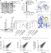
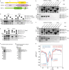
Fig. 1. Identification of the catalytic centre of TOFU-2.
a, Model of piRNA (21U RNA) formation in C. elegans. Individually transcribed piRNA precursors are stabilized by PETISCO. After the removal of the 5′-cap and two nucleotides, intermediates are loaded onto PRG-1, followed by trimming and 3′-end methylation. The nuclease that processes the 5′-end is currently unclear. b, Schematic of TOFU-1, TOFU-2 and SLFL-3/4, in comparison to rat SLFN13. The lines indicate low-complexity regions and the rectangles indicate the predicted folded domains. BD, bridging domain. c, Superposition of TOFU-1 and TOFU-2 SLFN domains onto the crystal structure of the N-terminal SLFN13 endoribonuclease domain (Protein Data Bank (PDB): 5YD0). Domains are coloured as in b. The magnified view shows the active site of SLFN13. Involved residues are shown as sticks. d, Label-free proteomic quantification of TOFU-2–HA and wild-type immunoprecipitates from young adult extracts. n = 4 biological replicates. The x axis shows the median fold enrichment of individual proteins, and the y axis shows −log10[P]. P values were calculated using Welch two-sided t-tests. The dashed lines represent enrichment thresholds at P = 0.05 and fold change > 2, curvature of enrichment threshold c = 0.05. The dots represent enriched (blue/red) or quantified (grey) proteins. Only uniquely matching peptides were used. e, Schematic of the mCherry–H2B piRNA sensor. f, Wide-field fluorescence microscopy analysis of adult hermaphrodites carrying the piRNA sensor in the following three genetic backgrounds: tofu-2(E216A) (top), prg-1(n4357) (middle) and wild type (bottom). Germlines are outlined by white dashed lines. Scale bar, 50 µm. A representative image from a series of ten is shown. g, Total mature piRNA levels (type 1) in wild-type and tofu-2(E216A)-mutant young adult hermaphrodites. n = 3 biological replicates. The red lines show the group means. P values were calculated using two-tailed unpaired t-tests. h, The relative abundance of type 1 piRNA precursors from individual loci in tofu-2(E216A)-mutant versus wild-type young adult hermaphrodites. n = 3 biological replicates. RPM, reads per million non-structural small RNA reads. Source data
Fig. 2
Fig. 2. TOFU-1, TOFU-2 and SLFL-3/4 form a mitochondria-bound complex.
a, Anti-GFP immunoprecipitation analysis of BmN4 cell lysates of cells that were transfected with the indicated constructs. eGFP–TOFU-2 was immunoprecipitated, followed by western blot detection of TOFU-1 (Flag), SLFL-3 (HA) or SLFL-4 (HA). Expression of 3×MYC–eGFP and of 3×Flag–mCherry served as negative controls. Note that low TOFU-2 levels in lanes 5 and 7 may limit the detection of interactions. All observations were performed at least in duplicate. IP, immunoprecipitation. b, Total mature piRNA levels (type 1) in young adult hermaphrodites of the indicated genotypes. n = 3 biological replicates. The red lines depict group means and P values were calculated using one-way analysis of variance (ANOVA) followed by Tukey’s honest significant difference (HSD) test (left) and two-tailed unpaired t-tests (right). The plot is based on two independent experiments (exp. 1 and 2). NS, non-significant. c, The relative abundance of type 1 piRNA precursors from individual loci in slfl-3−/−;slfl-4−/− mutant versus wild-type young adult hermaphrodites. n = 3 biological replicates. d, Single-plane confocal micrographs of BmN4 cells that were transfected with eGFP–TOFU-2 and full-length mCherry–SLFL-3 (top) or mCherry–SLFL-3(ΔTM) (bottom). TOFU-1 was also transfected but was not tagged with a fluorescent protein. Mitochondria were stained with Mito Tracker. Scale bars, 10 µm. The experiment was performed in duplicate; a representative image from a series of 20 is shown. e, AlphaFold2-predicted structure of a minimal trimeric TOFU-1–TOFU-2–SLFL-3 complex. The best of five predicted models is shown as a cartoon in two different orientations. TOFU-1 is shown in yellow, TOFU-2 in purple and SLFL-3 in green. The TOFU-2 active-site residues are shown as a stick representation and are magnified at the bottom right. Raw data are provided in Supplementary Fig. 1. Source data
Fig. 3
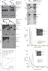
Fig. 3. PUCH is a cap- and sequence-specific endoribonuclease.
a, The sequence of the synthetic piRNA precursor used in the assay. The red line indicates the expected cleavage position. Both the precursor and expected cleavage product were run in the two left-most lanes of every gel to mark where these molecules are expected. b, In vitro cleavage assay of the piRNA precursor using anti-GFP immunoprecipitated material from BmN4 cell extracts. Cells were transfected with eGFP–TOFU-2, TOFU-1, SLFL-3 or SLFL-4 at various combinations, as indicated. All observations were performed at least in duplicate. nt, nucleotides. c, Cleavage assays with recombinant minimal PUCH (mini-PUCH) and different RNA substrates. E216A indicates the presence of TOFU-2 containing the catalytic E216A mutation. All observations were performed at least in duplicate. d, RNA obtained from a cleavage reaction (using either wild-type or TOFU-2(E216A)-mutant mini-PUCH) was ligated to a 10-nucleotide-long 5′OH-containing RNA adapter. The ligation product is indicated by an arrow. The experiment was performed in triplicate. e, In vitro cleavage assay on different types of RNA substrate using the PUCH complex retrieved from BmN4 cells by immunoprecipitation (IP). All observations were performed at least in duplicate. f,g, Cleavage assays with mini-PUCH and the indicated substrates. The experiment was performed in triplicate for f and once for g. Raw data are provided in Supplementary Fig. 1.
Fig. 4
Fig. 4. PETISCO is necessary for piRNA precursor accumulation in vivo and does not interfere with PUCH-mediated precursor cleavage.
a, The relative abundance of individual type 1 piRNA precursors in tofu-2(E216A) mutant (left) and tofu-2(E216A);pid-1(xf35) double-mutant (right) versus wild-type young adult hermaphrodites. n = 3 biological replicates. b, Total mature piRNA levels (type 1) in wild-type, tofu-2(E216A) mutant and tofu-2(E216A);pid-1(xf35) double-mutant young adult hermaphrodites. n = 3 biological replicates. The red lines show the group means. P values were calculated using one-way ANOVA followed by Tukey’s HSD test; the indicated P value relates to both mutant samples. NS, non-significant. c, In vitro piRNA precursor cleavage assays in the presence or absence of the PETISCO complex in a time series, in duplicate. In these experiments, PUCH was isolated from BmN4 cell extracts by immunoprecipitation (IP). d, Quantification of the cleavage reactions presented in c. Raw data are provided in Supplementary Fig. 1. Source data
Fig. 5
Fig. 5. TOFU-6 from PETISCO interacts with PUCH through TOFU-1.
a, The interaction between TOFU-1 and PETISCO was analysed using amylose pull-down assays. Purified MBP-tagged TOFU-182–373 was incubated with excess PETISCO. Input and elution fractions were analysed by SDS–PAGE followed by Coomassie staining. b, Purified TOFU-6eTUDOR, TOFU-1pep and a mixture thereof were analysed using size-exclusion chromatography. Chromatograms, TOFU-6eTUDOR (blue), TOFU-1pep (yellow) and TOFU-6eTUDOR + TOFU-1pep (black). Results from a and b were obtained in duplicates. c, The crystal structure of the TOFU-6eTUDOR–TOFU-1pep complex shown as a cartoon. The TOFU-6eTUDOR domain is shown in different shades of blue and TOFU-1pep in yellow. The magnified view shows the interaction interface; involved residues are shown as sticks. d, Western blot analysis of the expression levels of TOFU-1 and TOFU-6 for the indicated genotypes using anti-MYC and anti-H3 antibodies, followed by visualization using horseradish-peroxidase-linked secondary antibodies. The numbers indicate the approximate molecular mass (kDa). One out of two experiments is shown. e,f, Total mature (e) and precursor (f) type 1 piRNA levels in young adult hermaphrodites of indicated genotypes. n = 3 biological replicates. The red lines show the group means. P values were calculated using one-way ANOVA followed by Tukey’s HSD test. Note that mature and precursor reads derive from different libraries and their levels cannot be directly compared. NS, non-significant. g, The relative abundance of precursors from individual type 1 piRNA loci in young adult hermaphrodites of the indicated genotypes. n = 3 biological replicates. The underlying data are the same as in f. Raw data are provided in Supplementary Fig. 1. Source data
Extended Data Fig. 1
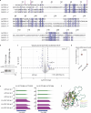
Extended Data Fig. 1. Mutation in the catalytic centre of TOFU-2 does not affect protein stability and interaction with TOFU-1.
a, Structure-based sequence alignment of the SLFN domains from TOFU-1, TOFU-2, SLFL-3 and SLFL-4. The acidic residues from the active site of TOFU-2 are highlighted with purple boxes and the residue number is indicated on the top. b, Extracts of young adult worms with genotype tofu-2::HA and tofu-2[E216A]::HA, were separated on SDS-PAGE. Western blot was probed using anti-HA and anti-H3 antibodies, followed by visualization with HRP. One of three experiments is shown. c, Volcano plot representing label-free proteomic quantification of TOFU-2[E216A]::HA and wild type immunoprecipitations from young adult extracts (n = 4 biological replicates). The X-axis represents the median fold enrichment of individual proteins in wild type (WT) versus the TOFU-2::HA mutant strain. The Y-axis indicates −log10(P-value) calculated using Welch two-sided t-test. Dashed lines represent enrichment thresholds at p-value = 0.05 and fold change > 2, c = 0.05. Each dot represents an enriched (blue/green) or quantified (grey/orange) proteins. The analysis was based on all peptides that matched to a given protein. In this experiment we could not detect unique peptides for SLFL-3 and SLFL-4. The lower enrichment of SLFL-3/4 in this experiment compared to the experiment shown in Fig. 1d most likely reflects experimental variations. PUCH stability is not affected by the TOFU-2[E216A] mutation as shown in Extended Data Fig. 5h. d, Total piRNA levels (type 2) in wild type and tofu-2[E216A]-mutant young adult hermaphrodites (n = 3 biological replicates). Red lines depict group means and P-values were calculated using two-tailed unpaired t-test. e, Genome browser tracks of two individual piRNA loci, displaying normalized read coverage in piRNA precursor libraries. The top three tracks (green) are from a wild type background, the bottom three tracks (purple) are from a tofu-2[E216A] mutant background. Note that mature piRNAs are severely depleted from precursor libraries, thus most of the depicted read coverage derive from piRNA precursors starting 2 nucleotides upstream of the 5′-ends of mature piRNAs which are indicated by a vertical line. f, Superposition of the AlphaFold predicted SLFN domains from TOFU-1, TOFU-2, SLFL-3 and SLFL-4. TOFU-1 is shown in yellow, TOFU-2 in purple and the paralogs SLFL-3/4 in different shades of green. Raw data are available in Supplementary Fig. 1. Source data
Extended Data Fig. 2
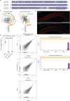
Extended Data Fig. 2. SLFL-3 and SLFL-4 are transmembrane proteins involved in piRNA biogenesis in C. elegans.
a, Sequence alignment of SLFL-3 and SLFL-4. The predicted C-terminal transmembrane helix is highlighted with a box. b, AlphaFold2 predicted structures of SLFL-3/4 shown as cartoon and coloured by pLDDT score, which reports on the model confidence. Dark blue indicates very high, light blue confident, yellow low, and orange very low model confidence. c, Widefield fluorescent microscopy of adult hermaphrodites carrying the mCherry::H2B-piRNA sensor in two genetic backgrounds: slfl-3(xf248) on top and wild type at the bottom. The germlines are outlined by a white dashed line. Scale bar – 50 µm. Representative image from a series of 10 is shown. d, Total piRNA levels (type 2) in young adult hermaphrodites of the indicated genotypes (n = 3 biological replicates). Red lines depict group means and P-values were calculated using one-way ANOVA test followed by a Tukey’s HSD test. e, Scatter plots depicting the relative abundance of type 1 piRNA precursors from individual loci in slfl4(-/-), slfl-3(ΔTM);slfl-4(-/-) and slfl-3(-/-) mutants versus wild type young adult hermaphrodites (n = 3 biological replicates). RPM: Reads per million non-structural sRNA reads. f, Prediction of transmembrane helices in SLFL-3 and SLFL-4 using TMHMM - 2.0. Source data
Extended Data Fig. 3
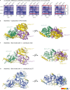
Extended Data Fig. 3. AlphaFold predicts a trimeric complex consisting of TOFU-1, TOFU-2 and either SLFL-3 or SLFL-4.
a, Predicted alignment error (PAE) plots for the five models predicted by Alphafold for full-length TOFU-1, TOFU-2, SLFL-3 and SLFL-4. The zoom-in highlights the predicted interaction between the SLFN domains of TOFU-2 and SLFL-3, suggesting that the TOFU-2 SPRY domain is no involved in complex formation. The expected position error in angstroms (Å) is colour coded where blue colour indicates low PAE (high confidence) and red colour indicates high PAE (low confidence). b, Predicted alignment error (PAE) plots for the five models predicted by Alphafold for core regions of TOFU-1, TOFU-1, SLFL-3 and SLFL-4. c, Schematic summary of the interaction results presented in a and b.
Extended Data Fig. 4
Extended Data Fig. 4. AlphaFold structure prediction of the trimeric complex consisting of TOFU-1, TOFU-2 and SLFL-3 shows convergence of models.
a-b, AlphaFold predicts a trimeric complex consisting of TOFU-1, TOFU-2 and SLFL-3. TOFU-1 residues 156-373, TOFU-2 residues 200-433 and SLFL-3 residues 103-300 were used for the prediction. The predicted alignment error (PAE) plots are shown in (a), the five superposed models are shown as cartoon in (b). TOFU-1 is coloured yellow, TOFU-2 purple and SLFL-3 green. The settings used for the prediction are shown on the top. The expected position error in angstroms (Å) is colour coded where blue colour indicates low PAE (high confidence) and red colour indicates high PAE (low confidence). c-d, The best of the five predicted models is coloured per chain (c) or per pLDDT score (d), which reports on the model confidence. Dark blue indicates very high, light blue confident, yellow low and orange very low model confidence.
Extended Data Fig. 5
Extended Data Fig. 5. Verification of SLFL-domain interactions obtained by AlphaFold using recombinant proteins.
a, Schematic domain organization of TOFU-1, TOFU-2 and SLFL-3. Lines indicate low-complexity regions and rounded rectangles indicate predicted folded domains. TM: transmembrane domain. b-c, A construct containing the TOFU-1 SLFN domain binds to the TOFU-2 SLFN domain while the TOFU-2 SPRY domain does not bind TOFU-1. Analysis of the interaction of different TOFU-1 constructs with the StrepII-tagged TOFU-2 SPRY domain in (b) and StrepII-tagged TOFU-2 SLFN domain in (c). The indicated constructs were co-expressed in E. coli and the StrepII-tagged bait was precipitated by Streptactin XT beads. Input and elution fractions were analysed by SDS-PAGE followed by Coomassie staining. Experiment is done in duplicate. d, SLFL-3 interacts with the TOFU-2 SLFN domain. Analysis of the interaction of different StrepII-tagged TOFU-2 constructs with the SLFL-3. The indicated constructs were co-expressed in E. coli and the StrepII-tagged bait was precipitated by Streptactin XT beads. Input and elution fractions were analysed by SDS-PAGE followed by Coomassie staining. Experiment is done in duplicate. e, TOFU-1, TOFU-2 and SLFL-3 form a trimeric complex. Different combinations of StrepII-tagged TOFU-1, TOFU-2 and SLFL-3 were co-expressed in E. coli and the StrepII-tagged bait was precipitated by Streptactin XT beads. Input and elution fractions were analysed by SDS-PAGE followed by Coomassie staining. Experiment is done in duplicate. f and g, Recombinant, purified mini PUCH from E. coli in the active form (f) and inactive form TOFU-2 E216A (g). PUCH purification was done once. h, The thermal stability of mini PUCH WT (shades of blue) and E216A mutant (shades of red) was assessed by differential scanning fluorimetry (DSF) for both samples in duplicates. They grey line indicates the buffer control. The first negative derivative of fluorescence intensity is plotted versus temperature. The two melting points (Tm) correspond to the two minima. RFU, relative fluorescence units. Raw data are available in Supplementary Fig. 1. Representative results are shown. Source data
Extended Data Fig. 6
Extended Data Fig. 6. Characterization of PUCH activity.
a-b, Cleavage assays using GFP-IP material from BmN4 cell extracts. Beads were washed with 1 mM EDTA. Subsequently, reactions were done in buffer containing the indicated divalent cations. Concentrations were 1 or 4 mM in a, 11 mM in b. ‘-‘ indicates that no divalent cations were added during the reaction. All observations were done at least in duplicate. c-d, In vitro piRNA precursor cleavage assay with either m7G-AAU or m7G-CAU substrate in a time-series with recombinant mini-PUCH. Observations were made in four experiments. e, Quantification of the cleavage reactions, presented in c and d. f, Cleavage reaction with recombinant mini-PUCH of AAU substrate in the presence of different cold RNA substrates as indicated. Experiment was done in duplicate. g, Electrophoretic mobility shift assay between PETISCO complex and piRNA precursor with various 5′-ends. Experiment was performed twice for m7G-capped and 5′-OH carrying substrate and once for 5′-end substrate. h, In vitro piRNA precursor cleavage kinetics in presence or absence of the PETISCO complex with recombinant mini-PUCH. Experiment had been performed twice. i, Quantification of the cleavage reactions, presented in h. Raw data are available in Supplementary Fig. 1. Representative results are shown. Source data
Extended Data Fig. 7
Extended Data Fig. 7. A peptide upstream of the TOFU-1 SLFN domain binds to the TOFU-6 eTUDOR domain.
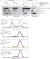
a-c, Analysis of the interaction of different TOFU-1 constructs with PETISCO and its subunits by amylose pull-down assays. Input and elution fractions were analysed by SDS-PAGE followed by Coomassie staining. a, Various purified MBP-tagged TOFU-1 truncations were incubated with excess PETISCO and precipitated using amylose beads. b, Purified MBP-tagged TOFU-182–373 was incubated with excess of the IFE-3/TOFU-6 and PID-3/ERH-2 subcomplexes precipitated using amylose beads. c, Purified MBP-tagged TOFU-182–373 was incubated with excess of the IFE-3/TOFU-6 subcomplex, the TOFU-6 RRM and the TOFU-6 eTUDOR domain and precipitated using amylose beads. d, Purified IFE-3/TOFU-6eTUDOR subcomplex, TOFU-182–172 and a mixture thereof were subjected to size exclusion chromatography. Chromatograms: IFE-3/TOFU-6eTUDOR (green), TOFU-182–172 (yellow) and IFE-3/TOFU-6eTUDOR + TOFU-182–172 (black). The inset shows a Coomassie-stained SDS polyacrylamide gel of the peak fractions from size exclusion chromatography. e, Purified TOFU-6eTUDOR, TOFU-182–172 and a mixture thereof were subjected to size exclusion chromatography. Chromatograms: TOFU-6eTUDOR (blue), TOFU-1182 (yellow) and TOFU-6eTUDOR + TOFU-1182 (black). The inset shows a Coomassie-stained SDS polyacrylamide gel of the peak fractions from size exclusion chromatography. Note: The chromatogram of TOFU-182–172 (yellow) is shown for comparison and is the same as shown in d. Also, the lanes of the polyacrylamide gel are derived from the same gel in as in d; thus lane 1 and the marker are identical for d and e. f, Binding of TOFU-6eTUDOR to TOFU-1pep measured by ITC. The binding affinity (Kd) and the stoichiometry (N) are the mean of three experiments and displayed error is the standard deviation. The experiment shows one of the three experiments as representative example. Raw data are available in Supplementary Fig. 1. Representative results are shown. All data from this figure were obtained at least in duplicate. Source data
Extended Data Fig. 8
Extended Data Fig. 8. Structure-based analysis of the TOFU-6:TOFU-1 interaction.
a, Pull-down assays with purified recombinant wild type and mutant MBP-TOFU-6eTUDOR domain and GST-tagged TOFU-182–172 and TOFU-182–113 constructs. MBP pull-down assays using MBP-TOFU-6eTUDOR domain constructs as bait are shown on the left, GST pull-down assays using GST-tagged TOFU-1 constructs as bait are shown on right. Input and elution fractions were analysed by SDS-PAGE followed by Coomassie staining. b-e, Analysis of the interaction between TOFU-1 and PETISCO by size exclusion chromatography. Purified recombinant wild type and mutant versions of TOFU-6eTUDOR and TOFU-182–172 and mixtures thereof were subjected to size exclusion chromatography. Chromatograms: TOFU-6eTUDOR (blue), TOFU-182–172 (yellow), TOFU-6eTUDOR V266E (pink), TOFU-182–172 L88R/L92R (violet); the mixture of the respective proteins is always shown in black. Note: Some chromatograms are shown several times in b-e for direct comparison. Raw data are available in Supplementary Fig. 1. Representative results are shown. All data from this Figure was obtained at least in duplicate. Source data
Extended Data Fig. 9
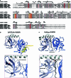
Extended Data Fig. 9. Comparison of the CeTOFU-6 eTUDOR domain to canonical eTUDOR domains.
a, Structure-based sequence alignment of experimentally determined eTUDOR domains. Ce, Caenorhabditis elegans; Dm, Drosophila melanogaster; Mm, Mus musculus and Hs, Homo sapiens. The PDB ID is given in brackets. The secondary structure elements are indicated above the sequence and the four residues forming the aromatic cage in canonical eTUDOR domains are highlighted by yellow boxes. b and c, Crystal structures of the C. elegans TOFU-6eTUDOR–TOFU-1pep and D. melanogaster PAPIeTUDOR–PIWIpep (PDB: 5ygd) complexes shown as cartoon. The eTUDOR domains are shown in different shades of blue, the TOFU-1pep in yellow and the PIWIpep containing the dimethyl-arginine residue in grey. The zoom-in view shows the region of the degenerated aromatic cage of TOFU-6eTUDOR (b) and the canonical aromatic cage of PAPIeTUDOR (c).

References

    1. Wells, J. N. & Feschotte, C. A field guide to eukaryotic transposable elements. Annu. Rev. Genet.54, 539–561 (2020). 10.1146/annurev-genet-040620-022145 - DOI - PMC - PubMed
    1. Ozata, D. M., Gainetdinov, I., Zoch, A., O’Carroll, D. & Zamore, P. D. PIWI-interacting RNAs: small RNAs with big functions. Nat. Rev. Genet.20, 89–108 (2019). 10.1038/s41576-018-0073-3 - DOI - PubMed
    1. Czech, B. et al. piRNA-guided genome defense: from biogenesis to silencing. Annu. Rev. Genet.52, 131–157 (2018). 10.1146/annurev-genet-120417-031441 - DOI - PMC - PubMed
    1. Cordeiro Rodrigues, R. J. et al. PETISCO is a novel protein complex required for 21U RNA biogenesis and embryonic viability. Genes Dev. 33, 857–870 (2019). 10.1101/gad.322446.118 - DOI - PMC - PubMed
    1. Jo, U. & Pommier, Y. Structural, molecular, and functional insights into Schlafen proteins. Exp. Mol. Med.54, 730–738 (2022). 10.1038/s12276-022-00794-0 - DOI - PMC - PubMed

Publication types

MeSH terms