. 2023 Dec;30(6):2760-2772.
doi: 10.1007/s12350-023-03371-8. Epub 2023 Sep 27.

Vascular adhesion protein-1-targeted PET imaging in autoimmune myocarditis

Affiliations

Vascular adhesion protein-1-targeted PET imaging in autoimmune myocarditis

Arghavan Jahandideh et al. J Nucl Cardiol. 2023 Dec.

Abstract

Background: Vascular adhesion protein-1 (VAP-1) is an adhesion molecule and primary amine oxidase, and Gallium-68-labeled 1,4,7,10-tetraazacyclododecane-N,N',N″,N‴-tetra-acetic acid conjugated sialic acid-binding immunoglobulin-like lectin 9 motif containing peptide ([68Ga]Ga-DOTA-Siglec-9) is a positron emission tomography (PET) tracer targeting VAP-1. We evaluated the feasibility of PET imaging with [68Ga]Ga-DOTA-Siglec-9 for the detection of myocardial lesions in rats with autoimmune myocarditis.

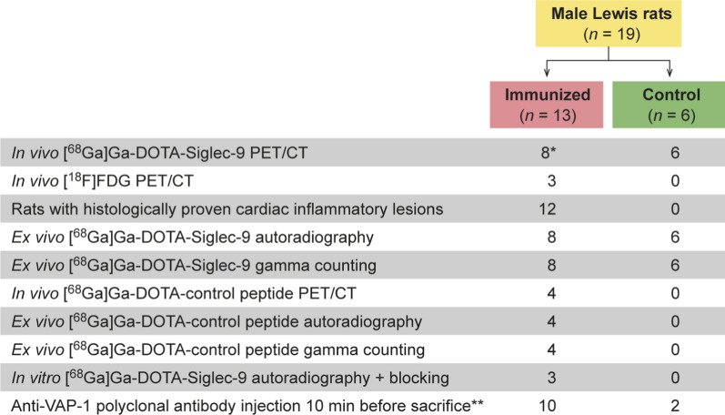
Methods: Rats (n = 9) were immunized twice with porcine cardiac myosin in complete Freund's adjuvant. Control rats (n = 6) were injected with Freund's adjuvant alone. On day 21, in vivo PET/computed tomography (CT) imaging with [68Ga]Ga-DOTA-Siglec-9 was performed, followed by ex vivo autoradiography, histology, and immunohistochemistry of tissue sections. In addition, myocardial samples from three patients with cardiac sarcoidosis were studied.

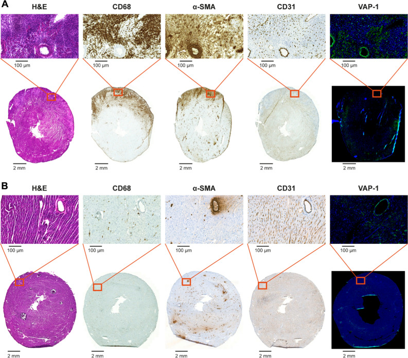
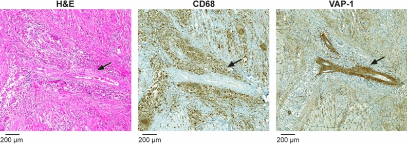
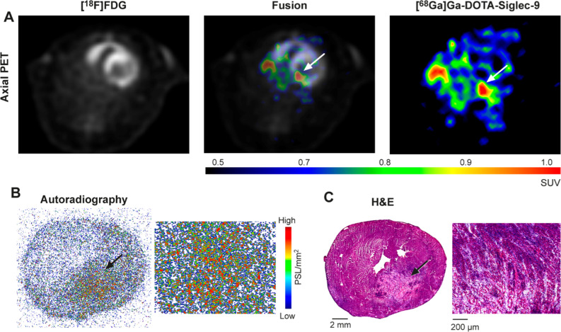
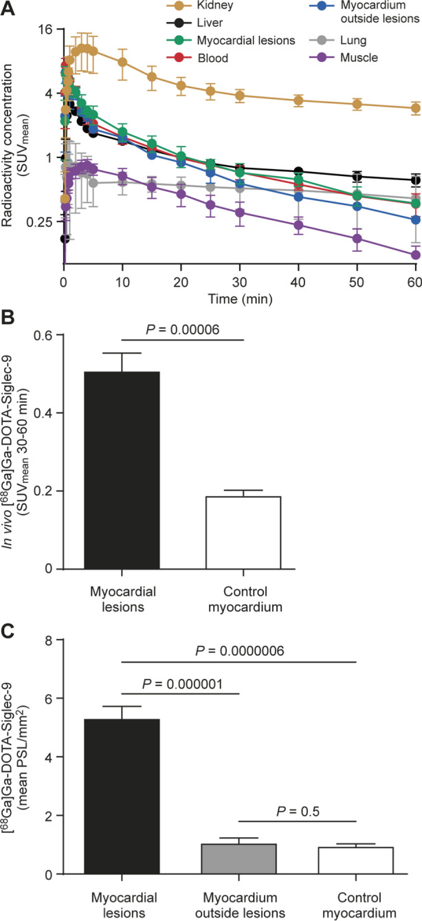
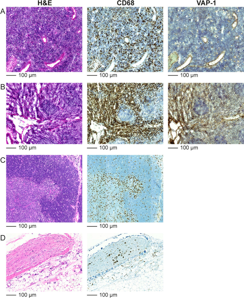
Results: [68Ga]Ga-DOTA-Siglec-9 PET/CT images of immunized rats showed higher uptake in myocardial lesions than in myocardium outside lesions (SUVmean, 0.5 ± 0.1 vs 0.3 ± 0.1; P = .003) or control rats (SUVmean, 0.2 ± 0.03; P < .0001), which was confirmed by ex vivo autoradiography of tissue sections. Immunohistochemistry showed VAP-1-positive staining in lesions of rats with myocarditis and in patients with cardiac sarcoidosis.

Conclusion: VAP-1-targeted [68Ga]Ga-DOTA-Siglec-9 PET is a potential novel technique for the detection of myocardial lesions.

Keywords: Experimental autoimmune myocarditis; Siglec-9; myocarditis; positron emission tomography; sarcoidosis; vascular adhesion protein-1.


Conflict of interest statement

Mikko Mäyränpää received consultancy or speaker fees from Boehringer Ingelheim, Bristol-Myers Squibb, MSD, Takeda, Bayer, Amgen, and Roche not related to the present study. Dr. Knuuti received consultancy fees from GE Healthcare and AstraZeneca and speaker fees from GE Healthcare, Bayer, Lundbeck, Boehringer-Ingelheim, Pfizer and Merck, outside of the submitted work. Sirpa Jalkanen owns stocks in Faron Pharmaceuticals Ltd. Antti Saraste received consultancy or speaker fees from Amgen, Astra Zeneca, Boehringer Ingelheim, Abbott, and Bayer not related to the present study. The authors report no other potential conflicts of interest relevant to this article.

Figures

Figure 1
Figure 1
A flow diagram of the study design and the numbers of animals used. *Four had a dynamic scan and the others a static scan. **A dose of 1 mg/kg of anti-VAP-1 polyclonal antibody was injected intravenously 10 minutes before sacrifice to detect surface-bound VAP-1 by immunofluorescence
Figure 2
Figure 2
(A) A myocardial lesion from a rat with autoimmune myocarditis identified by hematoxylin and eosin (H&E) staining. A consecutive cryosection stained with immunofluorescence shows intracellular and surface-bound vascular adhesion protein-1 (VAP-1). Rat received intravenous anti-VAP-1 antibody 10 minutes before sacrifice and staining was performed using only the secondary antibody. There are numerous CD68-positive macrophages in the inflammatory lesion as well as α-smooth muscle actin (α-SMA) on myofibroblasts. Staining with antibody against CD31 indicates the presence of vascular endothelial cells. (B) Histology and immunohistochemical and immunofluorescence staining of a control rat myocardium
Figure 3
Figure 3
Vascular adhesion protein-1 (VAP-1) is detected in a human coronary artery smooth muscle cells and in the endothelial cells of capillaries. Black arrow in hematoxylin and eosin (H&E) staining indicates the artery inside a sarcoid granuloma. Granuloma forming giant cells and macrophages around the artery (black arrow) are highlighted in CD68-staining. VAP-1 staining of consecutive section shows intensive VAP-1-staining in arterial medial smooth muscle cells and less intense staining in the capillary endothelial cells
Figure 4
Figure 4
(A) Representative in vivo PET images with [18F]FDG and [68Ga]Ga-DOTA-Siglec-9 tracers in a rat with autoimmune myocarditis. PET images at 30 to 60 minutes show focal [68Ga]Ga-DOTA-Siglec-9 uptake (white arrows) in the posterior left ventricle (LV) wall. (B) Ex vivo autoradiography of [68Ga]Ga-DOTA-Siglec-9 uptake shows co-localization with edematous and hemorrhagic myocardial lesion in the posterior wall of the LV, (C) which is confirmed by consecutive sections stained with hematoxylin and eosin (H&E)
Figure 5
Figure 5
Quantification of [68Ga]Ga-DOTA-Siglec-9 PET imaging. (A) Time-activity curves from rats with autoimmune myocarditis (n = 4) show that [68Ga]Ga-DOTA-Siglec-9 uptake (mean standardized uptake value, SUVmean) remains slightly higher in the myocardial lesions than in the blood (average of inferior vena cava and left ventricle). Bars indicate standard deviation. Note that the y-axis has a logarithmic scale. (B) The average myocardial [68Ga]Ga-DOTA-Siglec-9 uptake (SUVmean) 30 to 60 minutes after injection is higher in the myocardium of immunized rats than in the myocardium of control rats (n = 6). (C) Bars demonstrate [68Ga]Ga-DOTA-Siglec-9 uptake by ex vivo autoradiography as mean photo-stimulated luminescence per square millimeter (PSL/mm2) in myocardial lesions and myocardium outside lesions of immunized rats (n = 8), and myocardium of control rats (n = 6). Values are mean ± SD; unpaired t test for comparison of immunized rats’ myocardium and control myocardium, and paired t test for comparison of myocardial lesions and myocardium outside lesions
Figure 6
Figure 6
Hematoxylin and eosin (H&E), CD68 macrophage, and VAP-1 immunohistochemical staining of (A) spleen and (B) bone marrow cryosections from a rat with autoimmune myocarditis. Rat received intravenously anti-VAP-1 antibody 10 minutes before sacrifice and staining was performed using only the secondary antibody. H&E and CD68 macrophage staining of paraffin-embedded sections of (C) thymus and (D) white adipose tissue from a rat with autoimmune myocarditis

References

    1. Caforio AL, Pankuweit S, Arbustini E, Basso C, Gimeno-Blanes J, Felix SB, et al. Current state of knowledge on aetiology, diagnosis, management, and therapy of myocarditis: A position statement of the European Society of Cardiology Working Group on Myocardial and Pericardial Diseases. Eur Heart J. 2013;34:2636–2648. doi: 10.1093/eurheartj/eht210. - DOI - PubMed
    1. Kadkhodayan A, Chareonthaitawee P, Raman SV, Cooper LT. Imaging of inflammation in unexplained cardiomyopathy. JACC Cardiovasc Imaging. 2016;9:603–617. doi: 10.1016/j.jcmg.2016.01.010. - DOI - PubMed
    1. Friedrich MG, Sechtem U, Schulz-Menger J, Holmvang G, Alakija P, Cooper LT, et al. Cardiovascular magnetic resonance in myocarditis: A JACC white paper. J Am Coll Cardiol. 2009;53:1475–1487. doi: 10.1016/j.jacc.2009.02.007. - DOI - PMC - PubMed
    1. Youssef G, Leung E, Mylonas I, Nery P, Williams K, Wisenberg G, et al. The use of 18F-FDG PET in the diagnosis of cardiac sarcoidosis: A systematic review and metaanalysis including the Ontario experience. J Nucl Med. 2012;53:241–248. doi: 10.2967/jnumed.111.090662. - DOI - PubMed
    1. Tang R, Wang JT, Wang L, Le K, Huang Y, Hickey AJ, et al. Impact of patient preparation on the diagnostic performance of 18F-FDG PET in cardiac sarcoidosis: A systematic review and meta-analysis. Clin Nucl Med. 2016;41:e327–e339. doi: 10.1097/RLU.0000000000001063. - DOI - PubMed

Publication types

Substances