. 2023 Oct;12(10):e12363.
doi: 10.1002/jev2.12363.

Inhibition of extracellular vesicle-derived miR-146a-5p decreases progression of melanoma brain metastasis via Notch pathway dysregulation in astrocytes

Affiliations

Inhibition of extracellular vesicle-derived miR-146a-5p decreases progression of melanoma brain metastasis via Notch pathway dysregulation in astrocytes

Emma Rigg et al. J Extracell Vesicles. 2023 Oct.

Abstract

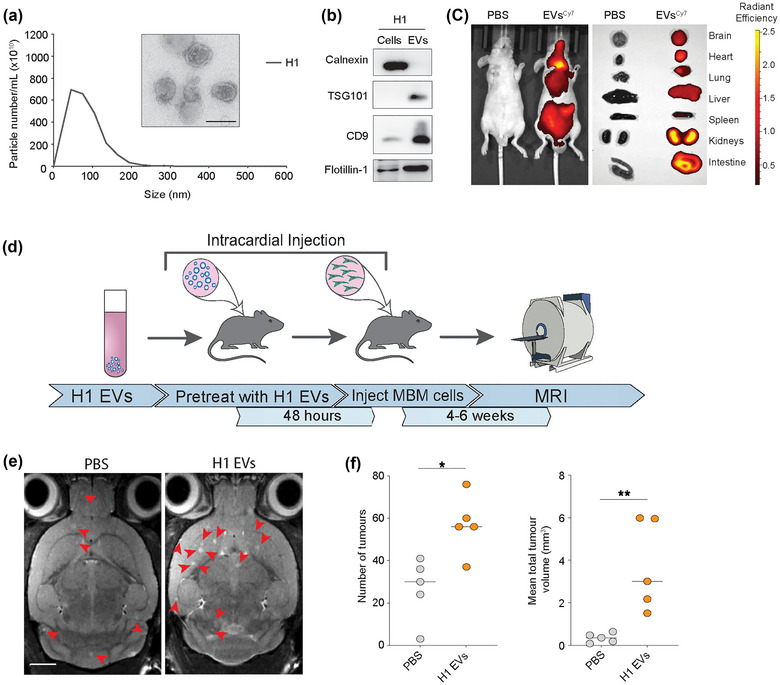
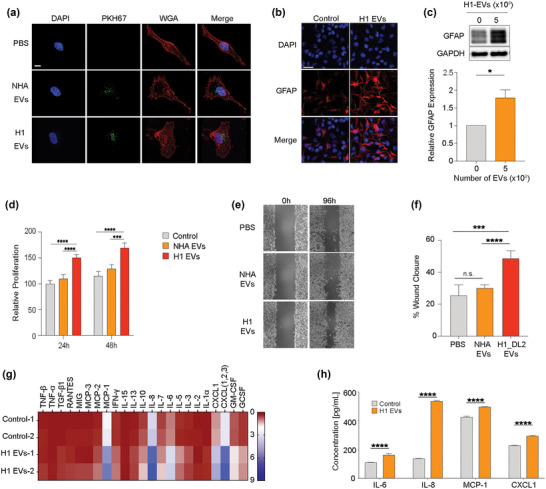
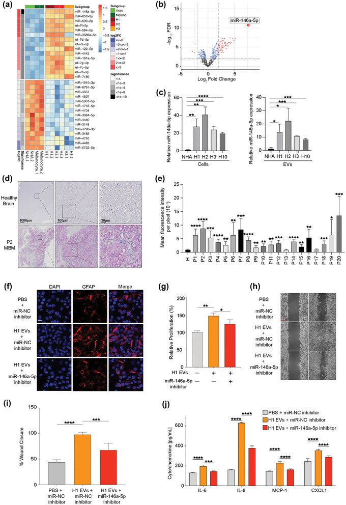
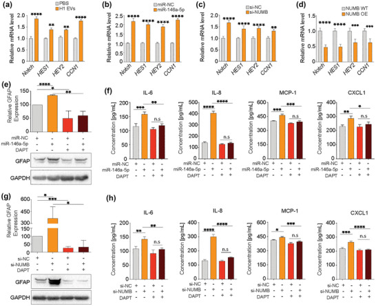
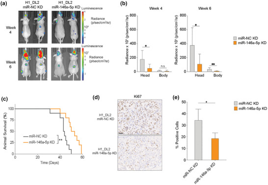
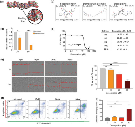
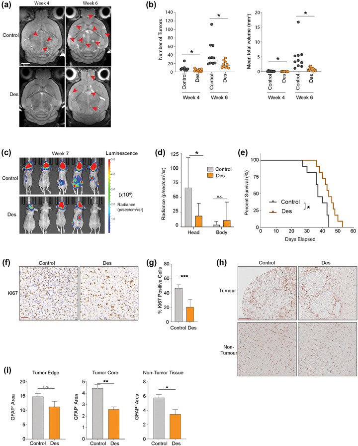
Melanoma has the highest propensity of all cancers to metastasize to the brain with a large percentage of late-stage patients developing metastases in the central nervous system (CNS). It is well known that metastasis establishment, cell survival, and progression are affected by tumour-host cell interactions where changes in the host cellular compartments likely play an important role. In this context, miRNAs transferred by tumour derived extracellular vesicles (EVs) have previously been shown to create a favourable tumour microenvironment. Here, we show that miR-146a-5p is highly expressed in human melanoma brain metastasis (MBM) EVs, both in MBM cell lines as well as in biopsies, thereby modulating the brain metastatic niche. Mechanistically, miR-146a-5p was transferred to astrocytes via EV delivery and inhibited NUMB in the Notch signalling pathway. This resulted in activation of tumour-promoting cytokines (IL-6, IL-8, MCP-1 and CXCL1). Brain metastases were significantly reduced following miR-146a-5p knockdown. Corroborating these findings, miR-146a-5p inhibition led to a reduction of IL-6, IL-8, MCP-1 and CXCL1 in astrocytes. Following molecular docking analysis, deserpidine was identified as a functional miR-146a-5p inhibitor, both in vitro and in vivo. Our results highlight the pro-metastatic function of miR-146a-5p in EVs and identifies deserpidine for targeted adjuvant treatment.

Keywords: brain metastasis; deserpidine; extracellular vesicles; melanoma; miR-146a-5p; normal human astrocytes (NHA).


Conflict of interest statement

The authors declare that there are no conflicts of interest.

Figures

FIGURE 1
FIGURE 1
MBM‐EVs contribute to increased metastatic burden. (a) Representative nanoparticle tracking analysis using Malvern Nanosight and transmission electron microscopy of H1‐EVs. Size range 50–200 nm. Scale bar = 100 nm. (b) Western blot analysis of EV‐characteristic markers on H1 cells and corresponding EVs. (c) In vivo and ex vivo NIR imaging of mice and harvested major organs after injection of Cy7 (excitation/emission: 745/820 nm) H1‐EVs or PBS control. (d) Schematic workflow of the exosome‐primed in vivo metastatic model. (e) Development of brain metastasis assessed by T2 weighted MRI at week 4 after priming with MBM‐derived EVs or PBS prior to intracardial injection of MBM H1_DL2 cells. Scale bar = 2 mm. (f) Quantification of the total number and volume of brain metastasis at week 4 in exosome‐primed animals compared to the control PBS group. n.s. = not significant, *p < 0.05, **p < 0.01.
FIGURE 2
FIGURE 2
MBM‐derived EVs induce activation in NHA. (a) NHA cultured with 5.0 × 109 EVs of PKH67‐stained (green) NHA‐ or H1‐EVs for 48 h to show uptake. Nuclei stained with DAPI (blue), membrane stained with WGA‐Texas Red (red). Magnification (100X). Scale bar = 10 μm. (b) Representative images of ICC‐stained NHA cells cultured with H1‐EVs or PBS control for 48 h. Merged images of DAPI (blue) and GFAP (red). Magnification 20X. Scale bar = 20 μm. (c) Western blot analysis of GFAP in NHA cells after culturing with 5.0 × 109 H1‐EVs or PBS for 48 h and subsequent quantification. (d) CCK8 proliferation assay of NHA cells cultured with 5.0 × 109 NHA‐ or H1‐EVs over 48 h. (e) Representative micrographs at the start and completion of a 96 h NHA wound healing assay co‐cultured with 5.0 × 109 NHA‐ or H1‐EVs. Scale bar: 300 μm. (f) Quantification of wound healing assay as percentage wound closure after 96 h. (g) Heatmap of a cytokine array of NHA conditioned media after culturing with 5.0 × 109 H1‐EVs or PBS for 48 h. (h) ELISA validation performed on NHA conditioned media after co‐culture with 5.0 × 109 H1‐EVs. ELISA of top four upregulated cytokines from cytokine array, IL‐6, IL‐8, MCP‐1 and CXCL1, were used. n.s.= not significant *p < 0.05, ***p < 0.001, ****p < 0.0001.
FIGURE 3
FIGURE 3
mir‐146a‐5p is significantly upregulated in H1‐EVs compared to normal melanocytes and astrocytes and induces similar responses in NHA cells as H1‐EVs. (a/b) Heatmap of the standardized expression for the top 15 up‐ and down‐regulated miRNAs (a) and a volcano‐plot (b) of differential miRNA expression in EVs derived from healthy (melanocytes and astrocytes) compared to MBM cell lines (H1, H2, H3). miR‐146a‐5p shows stable overexpression in MBM cell lines. (c) qPCR of miR‐146a‐5p expression in EVs and cells across multiple MBM cell lines (H1, H2, H3, H10) and normal cells (NHA). Cells normalized to endogenous control miR‐103, and EVs normalized to spike‐in C‐elegans miR‐39‐3p. (d) MiRNAscope in situ hybridization assay of miR‐146a‐5p expression in patient MBM samples compared to healthy brain controls. Red dots indicate successful binding of the miR‐146a‐5p probe, tissues counterstained with haematoxylin (purple). (e) Quantification of miR‐146a‐5p expression across patient samples compared to three pooled healthy brain controls. Data represented as mean fluorescence of 10 representative equal sized areas from each sample ± SE. (f) Representative images of ICC‐stained NHA cells cultured with H1‐EVs treated with miR‐146a‐5p inhibitor or miR‐NC (negative control). Merged images of DAPI (blue) and GFAP (red). Magnification 20X. Scale bar = 20 μm. (g) CCK8 proliferation assay of NHA cells after co‐culture with 5.0 × 109 H1‐EVs in the presence or absence of a miR‐146a‐5p inhibitor. (h) Representative micrographs at the beginning and completion of a 96 h NHA wound healing assay co‐cultured with H1‐EVs with or without miR‐146a‐5p or scramble inhibitor. Scale bar = 300 μm. (i) Quantification of wound healing assay as percentage wound closure after 96 h. J ELISA of IL‐6, IL‐8, MCP‐1 and CXCL1 levels from NHA cell conditioned media after co‐culture with 5.0 × 109 H1‐derived EVs in the presence or absence of miR‐146a‐5p inhibitor. *p < 0.05, **p < 0.01, ***p < 0.001, ****p < 0.0001.
FIGURE 4
FIGURE 4
miR‐146a‐5p increased NOTCH signalling through interaction with and downregulation of NUMB expression. (a) Heatmap of significantly differentially expressed mRNA (FDR < 0.05) from NHA cells treated with a miR‐146a‐5p mimic for 48 h and compared to a scrambled control (miR‐NC). (b) Significantly down‐regulated mRNA results were compared to three databases (TargetScan 7.1, miRBD and microT‐CDS) and 18 common genes were found as in situ projected binding partners. (c) mRNA‐pulldown assay followed by qPCR quantification of bound mRNA‐miR‐146a‐5p complexes from the common 18 binding partners. (d) qPCR of NUMB expression in NHA cells after co‐culture with 5.0 × 109 EVs from melanocytes, NHA or MBM cell lines (H1, H2 and H3), normalized with U6 expression. (e) WB of Numb protein expression in NHA cells after co‐culture with 5.0 × 109 EVs from melanocytes, NHA or MBM cell lines (H1, H2 and H3). (f) qPCR of NUMB expression in NHA cells after co‐culture with miR‐146a‐5p mimic or a scrambled control, normalized to U6 expression. (g) Western blot of Numb protein expression in NHA cells after co‐culture with miR‐146a‐5p mimic or a scrambled control. (h) Sequence of miR‐146a‐5p 3p binding site, and the comparison of NUMB 3′UTR‐wild type (WT) and mutated (MUT) region. (i) Dual luciferase assay on the WT or MUT 3′UTR region with miR‐146a‐5p or a scrambled control in NHA cells, represented as the relative firefly to Renilla luciferase activity. (j) ELISA of top four upregulated cytokines released by NHA cells with WT or overexpression of NUMB in the presence of miR‐146a‐5p mimic or control. n.s.= not significant, **p < 0.01, ***p < 0.001, ****p < 0.0001.
FIGURE 5
FIGURE 5
miR‐146a‐5p influences the activation of astrocytes through the upregulation of the Notch pathway via NUMB inhibition. (a–d) qPCR of relative mRNA expression of proteins involved in the Notch pathway from NHA cells after treatment with H1‐derived EVs or PBS control, miR‐146a‐5p mimic or a scrambled control, NUMB siRNA or a scrambled control, and NUMB OE or WT control. (e) Western blot analysis of GFAP expression in NHA cells after treatment with miR‐146a‐5p mimic and/or Notch inhibitor DAPT and associated quantification. (f) ELISA of top four upregulated cytokines released by NHA cells after treatment with miR‐146a‐5p mimic with and without the Notch pathway inhibitor DAPT. (g) Western blot analysis of GFAP expression in NHA cells after treatment with NUMB siRNA and/or DAPT associated quantification. (h) ELISA of top four up regulated cytokines released by NHA cells after treatment with NUMB siRNA and/or DAPT. All data displayed from three independent experiments. n.s.= not significant, *p < 0.05, **p < 0.01, ***p < 0.001, ****p < 0.0001.
FIGURE 6
FIGURE 6
miR‐146a‐5p knockdown in MBM cells significantly decreased tumour burden and increased survival in a mouse brain metastasis model. (a) 5 × 105 H1_DL2 miR‐NC KD (control) or H1_DL2 miR‐146a‐5pKD cells were injected intracardially into mice. Tumour burden was evaluated at week 4 and 6 with IVIS bioluminescent imaging. n = 10 mice per group. (b) Tumour distribution in head and body were quantified at week 4 and 6 by the average radiance in photons/s/cm2/steradia (p/s/cm2/sr). (c) Kaplan–Meier survival curves calculated for all animals in the treatment study. (d) Representative images of Ki67 staining of FFPE sections of brain tumour tissue from animal test subjects after sacrificing. Scale bar = 50 μm. (e) Quantification of Ki67 staining as a percentage of total cells in each tumour. n.s.= not significant, *p < 0.05, **p < 0.01.
FIGURE 7
FIGURE 7
Deserpidine reduces the expression of miR‐146a‐5p in vitro and reduces the viability and proliferation of MBM cells. (a) Schematic representation of the 3D structure of the miR‐146a‐5p binding site for high‐throughput molecular docking analysis. (b) Schematic representation of the top 3 binding partners of miR‐146a‐5p available for purchase and predicted to cross the blood brain barrier. (c) qPCR analysis of miR‐146a‐5p expression in H1 cells and EVs after deserpidine treatment. (d) Representative IC50 survival curve of H1 cells after treatment with increasing drug concentrations (0.01–150 μM) for 72 h and corresponding deserpidine IC50 doses of all MBM cell lines and NHA. (e) Representative images from wound healing assay of H1 cells after treatment with various concentrations of deserpidine and quantification of % wound closure at 72 h. Scale bar = 300 μm. (f) Annexin V‐FITC and PI staining to assess apoptosis by flow cytometry in H1 cells treated with increasing concentrations of deserpidine for 72 h and corresponding quantification of live cell and apoptotic cell populations (Q2 + Q3) represented as percentage of total cell population. All data displayed from three independent experiments. Quadrant 1: Live cells. Quadrant 2: Early apoptosis. Quadrant 3: Late apoptosis. Quadrant 4: Necrosis. n.s.= not significant, *p < 0.05, **p < 0.01, ****p < 0.0001. N = 3.
FIGURE 8
FIGURE 8
Deserpidine reduces tumour burden and increases mice overall survival in vivo. (a) Development of brain metastasis assessed by T2 weighted MRI at week 4 and 6 after intracardial injection of MBM H1_DL2 cells. Mice were either treated with 0.15 mg/kg deserpidine or solvent 3 days per week. n = 10 mice per group. (b) Quantification of the numbers and volumes of brain metastasis in 0.15 mg/kg deserpidine‐treated and control animals. (c) Development of tumour burden evaluated week 4 and 6 with IVIS bioluminescent imaging after i.c. injection of MBM H1_DL2 cells. Mice were either treated with 0.5 mg/kg deserpidine or solvent every 3 days. n = 10 mice per group. (d) tumour distribution in head and body were quantified by the average radiance in photons/s/cm2/steradia (p/s/cm2/sr). (e) Kaplan–Meier survival curves calculated for all animals in the second treatment study. (f) Representative images of Ki67 staining of formalin fixed paraffin embedded (FFPE) sections of brain tumour tissue from animal test subjects after sacrificing. Scale bar: 50 μm. (g) Quantification of Ki67 staining as a percentage of total cells in each tumour. (h) Representative images of GFAP staining of FFPE sections of brain tumour tissue and brain non‐tumour tissues from animal test subjects after sacrificing. Scale bar = 200 μm. (i) Quantification of GFAP staining as a percentage of intensity of GFAP in tumour edge, tumour core and non‐tumour tissue areas. Data are displayed as mean ± SEM. Groups were compared using the Welch t‐test. *p < 0.05, **p < 0.01, ***p < 0.001.

References

    1. Achrol, A. S. , Rennert, R. C. , Anders, C. , Soffietti, R. , Ahluwalia, M. S. , Nayak, L. , Peters, S. , Arvold, N. D. , Harsh, G. R. , Steeg, P. S. , & Chang, S. D. (2019). Brain metastases. Nature Reviews Disease Primers, 5, 5. - PubMed
    1. Alles, J. , Fehlmann, T. , Fischer, U. , Backes, C. , Galata, V. , Minet, M. , Hart, M. , Abu‐Halima, M. , Grässer, F. A. , Lenhof, H. P. , Keller, A. , & Meese, E. (2019). An estimate of the total number of true human miRNAs. Nucleic Acids Research, 47, 3353–3364. - PMC - PubMed
    1. Anvari, A. , Sasanpour, P. , & Rajabzadeh Kheradmardi, M. (2021). Radiotherapy and immunotherapy in melanoma brain metastases. Hematology/Oncology and Stem Cell Therapy, 16, 1–20. - PubMed
    1. Bartel, D. P. (2018). Metazoan microRNAs. Cell, 173, 20–51. - PMC - PubMed
    1. Bischof, K. , Knappskog, S. , Hjelle, S. M. , Stefansson, I. , Woie, K. , Salvesen, H. B. , Gjertsen, B. T. , & Bjorge, L. (2019). Influence of p53 isoform expression on survival in high‐grade serous ovarian cancers. Scientific Reports, 9, 5244. - PMC - PubMed

Publication types