Review
. 2023 Sep 5;12(18):2209.
doi: 10.3390/cells12182209.

Diverse Roles of Protein Palmitoylation in Cancer Progression, Immunity, Stemness, and Beyond

Affiliations
Review

Diverse Roles of Protein Palmitoylation in Cancer Progression, Immunity, Stemness, and Beyond

Mingli Li et al. Cells. .

Abstract

Protein S-palmitoylation, a type of post-translational modification, refers to the reversible process of attachment of a fatty acyl chain-a 16-carbon palmitate acid-to the specific cysteine residues on target proteins. By adding the lipid chain to proteins, it increases the hydrophobicity of proteins and modulates protein stability, interaction with effector proteins, subcellular localization, and membrane trafficking. Palmitoylation is catalyzed by a group of zinc finger DHHC-containing proteins (ZDHHCs), whereas depalmitoylation is catalyzed by a family of acyl-protein thioesterases. Increasing numbers of oncoproteins and tumor suppressors have been identified to be palmitoylated, and palmitoylation is essential for their functions. Understanding how palmitoylation influences the function of individual proteins, the physiological roles of palmitoylation, and how dysregulated palmitoylation leads to pathological consequences are important drivers of current research in this research field. Further, due to the critical roles in modifying functions of oncoproteins and tumor suppressors, targeting palmitoylation has been used as a candidate therapeutic strategy for cancer treatment. Here, based on recent literatures, we discuss the progress of investigating roles of palmitoylation in regulating cancer progression, immune responses against cancer, and cancer stem cell properties.

Keywords: ZDHHCs; cancer; cancer treatment; palmitoylation; protein post-translational modification; tumor.


Conflict of interest statement

The authors declare no conflict of interest.

Figures

Figure 1
Figure 1
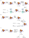
Reversible palmitoylation and questions to be addressed to understand palmitoylation. (a) Reversible process of protein palmitoylation. Palmitoyl transferase ZDHHCs transfers palmitoyl-CoA to the cysteine residue of proteins. By adding the lipid tail to proteins, palmitoylation increases the hydrophobicity of proteins and affects protein functions. Depalmitoylation is mediated by acyl-protein thioesterases. (b) When studies are focused on targeted proteins, six questions need to be addressed to understand how palmitoylation modifies protein functions: 1—if a specific protein is palmitoylated; 2—where the palmitoylation sites are or which cysteine residues are palmitoylated; 3—which ZDHHC is the main palmitoyl transferase responsible for the palmitoylation of the specific protein; 4—how palmitoylation affects functions of the specific protein; 5—if palmitoylation leads to pathological conditions or contributes to physiological conditions by modulating protein function; 6—how the palmitoylation is regulated. (c) When investigations are focused on palmitoyl transferase, four questions need to be addressed: 1—what the biological function of the ZDHHCs in the physiological or specific pathological contexts; 2—for a particular ZDHHC, what the substrates are; 3—if inhibitors can be developed to target the ZDHHCs; 4—how expression of ZDHHCs and how ZDHHCs-mediated palmitoylation are regulated.
Figure 2
Figure 2
Methods to detect palmitoylated proteins. (a) For ABE, free cysteines are blocked with NEM. Thioesters are then cleaved with NH2OH and newly generated cysteines are reacted with HPDP-Biotin. Following streptavidin bead enrichment, selectively captured proteins are eluted with reducing agents and then analyzed by Western blot or mass spectrometry. (b) Different from ABE, which uses NEM to block free cysteines, acyl-RAC exchange uses methyl methanethiosulfonate (MMTS) to block free cysteines. Further, acyl-RAC uses HPDP-Biotin; newly generated cysteines are reacted with thiol-sepharose resin but not HPDP-Biotin to label the newly generated cysteines after NH2OH cleavage. Taking advantage of the on-resin digestion availability, peptides with newly generated cysteines can be captured after elution for mass spectrometry. This allows identification of the specific sites that are palmitoylated. (c) With APE, after NH2OH cleavage, newly generated cysteines are reacted with 5 kDa methoxy-PEG-maleimide (mPEG-Mal). Due to the big size of mPEG-Mal, proteins which are labeled with mPEG-Mal migrate slower on the Western blot. Proteins with different numbers of mPEG-Mal can also be separated on the Western blot. This allows the identification of numbers of palmitoylation sites of specific proteins.
Figure 3
Figure 3
Roles of palmitoylation in modifying AKT, Wnt, and IGF-1 signaling. (a) For AKT signaling pathway, palmitoylation reduces mTOR stability, increases binding affinity of PCSK9 with PTEN, and prevents degradation of AKT. (b) For Wnt-mediated signaling, palmitoylation maintains plasma membrane localization of LRP6 and mediates CKAP4 translocation to the plasma membrane. (c) In IGF-1/IGF-1R signaling, palmitoylation mediates plasma membrane association of FLOT-1, an interacting protein of IGF-1R.
Figure 4
Figure 4
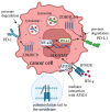
Roles of palmitoylation in regulating IFNγ/IFNGR1-mediated PD-L1/PD-1 signaling against cancer. Palmitoylation regulates this signaling in several layers. Specifically, palmitoylation mediates interaction between IFNGR1 and its binding partner AP3D1, promotes plasma membrane localization of PD-L1, and prevents degradation of PD-1. By doing so, palmitoylation positively contributes to the IFNγ/IFNGR1-mediated PD-L1/PD-1 signaling.
Figure 5
Figure 5
Regulation of the multipotency and proliferation of glioblastoma stem cells (GSCs) by palmitoylation. Palmitoylation regulates functions of GSK3β/EZH2, TGFBR/SMAD3, IL-6/GP130, and Oct4A and, thereby, plays critical roles in maintaining stem cell properties. In detail, palmitoylation increases GSK3β activity by blocking interaction of GSK3β and its upstream inhibitory kinases AKT and S6K, blocks EZH2 in methylating H3K27me3, mediates nuclei translocation of SMAD3, maintains plasma membrane location of GP130, and prevents degradation of OCT4A.
Figure 6
Figure 6
Palmitoylation negatively contributes to cancer. By reducing stability of AEG-1 (a) and translocating FLT3-ITD (b) from plasma membrane to ER, palmitoylation blocks the oncogenic signaling induced by AEG-1 and FLT3-ITD, respectively. On the other hand, by maintaining function of tumor suppressors, palmitoylation suppresses cancer. For example, palmitoylation helps MC1R in preventing melanomagenesis (c), maintaining the nuclear localization of TP53 (d), preventing degradation of SETD2 (e), and maintaining membrane localization of GNA13 (f).

References

    1. Braun P.E., Radin N.S. Interactions of lipids with a membrane structural protein from myelin. Biochemistry. 1969;8:4310–4318. doi: 10.1021/bi00839a014. - DOI - PubMed
    1. Schlesinger M.J., Magee A.I., Schmidt M.F. Fatty acid acylation of proteins in cultured cells. J. Biol. Chem. 1980;255:10021–10024. doi: 10.1016/S0021-9258(19)70417-7. - DOI - PubMed
    1. Schmidt M.F., Bracha M., Schlesinger M.J. Evidence for covalent attachment of fatty acids to Sindbis virus glycoproteins. Proc. Natl. Acad. Sci. USA. 1979;76:1687–1691. doi: 10.1073/pnas.76.4.1687. - DOI - PMC - PubMed
    1. Stoffyn P., Folch-Pi J. On the type of linkage binding fatty acids present in brain white matter proteolipid apoprotein. Biochem. Biophys. Res. Commun. 1971;44:157–161. doi: 10.1016/S0006-291X(71)80172-9. - DOI - PubMed
    1. Hayashi T. Post-translational palmitoylation of ionotropic glutamate receptors in excitatory synaptic functions. Br. J. Pharmacol. 2021;178:784–797. doi: 10.1111/bph.15050. - DOI - PubMed

Publication types