. 2023 Sep 7;12(18):2230.
doi: 10.3390/cells12182230.

New High-Affinity Thrombin Aptamers for Advancing Coagulation Therapy: Balancing Thrombin Inhibition for Clot Prevention and Effective Bleeding Management with Antidote

Affiliations

New High-Affinity Thrombin Aptamers for Advancing Coagulation Therapy: Balancing Thrombin Inhibition for Clot Prevention and Effective Bleeding Management with Antidote

Mohamad Ammar Ayass et al. Cells. .

Abstract

Thrombin is a key enzyme involved in blood clotting, and its dysregulation can lead to thrombotic diseases such as stroke, myocardial infarction, and deep vein thrombosis. Thrombin aptamers have the potential to be used as therapeutic agents to prevent or treat thrombotic diseases. Thrombin DNA aptamers developed in our laboratory exhibit high affinity and specificity to thrombin. In vitro assays have demonstrated their efficacy by significantly decreasing Factor II activity and increasing PT and APTT times in both plasma and whole blood. Aptamers AYA1809002 and AYA1809004, the two most potent aptamers, exhibit high affinity for their target, with affinity constants (Kd) of 10 nM and 13 nM, respectively. Furthermore, the in vitro activity of these aptamers displays dose-dependent behavior, highlighting their efficacy in a concentration-dependent manner. In vitro stability assessments reveal that the aptamers remain stable in plasma and whole blood for up to 24 h. This finding is crucial for their potential application in clinical settings. Importantly, the thrombin inhibitory activity of the aptamers can be reversed by employing reverse complement sequences, providing a mechanism to counteract their anticoagulant effects when necessary to avoid excessive bleeding. These thrombin aptamers have been determined to be safe, with no observed mutagenic or immunogenic effects. Overall, these findings highlight the promising characteristics of these newly developed thrombin DNA aptamers, emphasizing their potential for therapeutic applications in the field of anticoagulation therapy. Moreover, the inclusion of an antidote in the coagulation therapy regimen can improve patient safety, ensure greater therapeutic efficacy, and minimize risk during emergency situations.

Keywords: PE; VTE; aptamer; clot; coagulation cascade; thrombin; thrombosis.


Conflict of interest statement

The authors declare that they have no competing interest.

Figures

Figure 1
Figure 1
Illustration of the SELEX procedure used to develop the human alpha-thrombin-specific aptamers.A library of single-stranded DNA oligonucleotides (1015 different unique sequences) was used. Each unique sequence contains random bases (40 nt) flanked by two conserved primer binding sites, which are used for PCR amplification. Negative selection was performed using bare magnetic beads (no thrombin) to remove nonspecific interactions between DNA oligonucleotides and magnetic beads. Human alpha-thrombin native protein was immobilized on NHS-Activated Magnetic Beads. In the first positive selection step, the library was incubated with human thrombin protein immobilized on NHS-activated magnetic beads (1), the unbound sequences were separated from the bound ones (2), and target-bound sequences were eluted from target molecules and amplified by PCR using biotinylated reverse primer (3). The PCR product was pulled down using streptavidin beads, and the specific single stranded DNA was separated using sodium hydroxide buffer and utilized in the next round of selection (4). The process was repeated for nine rounds with increasing selection stringency by increasing the stringency of the washing buffer to 0.5 M NaCl and increasing the washing time. To enhance the specificity of the selected oligonucleotides, counter-selection was performed after the fourth round using thrombin depleted serum proteins immobilized on magnetic beads. A second counter-selection was performed after the sixth round to enrich the library with aptamers for the active site of thrombin. We used dabigatran, a drug that binds directly to active site of thrombin. Dabigatran (5 nmol) was applied to thrombin beads to block the thrombin active site. ssDNA from the sixth SELEX round were loaded onto the beads and the flow-through was collected, followed by three further rounds of traditional selection.
Figure 2
Figure 2
Binding of aptamers to thrombin. (A) Binding of the top nine enriched aptamer sequences to thrombin protein: thrombin protein was immobilized on a 96-well plate (see materials and method); the top nine enriched aptamers from the Next Generation Sequencing results were synthesized with a 3 biotin group and incubated with the immobilized thrombin in duplicates. Absorbance was measured after incubation with streptavidin horseradish peroxidase bound to the biotinylated aptamer in the presence of the TMB substrate. Each bar shows the average of duplicate measurements. (B) Competition assay for aptamer binding to thrombin with an excess of non-biotin labelled aptamers. The specific binding of four aptamers that displayed binding to thrombin was tested in the presence of a 100-fold excess of the corresponding nonlabeled aptamer. All three aptamers show specific binding to thrombin that can be competed out in the presence of nonlabeled aptamer. Each bar shows the average of duplicate measurements. (C) Secondary structure prediction for selected aptamers.
Figure 3
Figure 3
Effect of enriched aptamers on Factor II activity, Prothrombin time (PT), and Activated Partial Thromboplastin Time (APTT). Human plasma was incubated in the absence or presence of 2 µM of either Pradaxa or thrombin aptamers for 2 h at room temperature. (A) Inhibition of Factor II Activity by thrombin aptamers as compared to dabigatran was determined by measuring Factor II activity in citrated plasma. Increase of PT time (B) and APTT time (C) by thrombin aptamers as compared to dabigatran was measured in human citrated plasma. All measurements were conducted using an ACLTOP coagulation analyzer manufactured by Instrumentation Laboratory. The relative activity of Factor II, PT, and APTT time was determined by comparing the treated plasma sample to the untreated one. Each bar shows the average of duplicate measurements.
Figure 4
Figure 4
ELISA-based competition assay to determine the affinity constant (Kd) for the aptamers AYA1809002 and AYA1809004. The indicated concentration of either 5 Bioitn-AYA1809002 (A) or 5 Bioitn-AYA1809004 (B) was incubated with thrombin protein immobilized on a 96-well ELISA plate in the absence or presence of a 100-fold excess of non-biotinylated AYA1809002 or AYA1809004, respectively. Absorbance was measured after incubation with streptavidin horseradish peroxidase bound to the biotinylated aptamer in the presence of the TMB substrate. Each bar shows the average of duplicate measurements. The binding affinity of AYA1809002 to thrombin is estimated to be 10 nM, while that of AYA1809004 is estimated to be 13 nM.
Figure 5
Figure 5
Direct inhibition of thrombin activity by AYA1809002 (A) and AYA1809004 (B) in a dose-dependent manner. Different concentrations of aptamers, dabigatran at 2 µM, and buffer were added to their respective wells on the test plate. Subsequently, a thrombin enzyme mixture was added to each sample well. Following an incubation period of 15 min, a substrate mixture was introduced into each sample. Fluorescence measurements were taken using a SYNERGY/HTX multi-mode reader (BioTek) with excitation/emission wavelengths set at 360/460 nm. The measurements were recorded for 45 min at a temperature of 37 °C. Each bar shows the average of duplicate measurements.
Figure 6
Figure 6
Effect of AYA1809002 and AYA1809004 aptamers on Factor II activity and Prothrombin time (PT) in whole blood in a dose-dependent manner.Whole human blood collected in citrate-treated tubes was incubated in the absence or presence of different concentrations of thrombin aptamers. Dabigatran at a concentration of 1 µM was used as a control. For measurement of Factor II activity and PT time on ACLTOP coagulation analyzer, plasma was separated by centrifugation. Inhibition of Factor II activity by the thrombin aptamers AYA1809002 (A) and AYA1809004 (C) was determined as compared to non-treated blood. Increase of PT by thrombin aptamers AYA1809002 (B) and AYA1809004 (D) was measured as compared to non-treated blood. Each bar shows the average of duplicate measurements.
Figure 7
Figure 7
Stability of the selected aptamers in whole blood at room temperature. Citrated blood collected from a donor was incubated in the absence or presence of 1 µM of AYA1809002, AYA1809004, or Dabigatran for the indicated time. Factor II activity (A,C) and PT time (B,D) were measured for each sample. The relative activity was determined by dividing the measured activity of the treated whole blood sample by the activity of the corresponding untreated sample collected at the same time point. Each bar shows the average of duplicate measurements.
Figure 8
Figure 8
Restoration of Factor II Activity and PT Time using the reverse complement to AYA1809002 in whole blood. Citrated blood collected from a donor was incubated in the absence or presence of 1 µM AYA1809002 or dabigatran at room temperature. After 2 h, the indicated concentration of the reverse complement strand was added to the citrated blood sample. After an additional incubation period, plasma was collected per the existing protocol and Factor II activity (A) and PT time (B) were measured. Each bar shows the average of duplicate measurements.
Figure 9
Figure 9
Inhibition of clot-bound thrombin by the selected aptamers AYA1809002 (A) and AYA1809004 (B). Fibrin clots were formed from platelet-rich plasma. Different concentrations of aptamers, Dabigatran at 2 µM, and buffer were added to the respective wells containing the washed clots on the test plate. Following a 15 min incubation period, a substrate mixture was introduced into each sample. Fluorescence measurements were taken using a SYNERGY/HTX multi-mode reader (BioTek) with the excitation/emission wavelengths set at 360/460 nm. The measurements were recorded for 90 min at a temperature of 37 °C. Each bar shows the average of duplicate measurements.
Figure 10
Figure 10
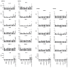
Immunogenic response of AYA1809002 and AYA1809004. Human PBMCs were stim- ulated with or without AYA1809002, AYA1809004, control aptamer, LPS, ODN 1826, and LPS+ODN 1826 at 37 °C for 24 and 72 h. The amount of cytokines secreted from the hPBMCs was assessed using a LEGENDplex Human Inflammation Kit. Soluble analytes were quantified using flow cytometry and analyzed with BioLegend’s LEGENDplexTM software. Each dot represents an individual donor (n = 4). Data are graphed as the mean value ± SEM. The p values were determined with one-way ANOVA, Dunnett’s multiple comparison test. * denotes p≤ 0.05, ** denotes p≤ 0.01, *** denotes p≤ 0.001. **** denotes p≤ 0.0001.
Figure 11
Figure 11
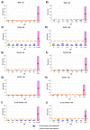
Microbial mutagenicity test for AYA1809002 (AJ). Assessment of the aptamer’s mutagenic potential involved subjecting S. typhimurium strains (TA98, TA100, TA1535, TA1537) and E. coli strains (wp2[pkM101] and wp2 uvrA) to varied concentrations of the aptamer. Positive and negative controls were included. The aptamer AYA1809002 was applied at concentrations ranging from 0.5 µM to 10 µM. This assay was conducted both in the presence and absence of metabolic activation, which was facilitated by liver homogenate S9. Results were obtained from three independent experiments, and the data are presented as the mean ± standard deviation (SD).
Figure 12
Figure 12
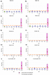
Microbial mutagenicity test for AYA1809004 (AJ). Assessment of the aptamer’s mutagenic potential involved subjecting S. typhimurium strains (TA98, TA100, TA1535, TA1537) and E. coli strains (wp2[pkM101] and wp2 uvrA) to varied concentrations of the aptamer. Positive and negative controls were included. The aptamer AYA1809004 was applied at concentrations ranging from 0.5 µM to 10 µM. This assay was conducted in both the presence and absence of metabolic activation, which was facilitated by liver homogenate S9. Results were obtained from three independent experiments, and the data are presented as the mean ± standard deviation (SD).

References

    1. World Thrombosis Day. 2023. [(accessed on 14 June 2023)]. Available online: https://www.worldthrombosisday.org/issue/thrombosis.
    1. Chen A., Stecker E., Warden B.A. Direct oral anticoagulant use: A practical guide to common clinical challenges. J. Am. Heart Assoc. 2020;9:e017559. doi: 10.1161/JAHA.120.017559. - DOI - PMC - PubMed
    1. Cera E. Thrombin as procoagulant and anticoagulant. J. Thromb. Haemost. 2007;5:196–202. doi: 10.1111/j.1538-7836.2007.02485.x. - DOI - PubMed
    1. Petrera N., Stafford A., Leslie B., Kretz C., Fredenburgh J., Weitz J. Long range communication between exosites 1 and 2 modulates thrombin function. J. Biol. Chem. 2009;284:25620–25629. doi: 10.1074/jbc.M109.000042. - DOI - PMC - PubMed
    1. Malovichko M., Sabo T., Maurer M. Ligand binding to anion-binding exosites regulates conformational properties of thrombin. J. Biol. Chem. 2013;288:8667–8678. doi: 10.1074/jbc.M112.410829. - DOI - PMC - PubMed

Publication types