. 2023 Sep 15;12(18):2282.
doi: 10.3390/cells12182282.

Complement C3 Reduces Apoptosis via Interaction with the Intrinsic Apoptotic Pathway

Affiliations

Complement C3 Reduces Apoptosis via Interaction with the Intrinsic Apoptotic Pathway

Zhou Fang et al. Cells. .

Abstract

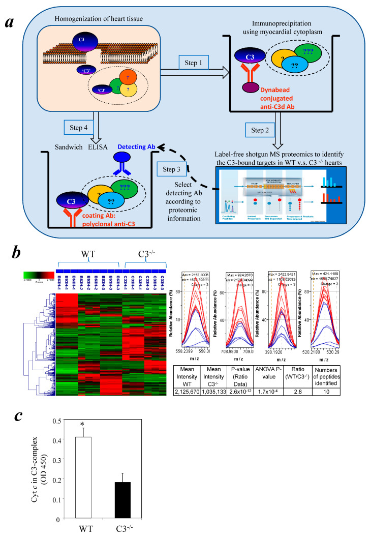
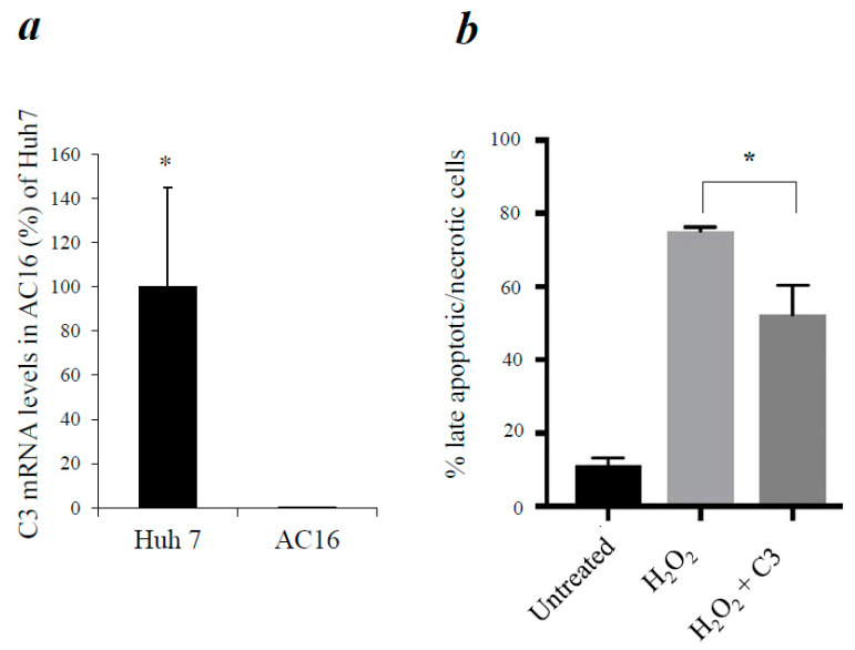
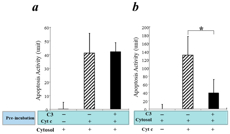
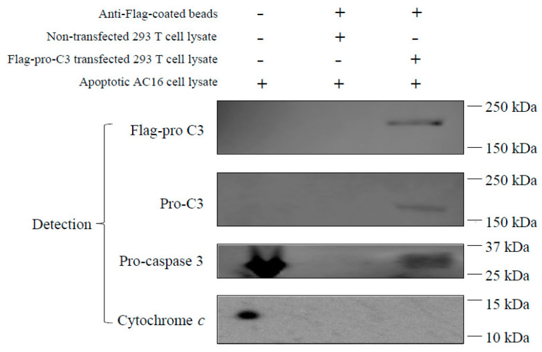
Myocardial ischemia/reperfusion (I/R) elicits an acute inflammatory response involving complement factors. Recently, we reported that myocardial necrosis was decreased in complement C3-/- mice after heart I/R. The current study used the same heart model to test the effect of C3 on myocardial apoptosis and investigated if C3 regulation of apoptosis occurred in human cardiomyocytes. Comparative proteomics analyses found that cytochrome c was present in the myocardial C3 complex of WT mice following I/R. Incubation of exogenous human C3 reduced apoptosis in a cell culture system of human cardiomyocytes that did not inherently express C3. In addition, human C3 inhibited the intrinsic apoptosis pathway in a cell-free apoptosis system. Finally, human pro-C3 was found to bind with an apoptotic factor, pro-caspase 3, in a cell-free system. Thus, we present firsthand evidence showing that C3 readily reduces myocardial apoptosis via interaction with the intrinsic apoptotic pathway.

Keywords: apoptosis; complement C3; cytochrome c; ischemia/reperfusion injury (IRI); pro-caspase 3.


Conflict of interest statement

The authors declare no conflict of interest.

Figures

Figure 1
Figure 1
Comparative proteomics identified cytochrome c in C3-containing complexes formed during myocardial I/R. (a) Schematic diagram depicts the steps of experimental approaches. Step 1: hearts were harvested and homogenized from WT and C3−/− mice, which were subjected to 1 h ischemia/3 h reperfusion; C3-containing complexes in heart lysates were immunoprecipitated with an anti-C3d Ab. Step 2: the C3-immunoprecipitated complexes were digested with trypsin and analyzed by label-free shotgun LC–mass spectrometry. Step 3: select detecting Ab according to proteomic information. Step 4: sandwich ELISA to confirm the specific protein in C3 complex. (b) Left panel: Chromatograms were recorded for each biological replicate in Resolution/Ion Mobility mode. Agglomerative hierarchical clusters of Z-score transformed intensity data were processed by the Elucidator program for all LC–MS chromatograms. Z-score coloration indicates protein abundance in WT compared with C3−/− samples (red, higher abundance; green, lower abundance; black, equal abundance). Right panel histograms: Comparison of cytochrome c peptide signals of specific m/z ratios between WT and C3−/− mice. Red and blue lines represent peptide signals from WT and C3−/− mice, respectively. At bottom-right is the summary table of the cytochrome c peptide signal comparisons of WT and C3−/− mice. (c) Cytochrome c is present in a cytosolic C3-binding complex following 1 h ischemia and 3 h reperfusion. Myocardial cytosolic factions were isolated. C3-binding complexes in the myocardial cytosolic fractions were captured with a polyclonal anti-C3 Ab bound to the surface of microplates. An anti-cytochrome c Ab was used to detect cytochrome c in C3-binding complexes (n = 3 mice/group). * p < 0.05.
Figure 2
Figure 2
Exposure to exogenous C3 reduced apoptosis in cardiomyocytes. (a) C3 mRNA levels in AC16 cardiomyocytes were measured by qPCR. Huh 7 human liver cell line was used as a positive control of C3 expression. Error bars indicate SEM; * p < 0.05. (b) AC16 cells were incubated with H2O2 for 60 min and then washed with PBS. Cells were then incubated in the presence or absence of purified C3 for 3 h. Apoptotic cell death was detected by FACS analyses of Annexin V-Propidium Iodide staining.
Figure 3
Figure 3
In a cell-free apoptosis system, C3 interacts with factor(s) downstream of cytochrome c and blocks cytochrome c-mediated apoptosis. (a) Purified C3 was pre-incubated with purified cytochrome c for one hour, followed by addition of purified cytosol from xenopus and incubation for one hour. Apoptosis activities were detected using substrate from the Caspase-3 Glo Apoptosis kit. (b) Purified C3 was pre-incubated with purified cytosol for one hour, followed by addition of cytochrome c and incubation for one hour. Apoptosis activities were detected as in (a). * p < 0.05.
Figure 4
Figure 4
Pro-C3 binding with pro-caspase 3 in a cell-free system. Lane 1: AC16 cells were treated with 500 mM H2O2 for 30 min. Cell lysate was prepared as described in Methods section. Lane 2: Anti-flag antibody coated beads were incubated with cell lysate of non-transfected 293 T cells (without Flag-pro-C3) and then with AC16 cell lysate. The pull-down materials were used for analyses. Lane 3: Anti-Flag antibody coated beads were incubated with cell lysate of transfected 293 T cells (containing Flag-pro-C3) and then with cell lysate of AC16 cells. The pull-down materials were then used for analyses.

Similar articles

Cited by

References

    1. Geisbrecht B.V., Lambris J.D., Gros P. Complement component C3: A structural perspective and potential therapeutic implications. Semin. Immunol. 2022;59:101627. doi: 10.1016/j.smim.2022.101627. - DOI - PMC - PubMed
    1. Carroll M.C., Isenman D.E. Regulation of humoral immunity by complement. Immunity. 2012;37:199–207. doi: 10.1016/j.immuni.2012.08.002. - DOI - PMC - PubMed
    1. Carroll M.V., Sim R.B. Complement in health and disease. Adv. Drug Deliv. Rev. 2011;63:965–975. doi: 10.1016/j.addr.2011.06.005. - DOI - PubMed
    1. Dunkelberger J.R., Song W.C. Complement and its role in innate and adaptive immune responses. Cell Res. 2010;20:34–50. doi: 10.1038/cr.2009.139. - DOI - PubMed
    1. Gorsuch W.B., Chrysanthou E., Schwaeble W.J., Stahl G.L. The complement system in ischemia-reperfusion injuries. Immunobiology. 2012;217:1026–1033. doi: 10.1016/j.imbio.2012.07.024. - DOI - PMC - PubMed

Publication types