. 2023 Sep 16;12(18):2294.
doi: 10.3390/cells12182294.

Anti-Inflammatory Effects of Synthetic Peptides Based on Glucocorticoid-Induced Leucine Zipper (GILZ) Protein for the Treatment of Inflammatory Bowel Diseases (IBDs)

Affiliations

Anti-Inflammatory Effects of Synthetic Peptides Based on Glucocorticoid-Induced Leucine Zipper (GILZ) Protein for the Treatment of Inflammatory Bowel Diseases (IBDs)

Musetta Paglialunga et al. Cells. .

Abstract

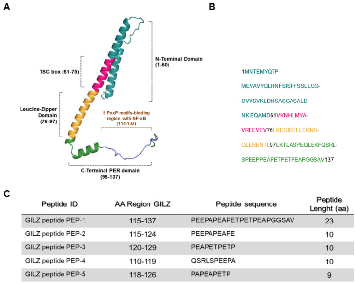
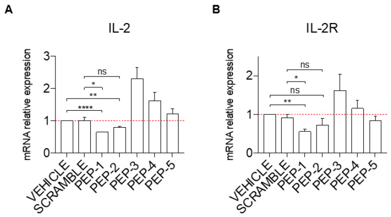
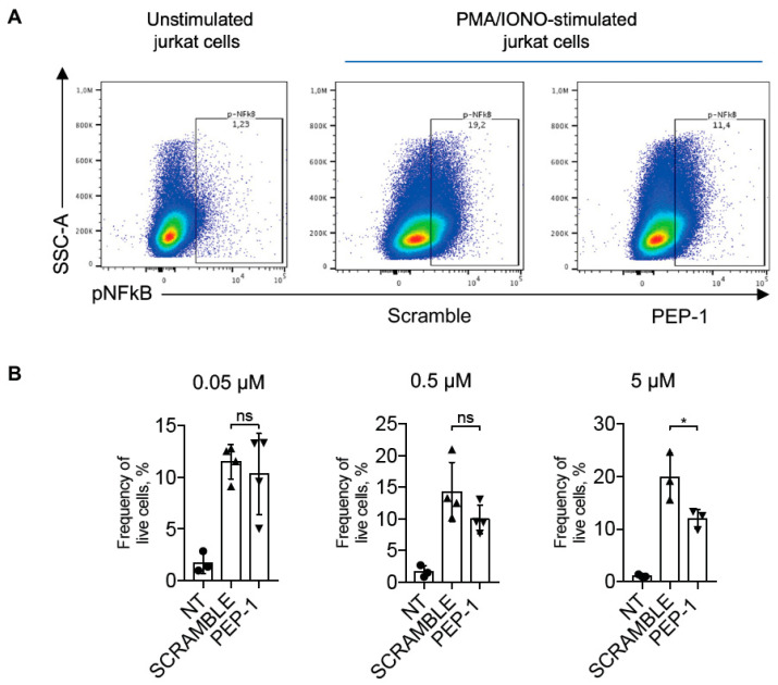
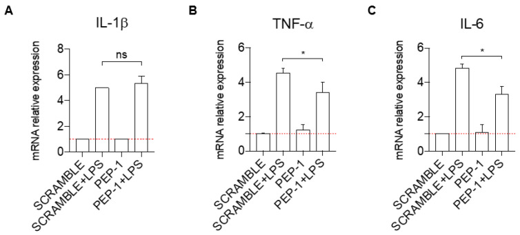
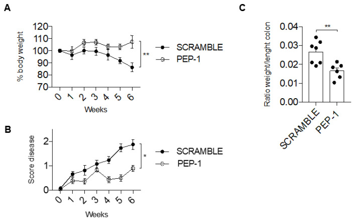
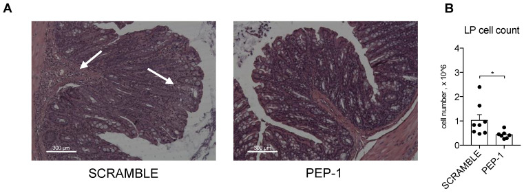
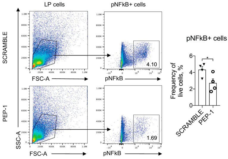
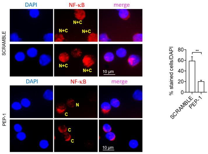
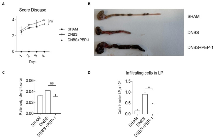
Glucocorticoids (GCs) are commonly used to treat autoimmune and inflammatory diseases, but their clinical effects and long-term use can lead to serious side effects. New drugs that can replace GCs are needed. Glucocorticoid-induced leucine zipper (GILZ) is induced by GCs and mediates many of their anti-inflammatory effects, such as inhibiting the pro-inflammatory molecule NF-κB. The GILZ C-terminal domain (PER region) is responsible for GILZ/p65NF-κB interaction and consequent inhibition of its transcriptional activity. A set of five short peptides spanning different parts of the PER region of GILZ protein was designed, and their anti-inflammatory activity was tested, both in vitro and in vivo. We tested the biological activity of GILZ peptides in human lymphocytic and monocytic cell lines to evaluate their inhibitory effect on the NF-κB-dependent expression of pro-inflammatory cytokines. Among the tested peptides, the peptide named PEP-1 demonstrated the highest efficacy in inhibiting cell activation in vitro. Subsequently, PEP-1 was further evaluated in two in vivo experimental colitis models (chemically induced by DNBS administration and spontaneous colitis induced in IL-10 knock-out (KO) mice (to assess its effectiveness in counteracting inflammation. Results show that PEP-1 reduced disease severity in both colitis models associated with reduced NF-κB pro-inflammatory activity in colon lamina propria lymphocytes. This study explored GILZ-based 'small peptides' potential efficacy in decreasing lymphocyte activation and inflammation associated with experimental inflammatory bowel diseases (IBDs). Small peptides have several advantages over the entire protein, including higher selectivity, better stability, and bioavailability profile, and are easy to synthesize and cost-effective. Thus, identifying active GILZ peptides could represent a new class of drugs for treating IBD patients.

Keywords: GILZ; IBDs; NF-κB; glucocorticoids; inflammation.


Conflict of interest statement

The authors declare no conflict of interest.

Figures

Figure 1
Figure 1
Predictive structure of GILZ protein. (A) A three-dimensional representation, created by COSMIC2 platform (https://cosmic-cryoem.org, accessed on 15 September 2023) and modified with PyMOL Molecular Graphics System (http://www.pymol.org/pymol accessed on 15 September 2023), of GILZ protein constituted by four domains: a N-terminal domain (NTD, 1–60 aa), TSC-box (61–75 aa), a leucine zipper (LZ, 76–97 aa), and a PER region spanning from 98 to 137 aa residues. (B) The primary murine sequence of GILZ protein. (C) The table represents sequences of five GILZ peptides spanning different portions of the PER region, which is responsible for GILZ-NF-κB protein-to-protein interaction [21].
Figure 2
Figure 2
PEP-1 decreases the mRNA expression level of IL-2 and IL-2Rα in Jurkat cells. The level of IL-2 (A) and IL-2Rα (B) mRNA relative expression was evaluated by qPCR in Jurkat cells treated or not with GILZ peptides (0.1 μM) for 1 h. Afterward, cells were activated with PMA and Ionomycin for 6 h and treated with acetonitrile alone (vehicle), scramble, or PEP peptides (0.1 μM), as indicated in the figure. Graphs represent the mean ± SEM of three independent experiments. * p < 0.05, ** p < 0.005, **** p < 0.0001, P-values were calculated according to unpaired Student’s t-test. ns: non-significant.
Figure 3
Figure 3
PEP-1 decreases pNF-κB/p65 activation in Jurkat cells. Representative dot plots of flow cytometry analysis of p-NF-κB/p65 in Jurkat cells stimulated or not with PMA and ionomycin for 15 min (A). Numbers within quadrants represent the frequency of the gated population of positive pNF-κB cells treated with scramble or PEP-1 peptide (5 μM), compared to the frequency of positive pNF-κB unstimulated Jurkat cells. After that, graphs represent the frequency of pNF-κB+ live Jurkat cells treated with different doses of PEP-1 at the concentration indicated in the above graphs (B). Graphs represent the mean of at least three independent experiments ± SD. * p < 0.05. p-values were calculated using the unpaired Student’s t-test. ns: non-significant.
Figure 4
Figure 4
PEP-1 decreases the mRNA expression level of pro-inflammatory cytokines in the THP-1 cell line. IL-1β (A), TNF-α (B), and IL-6 (C) mRNA relative expression was evaluated by qPCR for THP-1 cells activated with LPS, with GILZ peptide PEP-1 (5 μM) for 1 h. Data have been normalized, and activated THP-1 cells treated with the scrambled sequence of PEP-1 peptide were used as control. Graphs represent the mean ± SD of three independent experiments. * p < 0.05. p-values were calculated using the unpaired Student’s t-test. PMA: Phorbol Myristate Acetate. LPS: Lipopolysaccharide.
Figure 5
Figure 5
GILZ PEP-1 peptide treatment reduces the clinical signs of colitis in IL-10KO mice. After an intraperitoneal injection of PEP-1, or scrambled sequence (control) at week 7, mice were monitored daily for 6 weeks, and the percentage of body weight loss (A) was reported. Graph of total score disease (B) was documented, taking into consideration the stool consistency and rectal bleeding. Following the sixth week, mice were sacrificed to calculate the ratio between weight and length of the colon (C). Graphs represent mean ± SEM. * p < 0.05, ** p < 0.005. p-values were calculated using the unpaired Student’s t-test. Each dot represents an individual mouse (n = 8/group).
Figure 6
Figure 6
PEP-1 reduces leukocyte infiltration in the colon LP of IL-KO mice. Mice were sacrificed to estimate the infiltration of immune cells in the sub-mucosal tissues through histological examination by H&E staining (A). Lamina propria was infiltrated by inflammatory cells (arrowhead) in IL-10KO mice treated with scrambled peptide, unlike the result observed in mice treated with PEP-1. Subsequently, leukocytes infiltrated in LP were isolated, and the cell count was carried out (B) in mice treated with scramble or PEP-1. The graph represents mean ± SEM. * p < 0.05. p-values were calculated using the unpaired Student’s t-test. Each dot represents an individual mouse. (n = 8/group).
Figure 7
Figure 7
PEP 1 reduces p65/NF-κB phosphorylation in leukocytes infiltrating the colon LP of IL-10KO mice. Representative dot plots of flow cytometry analysis and frequency of cells positively stained for phosphorylated form of p65/NFκB (pNFκB+) among lymphocytes isolated from colon LP of IL-10KO mice treated with scramble or PEP-1 peptide. Numbers within quadrants represent the frequency of the gated population. Graphs represent mean ± SEM. * p < 0.05. p-values were calculated using the unpaired Student’s t-test. Each dot represents an individual mouse. (n = 8/group).
Figure 8
Figure 8
PEP 1 reduces NF-κB nuclear translocation in leukocytes infiltrating the colon LP of IL-10KO mice. Representative immunofluorescence pictures showing pNF-κB/p65 positive cells of isolated colon LP cells from IL-10KO mice; quantification of NF-κB nuclear translocation was calculated by measuring the NF-kB localization to the cytoplasmic region and normalized by counting the total number of cells stained with DAPI. “N + C” (yellow): NF-κB staining in both the nucleus and cytoplasm; “C” (yellow): NF-κB staining in the cytoplasm; “N” (yellow): NF-κB staining in the nucleus. Graph sideways: cell count by ImageJ software of cytoplasmic NF-κB positive cells compared to all DAPI positive cells (representing the whole number of cells present in the slides analyzed). The graph represents mean ± SD. ** p < 0.005. p-values were calculated using the unpaired Student’s t-test. (n = 8/group).
Figure 9
Figure 9
PEP-1 ameliorates clinical signs of colitis in DNBS-induced colitis mice. Before sacrifice, daily measurement of total disease score was conducted in SHAM and PEP-1 treated mice during DNBS-induced colitis (A). Following that, colon tissues from mice treated only with vehicle (SHAM), DNBS-induced colitis mice (DNBS), and DNBS-treated mice with PEP-1 injection (DNBS + PEP-1) were examined under a microscope (B), and the ratio weight/length of the colon was calculated (C); lymphocytes infiltrated in LP were isolated and counted from SHAM, DNBS-induced colitis mice, treated or not with PEP-1 (D). Graphs represent mean ± SEM. ** p < 0.005. p-values were calculated using the unpaired Student’s t-test. ns: non-significant. (n = 6/group).

References

    1. Diaz-Jimenez D., Kolb J.P., Cidlowski J.A. Glucocorticoids as Regulators of Macrophage-Mediated Tissue Homeostasis. Front. Immunol. 2021;12:669891. doi: 10.3389/fimmu.2021.669891. - DOI - PMC - PubMed
    1. Ronchetti S., Migliorati G., Bruscoli S., Riccardi C. Defining the role of glucocorticoids in inflammation. Clin. Sci. (Lond) 2018;132:1529–1543. doi: 10.1042/CS20171505. - DOI - PubMed
    1. Taves M.D., Ashwell J.D. Glucocorticoids in T cell development, differentiation and function. Nat. Rev. Immunol. 2021;21:233–243. doi: 10.1038/s41577-020-00464-0. - DOI - PubMed
    1. Bruscoli S., Febo M., Riccardi C., Migliorati G. Glucocorticoid Therapy in Inflammatory Bowel Disease: Mechanisms and Clinical Practice. Front. Immunol. 2021;12:691480. doi: 10.3389/fimmu.2021.691480. - DOI - PMC - PubMed
    1. Cain D.W., Cidlowski J.A. Immune regulation by glucocorticoids. Nat. Rev. Immunol. 2017;17:233–247. doi: 10.1038/nri.2017.1. - DOI - PMC - PubMed