Review
. 2023 Sep 19;12(18):2307.
doi: 10.3390/cells12182307.

Astrocytes Are a Key Target for Neurotropic Viral Infection

Affiliations
Review

Astrocytes Are a Key Target for Neurotropic Viral Infection

Maja Potokar et al. Cells. .

Abstract

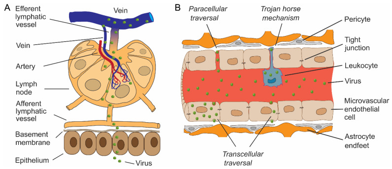
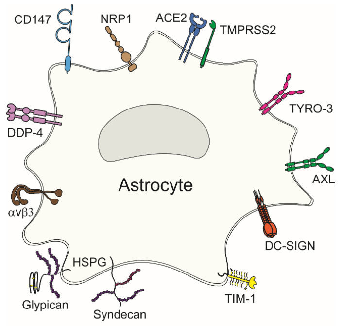
Astrocytes are increasingly recognized as important viral host cells in the central nervous system. These cells can produce relatively high quantities of new virions. In part, this can be attributed to the characteristics of astrocyte metabolism and its abundant and dynamic cytoskeleton network. Astrocytes are anatomically localized adjacent to interfaces between blood capillaries and brain parenchyma and between blood capillaries and brain ventricles. Moreover, astrocytes exhibit a larger membrane interface with the extracellular space than neurons. These properties, together with the expression of various and numerous viral entry receptors, a relatively high rate of endocytosis, and morphological plasticity of intracellular organelles, render astrocytes important target cells in neurotropic infections. In this review, we describe factors that mediate the high susceptibility of astrocytes to viral infection and replication, including the anatomic localization of astrocytes, morphology, expression of viral entry receptors, and various forms of autophagy.

Keywords: astrocytes; autophagy; cell metabolism; endosome; lysosome; oxidant species; viruses.


Conflict of interest statement

The funding sponsors had no role in the design of the study; in the collection, analyses, or interpretation of data; in the writing of the manuscript, and in the decision to publish the results.

Figures

Figure 1
Figure 1
Entry of viruses through the epithelial barrier, the lymphatic spread of viruses and neuroinvasion through the blood–brain barrier. (A) After entry through the epithelium, viruses may continue with subepithelial invasion by exploiting the network of lymphatics to enter lymph nodes. Viruses enter porous lymphatic capillaries and/or efferent lymphatic vessels to access the venous system. Circulating blood is the fastest and most effective way of viral dissemination to different tissues, including the central nervous system (CNS). (B) Entry into the delicate CNS parenchyma is restricted by an elaborate barrier network, most notably by the blood–brain barrier (BBB). Certain viruses can pass the BBB by the transcellular route by transferring through the lining of microvascular endothelial cells, where they may or may not replicate. In addition, viruses can pass the BBB by paracellular traversal between adjacent endothelial cells, with or without disruption of tight junctions. Viruses can also penetrate the BBB by transmigration within infected haematopoietic cells, known as the “Trojan horse” mechanism.
Figure 2
Figure 2
Viral entry receptors in astrocytes. Diverse plasma membrane receptors can mediate virus attachment to and entry into astrocytes. These include neuropilin-1 (NRP1), angiotensin-converting enzyme 2 (ACE2), type II transmembrane serine protease (TMPRSS2), TAM family receptors (TYRO-3 and AXL), dendritic cell (DC)-specific intercellular adhesion molecule-3-grabbing nonintegrin (DC-SIGN), T-cell Ig and mucin domain 1 (TIM-1), heparan sulphate proteoglycans (HSPGs) comprising syndecans and glypicans, αvβ3 integrin, dipeptidylpeptidase 4 (DPP4), and cluster of differentiation 147 (CD147).
Figure 3
Figure 3
Tick-borne encephalitis virus (TBEV) and West Nile virus (WNV), but not mosquito-only flavivirus (MOF), increase autophagy in human astrocytes. (a) Representative fluorescence micrographs documenting mock-infected human astrocytes (Control) and astrocytes after exposure to selected flaviviruses (TBEV, WNV, and MOF) for 48 h. Cells were expressing mRFP-EGFP-LC3, where LC3, a marker of autophagic compartments, is tandem fluorescent-tagged with mRFP (red fluorescence) and EGFP (green fluorescence). The pH of autophagosomes (AP) is close to neutral, which facilitates the fluorescence of both fluorophores, resulting in yellow objects. Fusion of AP with lysosomes yields autolysosomes (AL), i.e., organelles with acidic pH, where the EGFP fluorescence is quenched, resulting in red-only objects. Selected rectangular areas within the cells, enlarged at the corners (bottom, right), show superimposed images of mRFP and EGFP fluorescence. Arrows indicate AL. Adjacent, smaller panels display mRFP and EGFP fluorescence and co-localization masks (Col.mask) (co-localized objects correspond to AP) of the enlarged images. The white outlines in the large panels show the cell shape. (b) The total number of autophagic compartments (i) and the ratio of AL to AP (which is a measure of autophagic degradation activity) (ii) in mock-infected cells and after infection with TBEV, WNV, and MOF at 12, 24, and 48 h post-infection (hpi). Infection with TBEV increases the total number of autophagic compartments at all three time points tested, compared with mock-infected cells (i.e., controls at 48 hpi, which were confirmed to be comparable to controls at 12 and 24 hpi) (* p < 0.05, one-way ANOVA followed by Dunn’s test), but does not affect the AL:AP ratio (p  >  0.05, one-way ANOVA). WNV infection induces an increase in the total number of autophagic compartments at 48 hpi (* p <  0.05, one-way ANOVA followed by Dunn’s test) and does not affect the ratio AL:AP (p > 0.05, one-way ANOVA). MOF infection does not affect the number of autophagic compartments or the AL: AP ratio compared with mock-infected cells at any time point tested (p >  0.05, one-way ANOVA). (c) Diameter of AP and AL in mock-infected cells and 48 hpi with selected flaviviruses. Infection with TBEV, WNV and MOF does not affect the size of the autophagic compartment compared with mock-infected cells (p  >  0.05, one-way ANOVA). ALs are larger than APs in all experimental conditions (*** p < 0.001, Mann-Whitney U test). Full lines in the boxplots represent median values, and the dotted lines correspond to average values. The numbers below the boxplots are the number of cells (b) or compartments (c) analysed for each condition. Cells were infected with TBEV and MOF at an MOI of 0.1 and with WNV at an MOI of 1. Figure and figure legend reproduced from Tavcar Verdev et al. [46] with permission, licensed under a Creative Commons Attribution 4.0 International License (http://creativecommons.org/licenses/by/4.0 (accessed on 1 August 2023)).

Similar articles

Cited by

References

    1. Romero J.R., Newland J.G. Viral meningitis and encephalitis: Traditional and emerging viral agents. Semin. Pediatr. Infect. Dis. 2003;14:72–82. doi: 10.1053/spid.2003.127223. - DOI - PubMed
    1. Gora H., Smith S., Wilson I., Preston-Thomas A., Ramsamy N., Hanson J. The epidemiology and outcomes of central nervous system infections in Far North Queensland, tropical Australia; 2000–2019. PLoS ONE. 2022;17:e0265410. doi: 10.1371/journal.pone.0265410. - DOI - PMC - PubMed
    1. Ludlow M., Kortekaas J., Herden C., Hoffmann B., Tappe D., Trebst C., Griffin D.E., Brindle H.E., Solomon T., Brown A.S., et al. Neurotropic virus infections as the cause of immediate and delayed neuropathology. Acta Neuropathol. 2016;131:159–184. doi: 10.1007/s00401-015-1511-3. - DOI - PMC - PubMed
    1. Carroll D., Daszak P., Wolfe N.D., Gao G.F., Morel C.M., Morzaria S., Pablos-Mendez A., Tomori O., Mazet J.A.K. The Global Virome Project. Science. 2018;359:872–874. doi: 10.1126/science.aap7463. - DOI - PubMed
    1. Swanson P.A., II, McGavern D.B. Viral diseases of the central nervous system. Curr. Opin. Virol. 2015;11:44–54. doi: 10.1016/j.coviro.2014.12.009. - DOI - PMC - PubMed

LinkOut - more resources