. 2023 Sep 19;12(18):2315.
doi: 10.3390/cells12182315.

Characterisation of Lipoma-Preferred Partner as a Novel Mechanotransducer in Vascular Smooth Muscle Cells

Affiliations

Characterisation of Lipoma-Preferred Partner as a Novel Mechanotransducer in Vascular Smooth Muscle Cells

Alexandra Sporkova et al. Cells. .

Abstract

In arteries and arterioles, a chronic increase in blood pressure raises wall tension. This continuous biomechanical strain causes a change in gene expression in vascular smooth muscle cells (VSMCs) that may lead to pathological changes. Here we have characterised the functional properties of lipoma-preferred partner (LPP), a Lin11-Isl1-Mec3 (LIM)-domain protein, which is most closely related to the mechanotransducer zyxin but selectively expressed by smooth muscle cells, including VSMCs in adult mice. VSMCs isolated from the aorta of LPP knockout (LPP-KO) mice displayed a higher rate of proliferation than their wildtype (WT) counterparts, and when cultured as three-dimensional spheroids, they revealed a higher expression of the proliferation marker Ki 67 and showed greater invasion into a collagen gel. Accordingly, the gelatinase activity was increased in LPP-KO but not WT spheroids. The LPP-KO spheroids adhering to the collagen gel responded with decreased contraction to potassium chloride. The relaxation response to caffeine and norepinephrine was also smaller in the LPP-KO spheroids than in their WT counterparts. The overexpression of zyxin in LPP-KO VSMCs resulted in a reversal to a more quiescent differentiated phenotype. In native VSMCs, i.e., in isolated perfused segments of the mesenteric artery (MA), the contractile responses of LPP-KO segments to potassium chloride, phenylephrine or endothelin-1 did not vary from those in isolated perfused WT segments. In contrast, the myogenic response of LPP-KO MA segments was significantly attenuated while zyxin-deficient MA segments displayed a normal myogenic response. We propose that LPP, which we found to be expressed solely in the medial layer of different arteries from adult mice, may play an important role in controlling the quiescent contractile phenotype of VSMCs.

Keywords: LPP; VSMC; mechanosensitive genes; mechanotransduction; vascular remodelling.


Conflict of interest statement

All authors declare they have no financial interest to disclose that would be directly or indirectly related to the work submitted for publication.

Figures

Figure 1
Figure 1
Distribution of (a) LPP and (b) zyxin in cultured VSMCs isolated from the aorta of 3-month-old C57BL/6 mice. LPP and zyxin immunoreactivity is shown in red, and stress fibres stained with an anti-α-SMA antibody are shown in green. Nuclei were counterstained with DAPI (blue). (c) LPP staining in LPP-KO VSMCs to control for the specificity of the antibody, and (d) zyxin staining in the LPP-KO VSMCs (d). Representative images; the scale bar shown in d represents 20 µm.
Figure 2
Figure 2
Subcellular distribution of zyxin and LPP in cultured VSMCs (WT) in vitro. (a) Indirect immunofluorescence confocal laser scanning images of VSCM cultured for 48 h under static conditions (static), followed by 1 h or 8 h of exposure to cyclic stretching (13% elongation, 0.5 Hz). Inserts: Overlay of zyxin- or LPP-specific (purple) and α-smooth muscle actin (SMA)-specific (green) antibody staining; nuclei are additionally stained with DAPI (blue). Narrow arrow heads: focal adhesions, arrows: (position of) SMA-containing fibres; scale bar: 20 μm. (b) Representative Western blot analysis of cytosolic and nuclear fractions of VSMCs from 3-month-old WT mice pre-cultured for 48 h followed by 24 h incubation under static conditions or 8 and 24 h of exposure to cyclic stretching (13% elongation, 0.5 Hz), respectively. Alpha-tubulin and histone H3 (HH3) served as indicators for the purity of the subcellular fractions and as loading controls. (c) Statistical summary of 3 independent experiments with individual preparations of VSMCs isolated from the aorta of the 3-month-old WT mice. * p ˂ 0.05 as indicated.
Figure 3
Figure 3
Proliferation of WT and LPP-KO VSMCs. (a) The rate of proliferation was determined by counting the number of cells per optical field of view at 0 and 72 h. Each data point represents an individual VSMC’s preparation with >6 separate optical fields of view analysed. n = 4, * p ˂ 0.05 as indicated. (b) Percentage of WT and LPP-KO VSMCs grown in 3D spheroids expressing the proliferation marker Ki67. n = 4, * p < 0.05 as indicated. (c) Transient overexpression of zyxin and eGFP in LPP-KO VSMCs and their effect on the rate of proliferation. Each data point represents an individual VSMC’s preparation with >6 fields of view analysed. n = 5, * p < 0.05 as indicated.
Figure 4
Figure 4
LPP-KO VSMCs and WT VSMCs grown in 3D spheroids embedded in collagen gels form sprouts and invade the collagen gel. (a) Representative images, the scale bar represents 50 µm. (b) Summary of the average number of sprouts produced by individual LPP KO VSMC (n = 6, in red) and WT VSMC preparations (n = 4) with >10 spheroids per VSMC preparation analysed using the cellSens software. (c) Summary of the cumulative distance travelled by individual sprouts of a VSMC spheroid. n = 6 and n = 4 as in (b), * p < 0.05, ** p < 0.01 as indicated in (b,c) Transient overexpression of zyxin and eGFP in LPP-KO VSMCs and their effect on (d) the average number of sprouts and (e) cumulative sprout length. Each data point represents an individual VSMC preparation with >10 spheroids per VSMC preparation analysed. n = 3, * p ˂ 0.05 as indicated.
Figure 5
Figure 5
Comparison of the contractile properties of the VSMCs grown as 3D spheroids attached to a collagen gel. (a) The contractile response of the VSMC spheroids to potassium chloride (KCl, 60 mM) is calculated as reduction in the area of the collagen gel covered by the spheroids. (b) Relaxation response of the VSMC spheroids to caffeine (10 mM) and to norepinephrine (NE, 10 μM) (c). Each column represents the average responses of VSMC spheroids with >5 spheroids analysed per preparation and a total of 6 VSMCs preparations from individual animals per group (LPP KO or WT). * p ˂ 0.05 as indicated.
Figure 6
Figure 6
Immunofluorescence analysis of LPP and zyxin in 3rd-order mesenteric artery (MA) and femoral artery (FA) segments isolated from WT, LPP-KO or ZXY-KO mice. (ad) Representative images for LPP ((a,c); red) and zyxin ((b,d); red) in MA and FA segments isolated from WT mice. To properly localize both LIM domain proteins, an anti-α-SMA antibody was used to stain the VSMCs in the media ((eh), green) and an anti-CD31 antibody was used to visualize the endothelial cells lining the inner lumen of these segments ((ad), green). While there is a clear colocalization of zyxin with the endothelium (b,d), LPP solely colocalizes with the medial VSMCs (e,f) with which zyxin also colocalizes but to a smaller extent. Staining of both LIM domain proteins in MA segments isolated from the respective knockout mice confirmed the specificity of the antibodies used (i,j). LPP ((k), red) and zyxin ((m), red) were also stained in segments of the aorta isolated from WT mice. Here, co-staining of LPP and CD31 ((l), green) confirmed their mutually exclusive localization, whereas co-staining of zyxin and α-SMA ((n), green) revealed that zyxin is highly abundant in the media of this large conduit artery (yellow colour indicates prominent colocalization with the VSMCs in the media) as well as a distinct staining of the endothelium. Scale bars represent 10 μm in (ac,f) and 20 μm in (d,e,gn).
Figure 7
Figure 7
Comparison of the contractile response of 3rd-order mesenteric artery segments isolated from WT, LPP-KO or ZYX-KO mice to different vasoactive stimuli. The number of isolated perfused segments derived from individual mice is indicated on the graphs. Cumulative concentration–response curves for (a,b) phenylephrine (PE), (c,d) potassium chloride (KCl) and (e,f) endothelin-1 (ET-1).
Figure 8
Figure 8
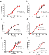
Comparison of the myogenic responses of 3rd-order mesenteric artery segments isolated from WT, LPP-KO or ZYX-KO mice. The number of isolated perfused segments derived from individual mice is indicated on the graphs. (a) Pressure–response curves for segments of (a) 3-month-old, (c) 6-month-old and (e) 12-month-old WT and LPP-KO mice; (b) 6-month-old and (d) 12-month-old WT and ZYX-KO mice; and (f) 6-month-old WT and LPP-KO mice made hypertensive using the DOCA-salt model of experimental hypertension. The area under the curve (AUC) was calculated for each individual group. * p ˂ 0.05, ** p ˂ 0.01 as indicated.
Figure 9
Figure 9
Susceptibility of the myogenic response of 3rd-order mesenteric artery segments isolated from WT or LPP-KO mice made hypertensive by employing the DOCA-salt model of experimental hypertension. The number of isolated perfused segments derived from individual mice is indicated on the graphs. Effect of the general gelatinase inhibitor GM6001 (0.5 µM) on (a) MA segments derived from (a) hypertensive WT and (b) LPP-KO mice and of the TRPC3 channel blocker Pyr 3 (3 µM) on MA segments isolated from (c) hypertensive WT and (d) LPP-KO mice. The area under the curve (AUC) was calculated for each individual group. * p ˂ 0.05, ** p ˂ 0.01 as indicated.
Figure 10
Figure 10
Passive pressure–diameter curves of 3rd-order MA segments isolated from WT, LPP-KO or ZYX-KO mice. The curves were generated in Ca2+-free physiological saline solution in the presence of EGTA. The number of isolated perfused segments derived from individual mice is indicated on the graph. ** p ˂ 0.01 as indicated.
Figure 11
Figure 11
Passive wall thickness of 3rd-order MA segments isolated from (a) 6- and (b) 12-month-old WT, LPP-KO or ZYX-KO mice. Wall thickness was determined at 80 mm Hg in Ca2+-free physiological saline solution in the presence of EGTA (passive distention). The number of isolated perfused segments derived from individual mice is indicated on the graph. * p ˂ 0.05 as indicated.

References

    1. Owens G.K., Kumar M.S., Wamhoff B.R., Franco P.N., Durrant L.M., Carreon D., Haddad E., Vergara A., Cascavita C., Obenaus A., et al. Molecular Regulation of Vascular Smooth Muscle Cell Differentiation in Development and Disease. Physiol. Rev. 2004;84:767–801. doi: 10.1152/physrev.00041.2003. - DOI - PubMed
    1. Gordon D., A Reidy M., Benditt E.P., Schwartz S.M. Cell proliferation in human coronary arteries. Proc. Natl. Acad. Sci. USA. 1990;87:4600–4604. doi: 10.1073/pnas.87.12.4600. - DOI - PMC - PubMed
    1. Shi N., Chen S.-Y. Smooth Muscle Cell Differentiation: Model Systems, Regulatory Mechanisms, and Vascular Diseases. J. Cell. Physiol. 2015;231:777–787. doi: 10.1002/jcp.25208. - DOI - PubMed
    1. Chakraborty R., Chatterjee P., Dave J.M., Ostriker A.C., Greif D.M., Rzucidlo E.M., Martin K.A. Targeting smooth muscle cell phenotypic switching in vascular disease. JVS Vasc. Sci. 2021;2:79–94. doi: 10.1016/j.jvssci.2021.04.001. - DOI - PMC - PubMed
    1. Geiger B., Spatz J.P., Bershadsky A.D. Environmental sensing through focal adhesions. Nat. Rev. Mol. Cell Biol. 2009;10:21–33. doi: 10.1038/nrm2593. - DOI - PubMed

Publication types