Review
. 2023 Sep 20;12(18):2318.
doi: 10.3390/cells12182318.

Biochemical and Molecular Pathways in Neurodegenerative Diseases: An Integrated View

Affiliations
Review

Biochemical and Molecular Pathways in Neurodegenerative Diseases: An Integrated View

Nitesh Sanghai et al. Cells. .

Abstract

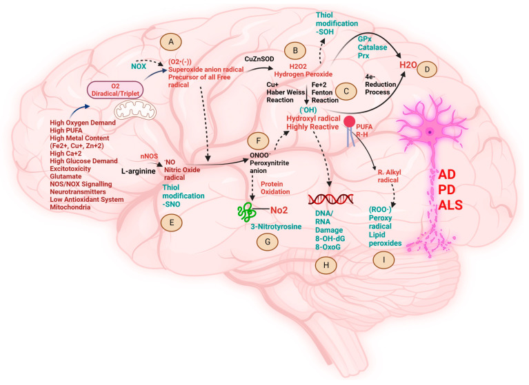
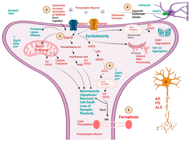
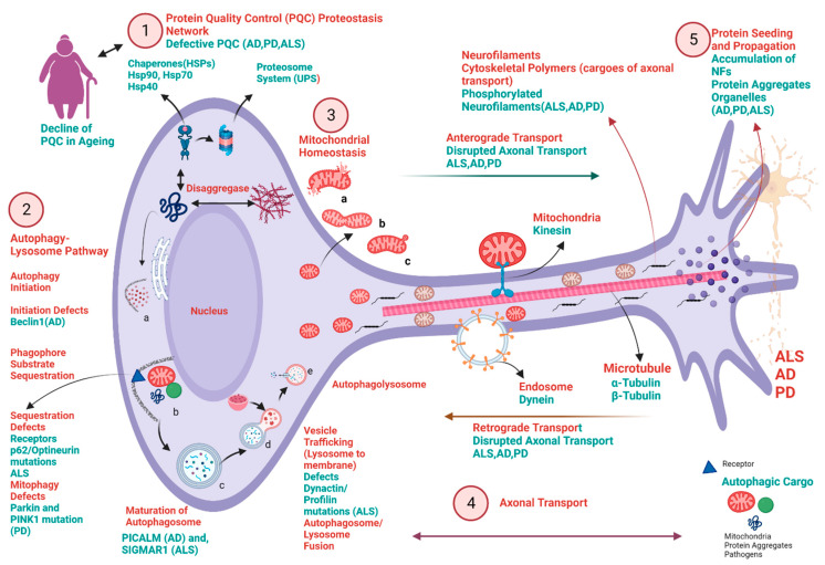
Neurodegenerative diseases (NDDs) like Alzheimer's disease (AD), Parkinson's disease (PD), and amyotrophic lateral sclerosis (ALS) are defined by a myriad of complex aetiologies. Understanding the common biochemical molecular pathologies among NDDs gives an opportunity to decipher the overlapping and numerous cross-talk mechanisms of neurodegeneration. Numerous interrelated pathways lead to the progression of neurodegeneration. We present evidence from the past pieces of literature for the most usual global convergent hallmarks like ageing, oxidative stress, excitotoxicity-induced calcium butterfly effect, defective proteostasis including chaperones, autophagy, mitophagy, and proteosome networks, and neuroinflammation. Herein, we applied a holistic approach to identify and represent the shared mechanism across NDDs. Further, we believe that this approach could be helpful in identifying key modulators across NDDs, with a particular focus on AD, PD, and ALS. Moreover, these concepts could be applied to the development and diagnosis of novel strategies for diverse NDDs.

Keywords: Alzheimer’s disease; Parkinson’s disease; ageing; amyotrophic lateral sclerosis; autophagy; calcium butterfly effect; chaperones; excitotoxicity; mitophagy; neurodegenerative diseases; neuroinflammation; oxidative stress; proteostasis.


Conflict of interest statement

The authors declare no conflict of interest.

Figures

Figure 1
Figure 1
Schematic presentation of various biochemical cross-talks and their detrimental manifestations (AI) in the brain provoked by oxidative stress and their implications in the progress of neurodegenerative diseases like Alzheimer’s disease (AD), Parkinson’s disease (PD), and amyotrophic lateral sclerosis (ALS). Brain is highly vulnerable to oxidative stress due to low regenerative capacity, enrichment of polyunsaturated fatty acids, high dependency on mitochondria for adenosine triphosphate (ATP) generation, elevated glucose demand, high concentration of metals like ferrous ion (Fe+2), cuprous ion (Cu+), zinc ion (Zn+2) and calcium ion (Ca+2), glutamate-induced excitotoxicity, high oxygen (O2) consumption, and relatively low antioxidant system. These multiple factors initiate various reaction pathways to create redox disbalance called oxidative and nitrosative stress in the brain, implicated in various NDDs. (A). The triplet unstable O2 undergoes reduction to produce the precursor of all radicals called superoxide anion radical (O2•−) via NAD(P)H oxidases (NOXs) pathway, i.e., one-electron trans-membrane transfer to (O2) [95]. (B). Antioxidant superoxide dismutase (SOD1) undergoes dismutation to scavenge (O2•−) to produce hydrogen peroxide (H2O2). (C). The weakly liganded (Fe+2) and (Cu+) undergo reduction to produce nature’s most vulnerable oxidant hydroxyl radical (HO) through Fenton’s reaction and Haber-Weiss reaction. (D). The final 4th electron reduction of H2O2 in the presence of antioxidants, like glutathione peroxidase (Gpx), catalase (cat), and peroxiredoxin system (Prx), forms water (H2O). (E). Overactivation of neuronal nitric oxide synthase (nNOS) produces nitric oxide (NO) radicals from L-arginine, which create nitrosative stress by modification of thiol group (SH) containing proteins. (F). Excessive superoxide anion radicals lead to inactivation of nitric oxide production and switch the biology to production of highly potent oxidant peroxynitrite anion (ONOO), which leads to the nitrosative stress by (SH) modification of free tyrosine (Tyr) residues to form 3-nitrotyrosine (3-NO2Tyr) (G), which act as a versatile biomarker of nitrosative stress and NDDs. (H). Highly reactive and mutagenic oxidant (HO) damages the nucleic acid deoxyribonucleic acid/ribonucleic acid (DNA/RNA) to form oxidative products 8-hydroxy-2′-deoxyguanosine(8-OHdG) and (8-OxoG), and acts as a universal biomarker for oxidative stress and NDDs (important to note that guanine is the most oxidation prone nucleobase because of low reduction potential [96]). Further, HO radical causes lipid peroxidation of lipid-rich neuronal membranes, resulting in the death of neurons. Lipid peroxides (ROO.) act as a biomarker of oxidative stress and NDDs. Created with BioRender.com (accessed on 19 September 2023).
Figure 2
Figure 2
Schematic presentation of various biochemical cross-talks, involving calcium ion (Ca+2), ferrous ion (Fe+2), and Zinc ion (Zn+2) implicated in the progress of neurodegenerative diseases like Alzheimer’s disease (AD), Parkinson’s disease (PD), and amyotrophic lateral sclerosis (ALS). (A). Excitotoxicity (neuronal death) is triggered by the excessive release of excitatory neurotransmitter glutamate (neurotoxic) from the presynaptic neuron and leads to activation of various biochemical cascades leading to neurotoxicity and hence, neuronal death. This process is initiated by the activation of the N-methyl-D-aspartic acid receptors (NMDAR) by excessive glutamate at postsynaptic neurons and thereby the release and accumulation of toxic intraneuronal Ca2+. (B). Glutamate-mediated excitotoxicity is increased because of the astrocyte-mediated downregulation of excitatory amino acid transporters 2 (EAAT2), which slows down the uptake of glutamate from the synaptic cleft and incites the excitotoxicity cascade. (C). Ca2+ overload initiates most of the deleterious downstream mechanisms of the cascade, through increasing Ca2+ overload in mitochondria, induction of proteases (calpains and caspases), decreasing the proton gradient (ΔpH), mitochondrial membrane potential (ΔΨm) and adenosine triphosphate (ATP), activation of phospholipase A2 (PLA2) pathway initiating downstream activation of arachidonic acid and prostaglandin E2 (PGE2), aggravation of mitochondrial and endoplasmic reticulum stress leading to superoxide dismutase (SOD1) and TAR DNA-binding protein (TDP-43) aggregation. (D). Surge of reactive oxygen species (ROS) like hydrogen peroxide (H2O2) and hydroxyl radical (HO) and reactive nitrogen species (RNS) like nitric oxide (NO) radical, formation of peroxynitrite anion (ONOO) increases the intraneuronal Zn2+ mobilization, which targets mitochondria and further exacerbates Ca2+ dysregulation and ROS production. (E). Ca+2 and Fe+2 dysregulation participates in the ferroptosis death of neurons. Iron dysregulation leads to Ca2+ dysregulation and vice versa. Excessive glutamate increases the Fe+2 intake inside the neurons, thereby leading to excitotoxicity and lipid peroxidation via Fenton’s reaction called Ferroptosis. Created with BioRender.com.
Figure 3
Figure 3
Schematic presentation of various common neuronal biochemical pathways perturbed or compromised in multiple neurodegenerative diseases, such as AD, PD, and ALS. The key points in the pathway and the selected disease-associated proteins are demonstrated in this picture. 1. Protein Quality Control (PQC) proteostasis network: molecular chaperones, including heat shock proteins (Hsp90, Hsp70, and Hsp40), regulate protein folding and maturation. Ubiquitin-proteosome system (UPS) is a crucial protein degradation pathway and is important for PQC and homeostasis. Any defect in the PQC leads to neurodegeneration (AD, PD, ALS). Decline of proteostasis is the hallmark of ageing and it decreases with age, leading to the accumulation of toxic and non-functional aggregates. 2. Autophagy-Lysosome Pathway (a,b,c,d,e.): Perturbations throughout the pathway, from initiation of autophagosome formation to degradation in the autolysosomes, have been suggested to be involved in neurodegenerative diseases like AD, PD, and ALS and further, could build an accumulation of pathogenic and toxic protein aggregates and defective mitochondria. a. Autophagy initiation defects due to decreased expression of protein Beclin1 in case of AD. b. Loss of sequestration into autophagosomes due to mutations in the gene-encoding p62/optineurin in the case of ALS, and mitophagy defects due to mutations in the gene-encoding protein PINK1/Parkin in the case of PD c. Defects in the maturation of autophagosome are due to decreasing expression of PICLAM protein in the case of AD, whereas mutation in SIGMAR1 gene in the case of ALS. c. Defects in vesicle trafficking (lysosome to membrane) are due to the mutations in the gene-encoding protein dynactin/profilin in case of ALS. 3. Dysregulation of mitochondrial quality control (MQC): including a (mitochondrial damage), b (mitochondrial fusion and fission dynamics), c (selective autophagy of mitochondria called mitophagy) results in decreased ATP production and dysfunctional proteostasis network. 4. Axonal transport defects in AD, PD, and ALS and underlying mechanisms: Defective axonal transport is due to perturbed anterograde and retrograde transport mechanisms involving mitochondrial kinesin and endosomal transport protein dynein. Further, disrupted neurofilament (NF) in forms of phosphorylated NF in the case of AD, PD, and ALS and microtubules (including α-Tubulin and β-Tubulin) are involved in the impairment of transport across neurons. 5. Protein Seeding and Propagation: Dysfunction of Intracellular propagation and seeding of toxic protein aggregates involved in the disease progression in case of AD, PD, and ALS. Created with BioRender.com.

References

    1. Kovacs G.G. Molecular pathology of neurodegenerative diseases: Principles and practice. J. Clin. Pathol. 2019;72:725–735. doi: 10.1136/jclinpath-2019-205952. - DOI - PubMed
    1. Wilson D.M., 3rd, Cookson M.R., Van Den Bosch L., Zetterberg H., Holtzman D.M., Dewachter I. Hallmarks of neurodegenerative diseases. Cell. 2023;186:693–714. doi: 10.1016/j.cell.2022.12.032. - DOI - PubMed
    1. Gitler A.D., Dhillon P., Shorter J. Neurodegenerative disease: Models, mechanisms, and a new hope. Dis. Model. Mech. 2017;10:499–502. doi: 10.1242/dmm.030205. - DOI - PMC - PubMed
    1. Gan L., Cookson M.R., Petrucelli L., La Spada A.R. Converging pathways in neurodegeneration, from genetics to mechanisms. Nat. Neurosci. 2018;21:1300–1309. doi: 10.1038/s41593-018-0237-7. - DOI - PMC - PubMed
    1. Cicero C.E., Mostile G., Vasta R., Rapisarda V., Signorelli S.S., Ferrante M., Zappia M., Nicoletti A. Metals and neurodegenerative diseases. A systematic review. Environ. Res. 2017;159:82–94. doi: 10.1016/j.envres.2017.07.048. - DOI - PubMed

LinkOut - more resources