Review
. 2023 Aug 24;12(9):1163.
doi: 10.3390/biology12091163.

A Story of Kinases and Adaptors: The Role of Lck, ZAP-70 and LAT in Switch Panel Governing T-Cell Development and Activation

Affiliations
Review

A Story of Kinases and Adaptors: The Role of Lck, ZAP-70 and LAT in Switch Panel Governing T-Cell Development and Activation

Luis M Fernández-Aguilar et al. Biology (Basel). .

Abstract

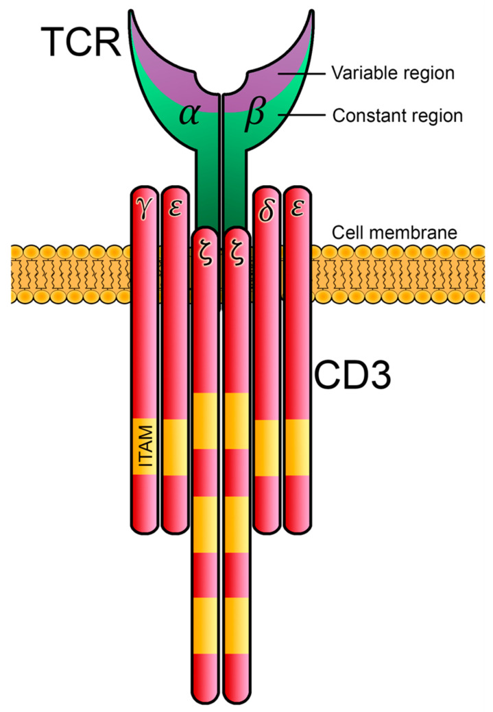
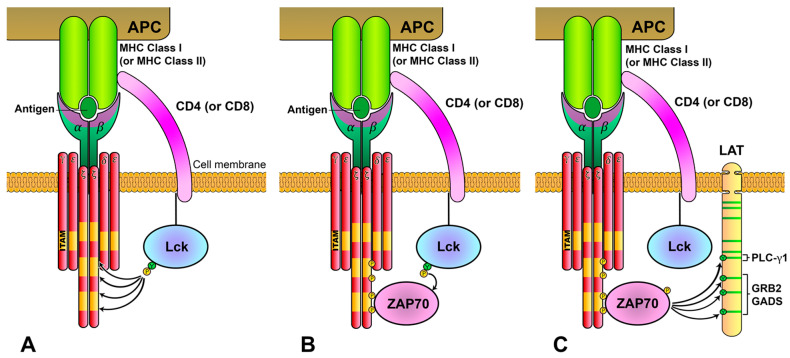
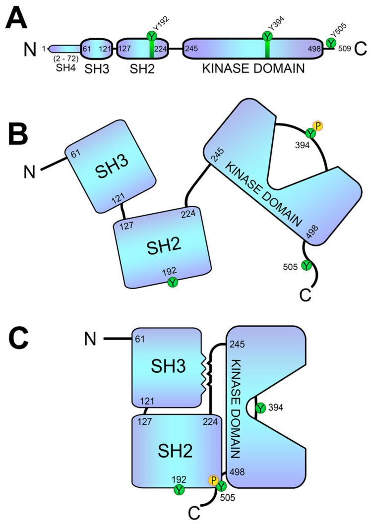
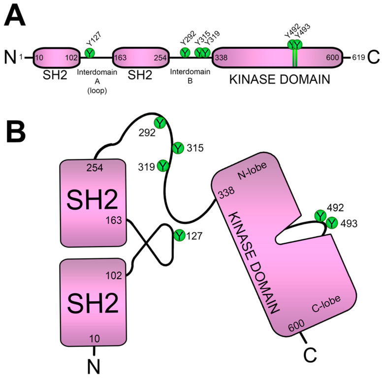
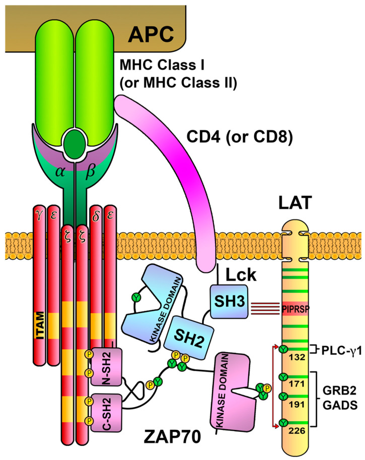
Specific antigen recognition is one of the immune system's features that allows it to mount intense yet controlled responses to an infinity of potential threats. T cells play a relevant role in the host defense and the clearance of pathogens by means of the specific recognition of peptide antigens presented by antigen-presenting cells (APCs), and, to do so, they are equipped with a clonally distributed antigen receptor called the T-cell receptor (TCR). Upon the specific engagement of the TCR, multiple intracellular signals are triggered, which lead to the activation, proliferation and differentiation of T lymphocytes into effector cells. In addition, this signaling cascade also operates during T-cell development, allowing for the generation of cells that can be helpful in the defense against threats, as well as preventing the generation of autoreactive cells. Early TCR signals include phosphorylation events in which the tyrosine kinases Lck and ZAP70 are involved. The sequential activation of these kinases leads to the phosphorylation of the transmembrane adaptor LAT, which constitutes a signaling hub for the generation of a signalosome, finally resulting in T-cell activation. These early signals play a relevant role in triggering the development, activation, proliferation and apoptosis of T cells, and the negative regulation of these signals is key to avoid aberrant processes that could generate inappropriate cellular responses and disease. In this review, we will examine and discuss the roles of the tyrosine kinases Lck and ZAP70 and the membrane adaptor LAT in these cellular processes.

Keywords: CD3; ITAMs; LAT; Lck; TCR; ZAP70; signaling.


Conflict of interest statement

The authors declare no conflict of interest. The funders had no role in the design of the study; in the collection, analyses or interpretation of the data; in the writing of the manuscript; or in the decision to publish the results.

Figures

Figure 1
Figure 1
Structure of the TCRαβ/CD3 complex. The T-cell receptor is constituted by heterodimers of α and β chains. The TCRα and TCRβ polypeptides have variable and constant immunoglobulin domains, and both chains are bound by covalent disulfide bonds. TCRαβ heterodimers and CD3 polypeptides are associated via non-covalent bonds. CD3 is constituted by CD3ε, CD3γ, CD3δ and ζ-chain polypeptides, organized as dimers (CD3εγ, CD3εδ and CD3ζζ). ITAMs are located in their intracellular tails, allowing for the transmission of information from the TCR inside the cell.
Figure 2
Figure 2
TCR/CD3-complex-mediated intracellular signaling. (A) TCR/CD3 complex recognizes antigens bound to MHC-I or MHC-II presented by an antigen-presenting cell (APC), which allows the activity of Lck kinase over the ITAMs located in CD3 ζ-chains. (B) Phosphorylation of ITAMs generates docking sites for ZAP70 kinase in the proximities of active Lck, which, in turn, phosphorylates tyrosines of ZAP70, activating its kinase domain. (C) Catalytically active ZAP70 preferentially phosphorylates tyrosines in the cytoplasmic tail of the LAT adaptor, generating binding sites for SH2 domains, leading to the formation of a signalosome.
Figure 3
Figure 3
Structure and conformations of the Lck kinase. (A) Linear sequence of Lck, showing its SH4, SH3, SH2 and catalytic domains. (B) Representation of the unfolded (open) Lck conformation. Tyrosine 505 (Y505) regulates conformational changes required for Lck activation. (C) Representation of the folded (closed) conformation of Lck. The phosphorylation of Y505 generates an SH2-binding site, causing a folded conformation in which the Y394 is not exposed. This closed conformation is further stabilized via the interaction of the SH3 domain with the proline motif located between the SH2 and kinase domains.
Figure 4
Figure 4
Structure of the ZAP70 kinase. (A) Linear sequence of ZAP70, showing its two tandem SH2 domains linked by interdomain A, and interdomain B that connects the second SH2 domain with the C-terminal kinase domain. (B) Representation of the ZAP70 conformation. The two SH2 domains bind specifically phosphorylated tyrosine residues in the CD3 ITAMs. Tyrosines 292, 315 and 319, responsible for ZAP70 enzymatic activity regulation, are located in interdomain B.
Figure 5
Figure 5
The LAT adaptor acts as a signaling scaffold. (A) Schematic representation of human LAT. LAT is an integral membrane adaptor with a short N-terminal extracellular region, a transmembrane region and a long cytoplasmic tail. This tail contains nine conserved tyrosine residues that are phosphorylated to give rise to new binding sites for the SH2 domains of proteins such as PLC-γ1, Grb2 and Gads. (B) LAT signalosome formation. The LAT signalosome is a multiprotein complex formed by LAT, PLC-γ1, Grb2, Gads and SLP-76.
Figure 6
Figure 6
Regulation of Lck activity via the PD-1/PAG pathway. (A) PAG is located in the T-cell membrane and has multiple tyrosine residues in their cytoplasmic region. (B) PD-L1 binding to PD-1 induces the phosphorylation of PAG tyrosines, generating binding sites for Csk. The SH2 domain of Csk binds to PAG-phosphorylated tyrosine 317, causing a conformational change that increases its kinase activity. Csk localizes close to the membrane and phosphorylates tyrosine 505 of Lck, inducing Lck to its inactive conformation.
Figure 7
Figure 7
Lck negative-feedback loop. TCR engagement activates intracellular signaling leading to ITAMs phosphorylation and ZAP70 recruitment. The activation of ZAP70 leads not only to downstream activation signaling, but also to the phosphorylation of Y192 of Lck, which inhibits its binding to CD45 phosphatase, allowing the phosphorylation of Y505 and promoting the closed Lck conformation.
Figure 8
Figure 8
Interactions connecting Lck, ZAP70 and LAT. The SH2 domain of Lck binds to the phosphorylated tyrosines in the interdomain B of ZAP70, and the SH3 domain of Lck binds to the proline-rich region of LAT (PIPRSP). This binding bridge generated by Lck localizes the ZAP70 kinase close to the LAT adaptor, favoring its tyrosine phosphorylation.
Figure 9
Figure 9
Slow phosphorylation of the sixth tyrosine of LAT regulates TCR signaling. The evolutionarily conserved glycine residue preceding the sixth tyrosine of LAT (Y132 in human LAT) makes it a worse substrate for ZAP70, leading to a slower phosphorylation of this residue with regard to the other C-terminal tyrosines. This slow kinetics phosphorylation is a molecular mechanism that supports the kinetic-proofreading (KPR) model of TCR ligand discrimination.

Similar articles

Cited by

References

    1. Chakraborty A.K., Weiss A. Insights into the initiation of TCR signaling. Nat. Immunol. 2014;15:798–807. doi: 10.1038/ni.2940. - DOI - PMC - PubMed
    1. Malissen B., Bongrand P. Early T cell activation: Integrating biochemical, structural, and biophysical cues. Annu. Rev. Immunol. 2015;33:539–561. doi: 10.1146/annurev-immunol-032414-112158. - DOI - PubMed
    1. Chitadze G., Oberg H.H., Wesch D., Kabelitz D. The Ambiguous Role of gammadelta T Lymphocytes in Antitumor Immunity. Trends Immunol. 2017;38:668–678. doi: 10.1016/j.it.2017.06.004. - DOI - PubMed
    1. Alcover A., Alarcon B., Di Bartolo V. Cell Biology of T Cell Receptor Expression and Regulation. Annu. Rev. Immunol. 2018;36:103–125. doi: 10.1146/annurev-immunol-042617-053429. - DOI - PubMed
    1. Schamel W.W., Alarcon B. Organization of the resting TCR in nanoscale oligomers. Immunol. Rev. 2013;251:13–20. doi: 10.1111/imr.12019. - DOI - PubMed