

Pegylated Liposomal Alendronate Biodistribution, Immune Modulation, and Tumor Growth Inhibition in a Murine Melanoma Model
- PMID: 37759709
- PMCID: PMC10527549
- DOI: 10.3390/biom13091309
Pegylated Liposomal Alendronate Biodistribution, Immune Modulation, and Tumor Growth Inhibition in a Murine Melanoma Model
Abstract
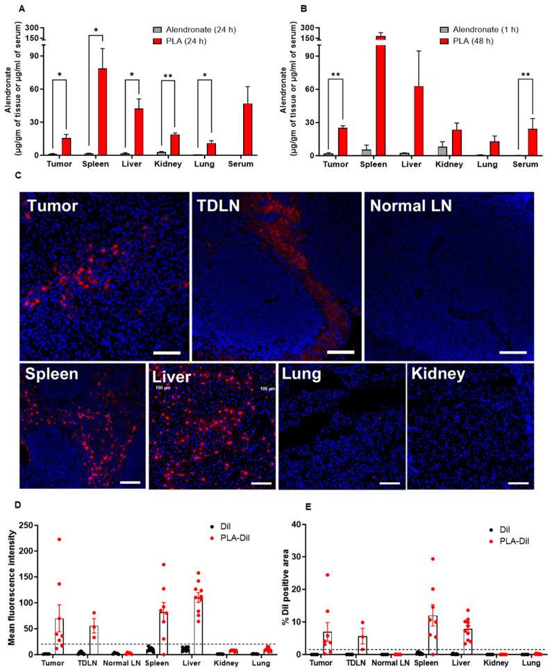
While tumor-associated macrophages (TAM) have pro-tumoral activity, the ablation of macrophages in cancer may be undesirable since they also have anti-tumoral functions, including T cell priming and activation against tumor antigens. Alendronate is a potent amino-bisphosphonate that modulates the function of macrophages in vitro, with potential as an immunotherapy if its low systemic bioavailability can be addressed. We repurposed alendronate in a non-leaky and long-circulating liposomal carrier similar to that of the clinically approved pegylated liposomal doxorubicin to facilitate rapid clinical translation. Here, we tested liposomal alendronate (PLA) as an immunotherapeutic agent for cancer in comparison with a standard of care immunotherapy, a PD-1 immune checkpoint inhibitor. We showed that the PLA induced bone marrow-derived murine non-activated macrophages and M2-macrophages to polarize towards an M1-functionality, as evidenced by gene expression, cytokine secretion, and lipidomic profiles. Free alendronate had negligible effects, indicating that liposome encapsulation is necessary for the modulation of macrophage activity. In vivo, the PLA showed significant accumulation in tumor and tumor-draining lymph nodes, sites of tumor immunosuppression that are targets of immunotherapy. The PLA remodeled the tumor microenvironment towards a less immunosuppressive milieu, as indicated by a decrease in TAM and helper T cells, and inhibited the growth of established tumors in the B16-OVA melanoma model. The improved bioavailability and the beneficial effects of PLA on macrophages suggest its potential application as immunotherapy that could synergize with T-cell-targeted therapies and chemotherapies to induce immunogenic cell death. PLA warrants further clinical development, and these clinical trials should incorporate tumor and blood biomarkers or immunophenotyping studies to verify the anti-immunosuppressive effect of PLA in humans.
Keywords: PD-1; cancer; immune checkpoint; immunotherapy; liposomal alendronate; melanoma; tumor.
Conflict of interest statement
The authors declare no potential conflict of interest.
Figures


Similar articles
-
Harnessing Nanomedicine to Potentiate the Chemo-Immunotherapeutic Effects of Doxorubicin and Alendronate Co-Encapsulated in Pegylated Liposomes.Pharmaceutics. 2023 Nov 9;15(11):2606. doi: 10.3390/pharmaceutics15112606. Pharmaceutics. 2023. PMID: 38004584 Free PMC article.
-
Comparative effects of free doxorubicin, liposome encapsulated doxorubicin and liposome co-encapsulated alendronate and doxorubicin (PLAD) on the tumor immunologic milieu in a mouse fibrosarcoma model.Nanotheranostics. 2022 Sep 1;6(4):451-464. doi: 10.7150/ntno.75045. eCollection 2022. Nanotheranostics. 2022. PMID: 36105861 Free PMC article.
-
Liposome-induced immunosuppression and tumor growth is mediated by macrophages and mitigated by liposome-encapsulated alendronate.J Control Release. 2018 Feb 10;271:139-148. doi: 10.1016/j.jconrel.2017.12.023. Epub 2017 Dec 23. J Control Release. 2018. PMID: 29277680 Free PMC article.
-
Repurposing amino-bisphosphonates by liposome formulation for a new role in cancer treatment.Semin Cancer Biol. 2021 Jan;68:175-185. doi: 10.1016/j.semcancer.2019.12.001. Epub 2019 Dec 23. Semin Cancer Biol. 2021. PMID: 31874280 Review.
-
Non-viral nano-immunotherapeutics targeting tumor microenvironmental immune cells.Biomaterials. 2019 Oct;219:119401. doi: 10.1016/j.biomaterials.2019.119401. Epub 2019 Jul 31. Biomaterials. 2019. PMID: 31398571 Review.
Cited by
-
Harnessing Nanomedicine to Potentiate the Chemo-Immunotherapeutic Effects of Doxorubicin and Alendronate Co-Encapsulated in Pegylated Liposomes.Pharmaceutics. 2023 Nov 9;15(11):2606. doi: 10.3390/pharmaceutics15112606. Pharmaceutics. 2023. PMID: 38004584 Free PMC article.
-
Immune Implications of Cholesterol-Containing Lipid Nanoparticles.ACS Nano. 2024 Oct 22;18(42):28480-28501. doi: 10.1021/acsnano.4c06369. Epub 2024 Oct 10. ACS Nano. 2024. PMID: 39388645 Review.
-
Nanodrug Delivery Systems for Myasthenia Gravis: Advances and Perspectives.Pharmaceutics. 2024 May 11;16(5):651. doi: 10.3390/pharmaceutics16050651. Pharmaceutics. 2024. PMID: 38794313 Free PMC article. Review.
-
Branched oncolytic peptides target HSPGs, inhibit metastasis, and trigger the release of molecular determinants of immunogenic cell death in pancreatic cancer.Front Mol Biosci. 2024 Oct 2;11:1429163. doi: 10.3389/fmolb.2024.1429163. eCollection 2024. Front Mol Biosci. 2024. PMID: 39417004 Free PMC article.
-
Advances in Materials Science for Precision Melanoma Therapy: Nanotechnology-Enhanced Drug Delivery Systems.Pharmaceutics. 2025 Feb 24;17(3):296. doi: 10.3390/pharmaceutics17030296. Pharmaceutics. 2025. PMID: 40142960 Free PMC article. Review.
References
-
- Luckman S.P., Hughes D.E., Coxon F.P., Russell R.G.G., Rogers M.J. Nitrogen-Containing Bisphosphonates Inhibit the Mevalonate Pathway and Prevent Post-Translational Prenylation of GTP-Binding Proteins, Including Ras. J. Bone Miner. Res. 1998;13:581–589. doi: 10.1359/jbmr.1998.13.4.581. - DOI - PubMed
Grants and funding
LinkOut - more resources
Full Text Sources
