Review
. 2023 Aug 31;13(9):1332.
doi: 10.3390/biom13091332.

Infection, Inflammation, and Immunity in Sepsis

Affiliations
Review

Infection, Inflammation, and Immunity in Sepsis

Undurti N Das. Biomolecules. .

Abstract

Sepsis is triggered by microbial infection, injury, or even major surgery. Both innate and adaptive immune systems are involved in its pathogenesis. Cytoplasmic presence of DNA or RNA of the invading organisms or damaged nuclear material (in the form of micronucleus in the cytoplasm) in the host cell need to be eliminated by various nucleases; failure to do so leads to the triggering of inflammation by the cellular cGAS-STING system, which induces the release of IL-6, TNF-α, and IFNs. These cytokines activate phospholipase A2 (PLA2), leading to the release of polyunsaturated fatty acids (PUFAs), gamma-linolenic acid (GLA), arachidonic acid (AA), eicosapentaenoic acid (EPA), and docosahexaenoic acid (DHA), which form precursors to various pro- and anti-inflammatory eicosanoids. On the other hand, corticosteroids inhibit PLA2 activity and, thus, suppress the release of GLA, AA, EPA, and DHA. PUFAs and their metabolites have a negative regulatory action on the cGAS-STING pathway and, thus, suppress the inflammatory process and initiate inflammation resolution. Pro-inflammatory cytokines and corticosteroids (corticosteroids > IL-6, TNF-α) suppress desaturases, which results in decreased formation of GLA, AA, and other PUFAs from the dietary essential fatty acids (EFAs). A deficiency of GLA, AA, EPA, and DHA results in decreased production of anti-inflammatory eicosanoids and failure to suppress the cGAS-STING system. This results in the continuation of the inflammatory process. Thus, altered concentrations of PUFAs and their metabolites, and failure to suppress the cGAS-STING system at an appropriate time, leads to the onset of sepsis. Similar abnormalities are also seen in radiation-induced inflammation. These results imply that timely administration of GLA, AA, EPA, and DHA, in combination with corticosteroids and anti-IL-6 and anti-TNF-α antibodies, may be of benefit in mitigating radiation-induced damage and sepsis.

Keywords: cGAS-STING system; cytokines; eicosanoids; inflammation; lipoxins; radiation; resolvins; sepsis; wound healing.


Conflict of interest statement

The author declares no conflict of interest.

Figures

Figure 1
Figure 1
Scheme showing metabolism of essential fatty acids (EFAs) and the effect of corticosteroids on the same. Note the differences in the actions of corticosteroids (dexamethasone) and radiation. Corticosteroids inhibit desaturases, and PLA2, COX-2, and LOX enzymes, whereas radiation blocks only desaturases, but activates PLA2, COX-2, and LOX enzymes. Radiation enhances the formation of PGs, LTs, and TXs, but blocks the formation of LXA4, resolvins, protectins, and maresins by inducing deficiency of AA/EPA/DHA, whereas corticosteroids block the formation of PGS, LTs, TXs, and LXA4, resolvins, protectins, and maresins by inhibiting PLA2, COX-2, and LOX enzymes, and induces deficiency of AA/EPA/DHA by inhibiting desaturases. Thus, corticosteroids are potent inhibitors of inflammation but also block resolution of inflammation, whereas radiation is a potent inducer of inflammation and interferes with the inflammation resolution process.
Figure 2
Figure 2
Scheme of formation of MN cells. All agents that damage DNA can cause the formation of MN cells. Microbes, when they enter the cell, shed their DNA/RNA; such cytoplasmic nuclear material is recognized by the cGAS-STING system, leading to an inflammatory response, as evidenced by enhanced levels of IL-6, TNF-α, HMGB1, IFNs, and ROS, and the increased formation of lipid peroxides, both in the plasma and in the cells (see also Figure 3).
Figure 3
Figure 3
Scheme showing formation of MN cells and activation of the cGAS-STING pathway and NF-κB. Cytokines activate the PLA2 enzyme, resulting in the release of PUFAs from the cell membrane. PUFAs (especially AA) are converted to PGE2, LXA4, and other respective metabolites that modulate the production of IL-6, TNF, and IFN. PUFAs, PGE2, and LXA4 suppress NF-kB activation and, thus, suppress the inflammatory process. LXA4 and PGE2 suppress the cGAS-STING pathway. IL-6, TNF, and IFN-γ augment PGE2 formation, which has both pro- and anti-inflammatory actions. Once the inflammation process reaches its threshold, PGE2 triggers the production of LXA4, which initiates inflammation resolution. Increased formation of LXA4 is due to the activation of 5- and 12-LOX enzymes induced by PGE2, which redirects AA to generate LXA4. The cGAS-STING pathway is suppressed by LXA4/AA/EPA/DHA. Thus, the availability of adequate amounts of AA/EPA/DHA and the formation of LXA4, resolvins, protectins, and maresins suppress the inflammatory process.
Figure 4
Figure 4
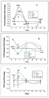
Effect of whole-body lethal radiation and GLA treatment on the expressions of delta-6-desaturase (A), COX-2 (C), and 5-LOX (B) enzymes. This data is taken from reference no. [80]. Effect of GLA and irradiation (7.5 Gy) on genes associated with PUFA metabolism in duodenum tissue on day1, day3, day 7 and day14. Values (n = 3) expressed as mean ± SEM. a,b,c p < 0.01 when compared with control, GLA and irradiation.
Figure 5
Figure 5
Effect of radiation and GLA on plasma levels of PGE2 (A), LTE4 (B), and LXA4 (C). Note the dynamic equilibrium among these eicosanoids from time to time. Estimation of PUFA metabolites PG E2 (A), LT E4 (B) and LXA4 (C) levels by ELISA method. Mouse were pretreated with 100 μg/kg GLA at 48, 24 and 1 h prior to irradiation and were subjected to total body irradiation of 7.5 Gy (at 1 Gy/min) and duodenum samples were obtained on day 1, day 3, day 7 and day 14 post-irradiation. All the values are expressed as mean ± SEM (n = 3). Statistical significance was calculated using t-test and p-value (<0.001) represented as a,b,c when compared to control, GLA and IR alone respectively. C- Control, GLA- Gamma-linolenic acid, IR- Irradiation (7.5 Gy).
Figure 6
Figure 6
Effect of radiation and GLA on plasma levels of HMGB1 (A), TNF (B), IL-6 (C), and IL-10 (D). Estimation of cytokines HMGB1 (A),TNF-α (B), IL-6 (C) and IL-10 (D) levels by ELISA method. Mouse were pretreated with 100 μg/kg GLA at 48, 24 and 1 h prior to irradiation and were subjected to total body irradiation of 7.5 Gy (at 1 Gy/min) and duodenum samples were obtained on day 1, day 3, day 7 and day 14 post-irradiation. All the values are expressed as mean ± SEM (n = 3). Statistical significance was calculated using t-test and p-value (<0.05) represented as a,b,c when compared to control, GLA and IR alone respectively. This data is from reference no. [80].

References

    1. Singer M., Deutschman C.S., Seymour C.W., Shankar-Hari M., Annane D., Bauer M., Bellomo R., Bernard G.R., Chiche J.-D., Coopersmith C.M., et al. The Third International Consensus Definitions for Sepsis and Septic Shock (Sepsis-3) JAMA. 2016;315:801–810. doi: 10.1001/jama.2016.0287. - DOI - PMC - PubMed
    1. Toti P., De Felice C., Occhini R., Schuerfeld K., Stumpo M., Epistolato M.C., Vatti R., Buonocore G. Spleen depletion in neonatal sepsis and chorioamnionitis. Am. J. Clin. Pathol. 2004;122:765–771. doi: 10.1309/RV6E9BMC9954A2WU. - DOI - PubMed
    1. Monneret G. How to identify systemic sepsis-induced immunoparalysis. Adv. Sepsis. 2005;4:42–49.
    1. Monneret G., Venet F., Pachot A., Lepape A. Monitoring Immune Dysfunctions in the Septic Patient: A New Skin for the Old Ceremony. Mol. Med. 2008;14:64–78. doi: 10.2119/2007-00102.Monneret. - DOI - PMC - PubMed
    1. Arbour N.C., Lorenz E., Schutte B.C., Zabner J., Kline J.N., Jones M., Frees K., Watt J.L., Schwartz D.A. TLR4 mutations are associated with endotoxin hyporesponsiveness in humans. Nat. Genet. 2000;25:187–191. doi: 10.1038/76048. - DOI - PubMed