Review
. 2023 Sep 2;13(9):1339.
doi: 10.3390/biom13091339.

Recent Advances in SARS-CoV-2 Main Protease Inhibitors: From Nirmatrelvir to Future Perspectives

Affiliations
Review

Recent Advances in SARS-CoV-2 Main Protease Inhibitors: From Nirmatrelvir to Future Perspectives

Andrea Citarella et al. Biomolecules. .

Abstract

The main protease (Mpro) plays a pivotal role in the replication of severe acute respiratory syndrome coronavirus 2 (SARS-CoV-2) and is considered a highly conserved viral target. Disruption of the catalytic activity of Mpro produces a detrimental effect on the course of the infection, making this target one of the most attractive for the treatment of COVID-19. The current success of the SARS-CoV-2 Mpro inhibitor Nirmatrelvir, the first oral drug for the treatment of severe forms of COVID-19, has further focused the attention of researchers on this important viral target, making the search for new Mpro inhibitors a thriving and exciting field for the development of antiviral drugs active against SARS-CoV-2 and related coronaviruses.

Keywords: COVID-19; Paxlovid; SARS-CoV-2 Mpro; coronavirus; nirmatrelvir; peptidomimetics; protease inhibitors.


Conflict of interest statement

The authors declare no conflict of interest.

Figures

Figure 1
Figure 1
(A) Crystal structure of SARS-CoV-2 Mpro (PDB: 7ALH) enlightening the two monomers (monomer A is depicted in orange, monomer B in cyan); (B) crystal structure of SARS-CoV-2 Mpro monomer (PDB: 1P9S) enlightening the three domains (domain I is depicted in blue, domain II in green, domain III in orange).
Figure 2
Figure 2
Active site of the SARS-CoV-2 Mpro with position of the catalytic dyad, oxyanion hole and subsites S1′–S5.
Figure 3
Figure 3
Schematic representation of the allosteric sites of Mpro. The allosteric sites are located near the dimerization site (allosteric site 1) or between domains II and III (allosteric site 2). The active site in each monomer is indicated with a yellow circle.
Figure 4
Figure 4
Chemical structure and biological activity of Pfizer inhibitors Nirmatrelvir, 2 and 3.
Figure 5
Figure 5
Chemical structure and biological activity of compounds derived from 2.
Figure 6
Figure 6
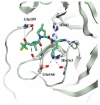
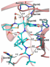
Crystal structure of the complex of SARS-CoV-2 Mpro/Nirmatrelvir (PDB: 7MLF).
Figure 7
Figure 7
Chemical structure and biological activity of compounds 8 and 9.
Figure 8
Figure 8
Chemical structure of compounds 10 and 11. The part marked with dotted circle highlights the modifications with respect to Nirmatrelvir.
Figure 9
Figure 9
Chemical structure and biological activity of Boceprevir, Narlaprevir, GC-376, 12 and 13. The colors highlight the chemical features that the authors work on for the design of new compounds.
Figure 10
Figure 10
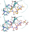
(A) X-ray crystal structures of SARS-CoV-2 Mpro in complex with 12 (PDB: 7TEH); (B) X-ray crystal structures of SARS-CoV-2 Mpro in complex with 13 (PDB:7TFR).
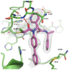
Figure 11
Figure 11
Chemical structure and biological activity of compounds 14 and 15. The part marked with dotted circle highlights the isoelectronic replacement of the nitrile group.
Figure 12
Figure 12
Chemical structure and biological activity of the first aldehyde derivatives (1618) as inhibitors of SARS-CoV-2 Mpro.
Figure 13
Figure 13
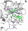
(A) Chemical structure and biological activity of compounds 19 and 20; (B) X-ray structure of SARS-CoV-2 Mpro in complex with compound 19 (PDB: 6WTJ).
Figure 14
Figure 14
(A) Chemical structure and biological activity of compounds 21 and 22. Those parts marked with colors indicate modifications with respect to the lead structure 20; (B) X-ray structure of SARS-CoV-2 Mpro in complex with compound 21 (PDB: 7LCO).
Figure 15
Figure 15
(A) Chemical structure and biological activity of compounds 23 and 24. Those parts marked with colors indicate modifications with respect to the lead structure 20; (B) X-ray structure of SARS-CoV-2 Mpro in complex with compound 24 (PDB: 7TIZ).
Figure 16
Figure 16
Chemical structure of Telaprevir and chemical structure and biological activity of compounds 2527.
Figure 17
Figure 17
X-ray structure of SARS-CoV-2 Mpro in complex with compound 26 (PDB: 7D3I).
Figure 18
Figure 18
Chemical structure and biological activity of compound 28.
Figure 19
Figure 19
Chemical structure and biological activity of compounds 29 and 30.
Figure 20
Figure 20
(A) X-ray structure of SARS-CoV-2Mpro in complex with compound 29 (PDB: 7LIH); (B) X-ray structure of SARS-CoV-2Mpro in complex with compound 30 (PDB: 7LIY).
Figure 21
Figure 21
Chemical structure and biological activity of compounds 3133.
Figure 22
Figure 22
(A) Chemical structure and biological activity of compound 34; (B) X-ray structure of SARS-CoV-2 Mpro in complex with 34 (PDB: 7AKU).
Figure 23
Figure 23
Chemical structure and biological activity of compounds 3539.
Figure 24
Figure 24
(A) Chemical structure and biological activity of compounds 40 and 41; (B) X-ray structures of SARS-CoV Mpro in complex with compound 40 (PDB: 7RVQ).
Figure 25
Figure 25
Chemical structure and biological activity of compounds 42, 43 and 44.
Figure 26
Figure 26
X-ray structure of SARS-CoV-2 Mpro in complex with compound 43 (PDB: 7MBI).
Figure 27
Figure 27
Chemical structure and biological activity of compounds 45, 46 and 47.
Figure 28
Figure 28
X-ray structure of SARS-CoV-2 Mpro in complex with 46 (PBD: 8DOX).
Figure 29
Figure 29
Chemical structure and biological activity of compound 48.
Figure 30
Figure 30
Chemical structure and biological activity of compound 49.
Figure 31
Figure 31
X-ray/neutron (XN) crystal structure of SARS-CoV-2 Mpro in complex with 49 (PDB: 7TD1).
Figure 32
Figure 32
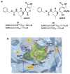
(A) Chemical structure and biological activity of compound 50; (B) predicted binding mode of 50 with SARS-CoV-2Mpro.
Figure 33
Figure 33
Chemical structure and biological activity of compounds 51 and 52.
Figure 34
Figure 34
Chemical structure and biological activity of compound 53.
Figure 35
Figure 35
(A) Chemical structure and biological activity of compounds 54 and 55; (B) X-ray structure of SARS-CoV-2 Mpro in complex with compound 55 (PDB: 7MLF).
Figure 36
Figure 36
Chemical structure and biological activity of compounds 5660.
Figure 37
Figure 37
X-ray structure of SARS-CoV-2 Mpro in complex with compound 57 (PDB: 7RN1).
Figure 38
Figure 38
(A) Chemical structure and biological activity of compound 61; (B) docking of SARS-CoV-2 Mpro in complex with compound 61.
Figure 39
Figure 39
(A) Chemical structure and biological activity of compound 62; (B) X-ray structure of SARS-CoV-2 Mpro in complex with compound 62 (PBD: 7VVT).
Figure 40
Figure 40
Chemical structure and biological activity of compounds 6366.
Figure 41
Figure 41
X-ray structure of SARS-CoV-2 Mpro in complex with compound 66 (PDB: 8B56).
Figure 42
Figure 42
Chemical structure and biological activity of compounds 6769.
Figure 43
Figure 43
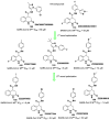
Structure and biological activity of 70 and resolution of the two diastereomers.
Figure 44
Figure 44
(A) Chemical structure and biological activity of compound 71; (B) X-ray structure of SARS-CoV-2 Mpro in complex with 71 (PDB: 7FAZ).
Figure 45
Figure 45
(A) Chemical structure and biological activity of compound 72; (B) X-ray structure of SARS-CoV-2 Mpro with 72 (PDB: 8IGN).
Figure 46
Figure 46
(A) Chemical structure and biological activity of compound 73; (B) X-ray crystal structure of SARS-CoV-2 Mpro in complex with 73 (PDB: 8HHT).
Figure 47
Figure 47
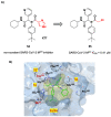
(A) Chemical structure and biological activity of compound 74; (B) X-ray crystal structure of SARS-CoV-2 Mpro in complex with 74 (PDB: 8HHU).
Figure 48
Figure 48
(A) Chemical structure and biological activity of compounds 75 and 76; (B) X-ray structure of SARS-CoV-2 Mpro in complex with 75 (PDB: 6LU7).
Figure 49
Figure 49
(A) Chemical structure and biological activity of compound 77; (B) X-ray structure of SARS-CoV-2 Mpro in complex with 77 (PDB: 7JT7).
Figure 50
Figure 50
Chemical structure and biological activity of compounds 78 and 79.
Figure 51
Figure 51
Chemical structure and biological activity of compounds 8082.
Figure 52
Figure 52
(A) Chemical structure and biological activity of compounds 83 and 84; (B) X-ray structure of SARS-CoV-2Mpro in complex with 83 (PDB: 7MB0).
Figure 53
Figure 53
Chemical structure and biological activity of compounds 8588.
Figure 54
Figure 54
(A) Chemical structure and biological activity of compounds 89 and 90; (B) X-ray structure of SARS-CoV-2 Mpro in complex with compound 89 (PDB: 7MLG).
Figure 55
Figure 55
Chemical structure and biological activity of compounds 91 and 92.
Figure 56
Figure 56
(A) Chemical structure and biological activity of compound 93; (B) X-ray crystal structure of SARS-CoV-2 Mpro in complex with 93 (PDB: 7B3E).
Figure 57
Figure 57
Chemical structure and biological activity of compounds 9496.
Figure 58
Figure 58
(A) Chemical structure and biological activity of compounds 9799; (B) X-ray crystal structure of SARS-CoV-2 Mpro in complex with 97 (PDB: 7RBZ).
Figure 59
Figure 59
Chemical structure and biological activity of compounds 100 and 101.
Figure 60
Figure 60
(A) Chemical structure and biological activity of compound 102; (B) X-ray crystal structure of SARS-CoV-2 Mpro in complex with 102 (PDB: 7JT0).
Figure 61
Figure 61
(A,C) Chemical structure and biological activity of compounds 103109; (B) X-ray crystal structure of SARS-CoV-2 Mpro in complex with 103 (PDB: 7X6K).
Figure 62
Figure 62
Chemical structure and biological activity of compounds 110114.
Figure 63
Figure 63
Chemical structure and biological activity of compounds 115119.
Figure 64
Figure 64
Chemical structure and biological activity of compounds 120124.
Figure 65
Figure 65
Chemical structure and biological activity of Ebsulfur, 126 and 127.
Figure 66
Figure 66
Chemical structure and biological activity of compounds 128134.
Figure 67
Figure 67
Chemical structure and biological activity of compounds 135137.
Figure 68
Figure 68
(A) Chemical structure and biological activity of the compound 54; (B) X-ray crystal structure of SARS-CoV-2 Mpro in complex with 54 (PDB: 6W63), highlighting binding site interactions.
Figure 69
Figure 69
(A) Chemical structure and biological activity of the compound 56; (B) X-ray crystal structure of SARS-CoV-2 Mpro in complex with 56 (PDB: 7KX5).
Figure 70
Figure 70
(A) Chemical structure and biological activity of compound 63; (B) X-ray structure of SARS-CoV-2 Mpro in complex with 63 (PDB: 7LTJ).
Figure 71
Figure 71
(A) Chemical structure and biological activity of compound 64; (B) X-ray structure of SARS-CoV-2 Mpro in complex with 64 (PDB: 8ACL).
Figure 72
Figure 72
(A) Chemical structure and biological activity of compounds 138140; (B) X-ray crystal structure of SARS-CoV-2 Mpro in complex with 140 (PDB: 7VU6).
Figure 73
Figure 73
(A) Chemical structure and biological activity of compounds 141 and 142; (B) docking of SARS-CoV-2 Mpro in complex with 141; (C) docking of SARS-CoV-2 Mpro in complex with 142.
Figure 74
Figure 74
Chemical structure and biological activity of compounds 143 and 144.
Figure 75
Figure 75
(A) Chemical structure and biological activity of compounds 145 and 146. (B) Docking of SARS-CoV-2 Mpro in complex with 145; (C) docking of SARS-CoV-2 Mpro in complex with 146.
Figure 76
Figure 76
Chemical structure and biological activity of compounds 147149.
Figure 77
Figure 77
Chemical structure and biological activity of compounds 150158.
Figure 78
Figure 78
X-ray crystal structure of SARS-CoV-2 Mpro in complex with 155 (PDB:7P2G).
Figure 79
Figure 79
(A) Chemical structure and biological activity of compounds 159161. (B) X-ray crystal structure of 161 in complex with SARS-CoV-2 Mpro (PDB: 8I4S).
Figure 80
Figure 80
(A) Chemical structure and biological activity of compound 159; (B) SARS-CoV-2 Mpro/162 complex suggested by molecular docking.
Figure 81
Figure 81
(A) Chemical structure and biological activity of compound 163. (B) Docking of SARS-CoV-2 Mpro in complex with 163.
Figure 82
Figure 82
(A) Chemical structure and biological activity of compounds 164; (B) X-ray crystal structure of SARS-CoV-2 Mpro in complex with 164 (PDB: 7CA8).
Figure 83
Figure 83
Chemical structure and biological activity of compound 165 and 166.
Figure 84
Figure 84
(A) Chemical structure and biological activity of compound 167; (B) X-ray crystal structure of SARS-CoV-2 Mpro in complex with 167 (PDB: 7AXM).
Figure 85
Figure 85
Chemical structure and biological activity of compounds 168 and 169.
Figure 86
Figure 86
(A) Chemical structure and biological activity of compound 170; (B) X-ray crystal structure of SARS-CoV-2 Mpro in complex with 170 (PDB: 7AGA).
Figure 87
Figure 87
Chemical structure and biological activity of compounds 171173.
Figure 88
Figure 88
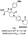
Chemical structure and biological activity of compound 174.

Similar articles

Cited by

References

    1. von Delft A., Hall M.D., Kwong A.D., Purcell L.A., Saikatendu K.S., Schmitz U., Tallarico J.A., Lee A.A. Accelerating Antiviral Drug Discovery: Lessons from COVID-19. Nat. Rev. Drug Discov. 2023;22:585–603. doi: 10.1038/s41573-023-00692-8. - DOI - PMC - PubMed
    1. Citarella A., Scala A., Piperno A., Micale N. SARS-CoV-2 Mpro: A Potential Target for Peptidomimetics and Small-Molecule Inhibitors. Biomolecules. 2021;11:607. doi: 10.3390/biom11040607. - DOI - PMC - PubMed
    1. Mahase E. COVID-19: Pfizer’s Paxlovid Is 89% Effective in Patients at Risk of Serious Illness, Company Reports. BMJ. 2021;375:n2713. doi: 10.1136/bmj.n2713. - DOI - PubMed
    1. Chia C.S.B., Xu W., Shuyi Ng P. A Patent Review on SARS Coronavirus Main Protease (3CL pro) Inhibitors. ChemMedChem. 2022;17:e202100576. doi: 10.1002/cmdc.202100576. - DOI - PMC - PubMed
    1. Capasso C., Nocentini A., Supuran C.T. Protease Inhibitors Targeting the Main Protease and Papain-like Protease of Coronaviruses. Expert Opin. Ther. Pat. 2021;31:309–324. doi: 10.1080/13543776.2021.1857726. - DOI - PubMed

MeSH terms