. 2023 Sep 2;13(9):1341.
doi: 10.3390/biom13091341.

Role of Lipids and Divalent Cations in Membrane Fusion Mediated by the Heptad Repeat Domain 1 of Mitofusin

Affiliations

Role of Lipids and Divalent Cations in Membrane Fusion Mediated by the Heptad Repeat Domain 1 of Mitofusin

Anaïs Vlieghe et al. Biomolecules. .

Abstract

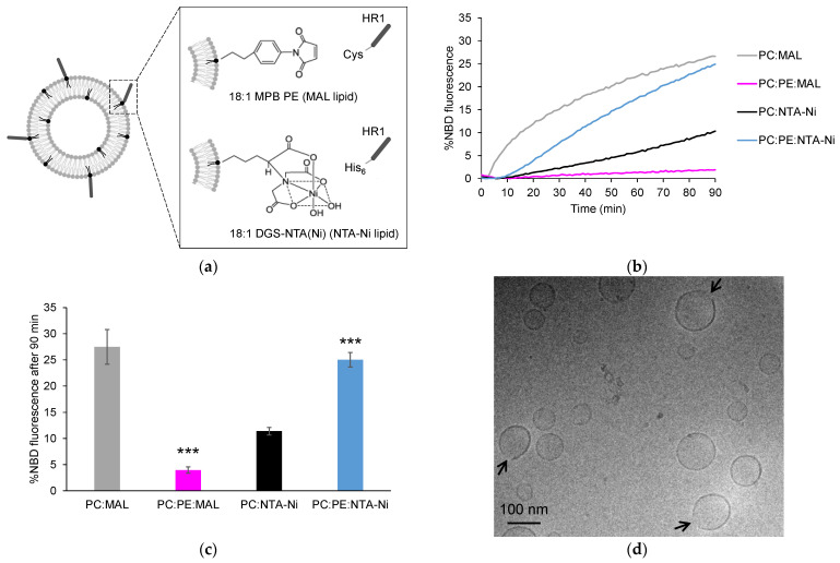
Mitochondria are highly dynamic organelles that constantly undergo fusion and fission events to maintain their shape, distribution and cellular function. Mitofusin 1 and 2 proteins are two dynamin-like GTPases involved in the fusion of outer mitochondrial membranes (OMM). Mitofusins are anchored to the OMM through their transmembrane domain and possess two heptad repeat domains (HR1 and HR2) in addition to their N-terminal GTPase domain. The HR1 domain was found to induce fusion via its amphipathic helix, which interacts with the lipid bilayer structure. The lipid composition of mitochondrial membranes can also impact fusion. However, the precise mode of action of lipids in mitochondrial fusion is not fully understood. In this study, we examined the role of the mitochondrial lipids phosphatidylethanolamine (PE), cardiolipin (CL) and phosphatidic acid (PA) in membrane fusion induced by the HR1 domain, both in the presence and absence of divalent cations (Ca2+ or Mg2+). Our results showed that PE, as well as PA in the presence of Ca2+, effectively stimulated HR1-mediated fusion, while CL had a slight inhibitory effect. By considering the biophysical properties of these lipids in the absence or presence of divalent cations, we inferred that the interplay between divalent cations and specific cone-shaped lipids creates regions with packing defects in the membrane, which provides a favorable environment for the amphipathic helix of HR1 to bind to the membrane and initiate fusion.

Keywords: Mitofusin; amphipathic helix; divalent cations; fusion; lipid packing defects; membrane; mitochondria.


Conflict of interest statement

The authors declare no conflict of interest.

Figures

Figure 1
Figure 1
Influence of lipid anchors and PE on HR1-mediated fusion. (a) The peptides used in this study were HR1 fragments of Mfn1 with a Cys or a His6 tag at their C-terminus, allowing for their chemical coupling to the liposomes functionalized with either 18:1 MPB PE (MAL lipids) or 18:1 DGS-NTA(Ni) (NTA-Ni lipids), respectively. (b) Representative kinetics of a FRET-based lipid mixing assay between the liposomes containing 5 mol% of either MAL or NTA-Ni lipids in their membrane, along with 95 mol% PC or 65 mol% PC and 30 mol% PE. The fusion reaction was initiated by adding HR1-Cys or HR1-His6 peptides at t = 0, with 25 µM HR1 and 500 µM lipids in the reaction mix. The control experiments in which the buffer alone was added in place of HR1 are presented in Figure S1. (c) Average extent of lipid mixing observed after a 90-min period, based on the data from the n = 7 to 21 independent kinetics experiments, similar to the one presented in panel (b). The error bars represent the standard errors of the mean. Statistical comparisons were performed using two-sample t-tests against the condition without PE (*** p < 0.001). (d) Cryo-EM picture of the liposomes composed of 95 mol% PC and 5 mol% MAL after a 1-h incubation at 37 °C with HR1-Cys peptides (12.5 µM peptides and 500 µM lipids).
Figure 2
Figure 2
Surface density of HR1 on the liposome membrane. (a) Liposomes with the same lipid compositions as shown in Figure 1 were incubated with HR1-Cys or HR1-His6 peptides (500 µM lipids and 25 µM peptides) at 37 °C for 1 h (same color code as in Figure 1). The reaction mixes were separated using a discontinuous nycodenz gradient to distinguish the HR1-bound liposomes from unbound HR1. The protein and lipid recoveries in the floated samples were estimated using SDS-PAGE stained with Coomassie upon comparison with the non-floated samples. (b) The HR1-to-lipid ratios in the liposome membrane were estimated from the n = 3 to 5 independent experiments. The error bars represent the standard errors of the mean. Statistical comparisons were performed using two-sample t-tests against the condition without PE (n.s. p > 0.05; *** p < 0.001).
Figure 3
Figure 3
Effect of CL and divalent cations on HR1-mediated fusion. (a) Representative kinetics of a FRET-based lipid mixing assay between the liposomes containing 5 mol% NTA-Ni lipids in their membrane, along with different lipid compositions: 95 mol% PC (black), 90 mol% PC and 5 mol% CL (light green), or 75 mol% PC and 20 mol% CL (dark green). The fusion reaction was initiated by adding HR1-His6 peptides at t = 0 in the absence or presence of the divalent cations Ca2+ or Mg2+ (500 µM lipids, 25 µM peptides and 1 mM cations). The control experiments with the buffer alone instead of HR1 are presented in Figure S1. (b) Average extent of lipid mixing observed after a 90-min period, based on the data from the n = 3 to 21 independent kinetics experiments, similar to the one presented in panel (a). The error bars represent the standard errors of the mean. Statistical comparisons were performed using two-sample t-tests against the condition without CL and with the same ionic composition (** p < 0.01; *** p < 0.001).
Figure 4
Figure 4
Effect of PA and divalent cations on HR1-mediated fusion. (a) Representative kinetics of a FRET-based lipid mixing assay between liposomes containing 5 mol% NTA-Ni lipids in their membrane, along with different lipid compositions: 95 mol% PC (black), 85 mol% PC and 10 mol% PA (yellow), or 65 mol% PC and 30 mol% PA (red). The fusion reaction was initiated by adding HR1-His6 peptides at t = 0 in the absence or presence of the divalent cations Ca2+ or Mg2+ (500 µM lipids, 25 µM peptides and 1 mM cations). The control experiments with the buffer alone instead of HR1 are presented in Figure S1. (b) Average extent of lipid mixing observed after a 90-min period, based on the data from the n = 7 to 21 independent kinetics experiments, similar to the one presented in panel (a). The error bars represent the standard errors of the mean. Statistical comparisons were performed using two-sample t-tests against the condition without PA and with the same ionic composition (n.s. p > 0.05; * p < 0.05; ** p < 0.01; *** p < 0.001).
Figure 5
Figure 5
Effect of PE and PA on HR1-mediated hemifusion. (a) Hemifusion events were quantified using the sodium dithionite assay. Fluorescent donor liposomes were pre-treated with sodium dithionite to eliminate the NBD fluorescence of their outer leaflet, allowing for only full fusion events to lead to fluorescence dequenching in the FRET-based lipid mixing assay. (b) The percentage of liposomes that underwent hemifusion after 90 min was determined by comparing the fluorescence dequenching signals obtained with or without prior sodium dithionite treatment (n = 3 to 11 independent experiments; error bars represent standard errors of the mean). Statistical comparisons were performed using two-sample t-tests against the condition without PE or PA and with the same ionic composition (n.s. p > 0.05; * p < 0.05; ** p < 0.01; *** p < 0.001).

Similar articles

Cited by

References

    1. Liesa M., Palacín M., Zorzano A. Mitochondrial dynamics in mammalian health and disease. Physiol. Rev. 2009;89:799–845. doi: 10.1152/physrev.00030.2008. - DOI - PubMed
    1. Chan D.C. Mitochondrial fusion and fission in mammals. Annu. Rev. Cell Dev. Biol. 2006;22:79–99. doi: 10.1146/annurev.cellbio.22.010305.104638. - DOI - PubMed
    1. Rojo M., Legros F., Chateau D., Lombès A. Membrane topology and mitochondrial targeting of mitofusins, ubiquitous mammalian homologs of the transmembrane GTPase Fzo. J. Cell Sci. 2002;115:1663–1674. doi: 10.1242/jcs.115.8.1663. - DOI - PubMed
    1. Eura Y., Ishihara N., Yokota S., Mihara K. Two mitofusin proteins, mammalian homologues of FZO, with distinct functions are both required for mitochondrial fusion. J. Biochem. 2003;134:333–344. doi: 10.1093/jb/mvg150. - DOI - PubMed
    1. Koshiba T., Detmer S.A., Kaiser J.T., Chen H., McCaffery J.M., Chan D.C. Structural basis of mitochondrial tethering by mitofusin complexes. Science. 2004;305:858–862. doi: 10.1126/science.1099793. - DOI - PubMed

Publication types

LinkOut - more resources